. 2012 Oct 16;109(42):16888-93.
doi: 10.1073/pnas.1208468109. Epub 2012 Oct 1.

Modification of RelA by O-linked N-acetylglucosamine links glucose metabolism to NF-κB acetylation and transcription

Affiliations

Modification of RelA by O-linked N-acetylglucosamine links glucose metabolism to NF-κB acetylation and transcription

David F Allison et al. Proc Natl Acad Sci U S A. .

Abstract

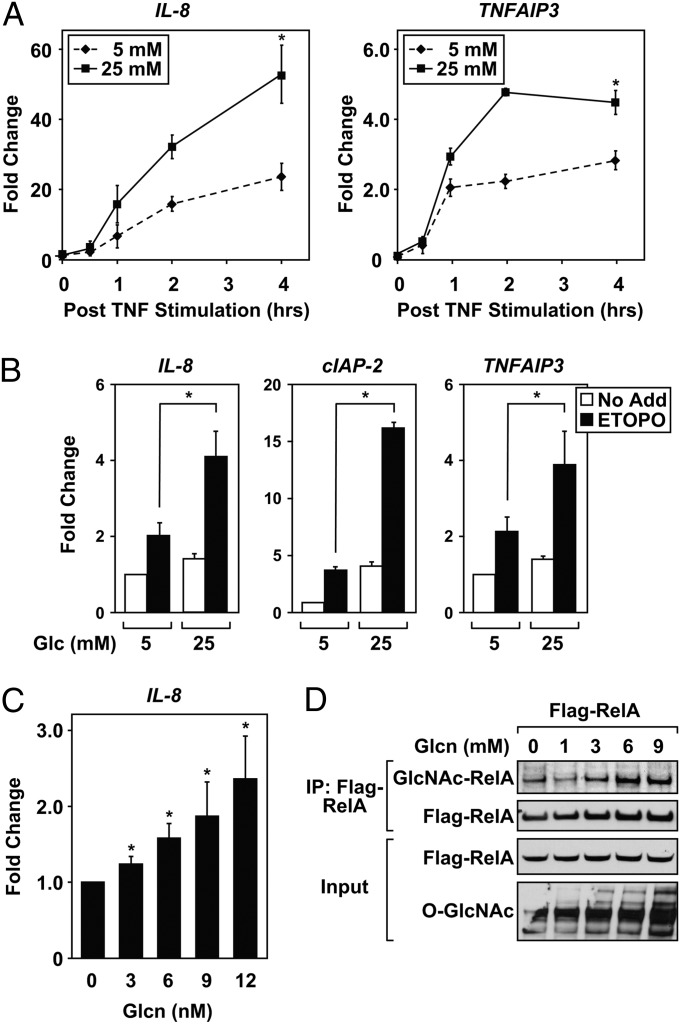
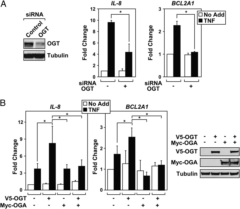
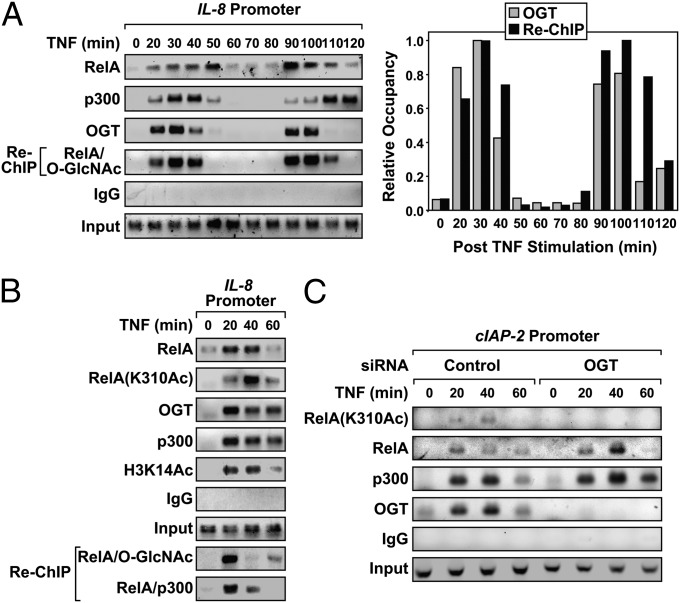
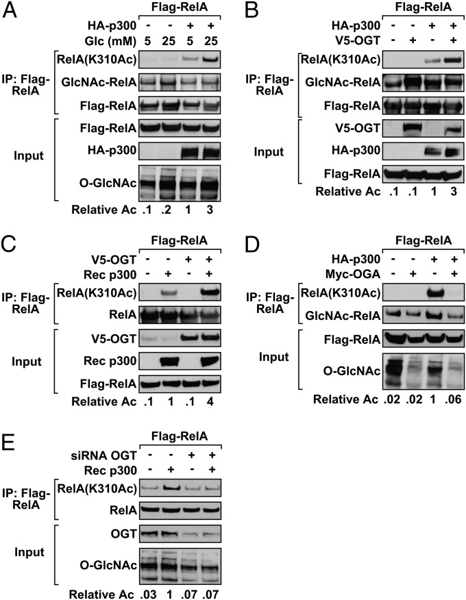
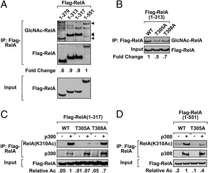
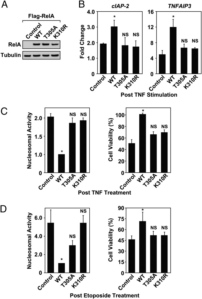
The molecular mechanisms linking glucose metabolism with active transcription remain undercharacterized in mammalian cells. Using nuclear factor-κB (NF-κB) as a glucose-responsive transcription factor, we show that cells use the hexosamine biosynthesis pathway and O-linked β-N-acetylglucosamine (O-GlcNAc) transferase (OGT) to potentiate gene expression in response to tumor necrosis factor (TNF) or etoposide. Chromatin immunoprecipitation assays demonstrate that, upon induction, OGT localizes to NF-κB-regulated promoters to enhance RelA acetylation. Knockdown of OGT abolishes p300-mediated acetylation of RelA on K310, a posttranslational mark required for full NF-κB transcription. Mapping studies reveal T305 as an important residue required for attachment of the O-GlcNAc moiety on RelA. Furthermore, p300 fails to acetylate a full-length RelA(T305A) mutant, linking O-GlcNAc and acetylation events on NF-κB. Reconstitution of RelA null cells with the RelA(T305A) mutant illustrates the importance of this residue for NF-κB-dependent gene expression and cell survival. Our work provides evidence for a unique regulation where attachment of the O-GlcNAc moiety to RelA potentiates p300 acetylation and NF-κB transcription.


Conflict of interest statement

The authors declare no conflict of interest.

Figures

Fig. 1.
Fig. 1.
Flux through the HBP enhances NF-κB transcription. (A) HEK 293T cells were treated with TNF (1 ng/mL) after overnight incubation in serum-free DMEM containing either 5 mM or high (25 mM) glucose concentrations. Expression of IL-8 (Left) and TNFAIP3 (Right) was quantified by using QRT-PCR, and samples were normalized to GAPDH levels. Fold change values represent comparison with unstimulated cells incubated in 5 mM glucose. (B) HEK 293T cells were cultured overnight in either 5 mM or 25 mM glucose DMEM, and the next day, cells were treated with etoposide (100 μM) for 4 h. QRT-PCR demonstrates elevated NF-κB responsive transcripts in high glucose, compared with cells cultured in 5 mM glucose. Fold change values represent comparison with untreated cells incubated in 5 mM glucose. (C) HEK 293T cells were cultured overnight in 5 mM glucose DMEM. Cells were then exposed to glucosamine (Glcn) for 2 h, stimulated with TNF (1 ng/mL) for 4 h, and QRT-PCR was performed. Fold change represents values obtained from TNF-stumulated cells incubated in the absence of Glcn. (D) HEK 293T cells transfected with plasmids encoding Flag-RelA were incubated in 5 mM glucose media overnight and subsequently exposed to Glcn for 4 h. Flag-RelA was immunoprecipitated, and O-GlcNAc-modified RelA was analyzed by immunoblotting. Total proteins were analyzed for the O-GlcNAc modification by immunoblot. QRT-PCR results in AC are a calculated mean ± SD, *P < 0.05, n = 3. Immunoblots are a representative example from at least three independent experiments.
Fig. 2.
Fig. 2.
NF-κB transcription requires OGT. (A) HEK 293T cells were transfected with siRNA targeting either OGT or nonspecific control. After knockdown, protein levels of OGT were analyzed by immunoblot (Left), using α-tubulin as a loading control. HEK 293T cells were subjected to OGT knockdown, serum starved overnight, and treated with TNF (1 ng/mL) for 4 h. Expression of IL-8 or BCL2A1 was quantified by QRT-PCR (Right), and samples were normalized to GAPDH levels. Expression is represented as fold change compared with the unstimulated control knockdown. (B) OGT and OGA were transfected into HEK 293T cells alone or in tandem. After transfection, cells were treated with TNF (1 ng/mL) for 4 h. Expression of IL-8 or BCL2A1 expression was quantified by QRT-PCR (Left). Represented fold change values are compared with the unstimulated vector control. Immunoblots confirm the expression of V5-tagged OGT and Myc-tagged OGA (Right), compared with the α-tubulin loading control. Data are a calculated mean ± SD, *P < 0.05, n = 3.
Fig. 3.
Fig. 3.
NF-κB–regulated promoters require OGT for RelA acetylation. (A) DU145 cells were treated with TNF (10 ng/mL) over a 2-h time course, and chromatin immunoprecipitation (ChIP) and Re-ChIP assays were performed. Control IgG antibody was used as a negative IP control, and inputs served as a loading control. The relative IL-8 promoter occupancy was determined after normalization to the maximum level of chromatin-bound OGT or RelA/O-GlcNAc Re-ChIP complexes arbitrarily adjusted to one (Right). (B) DU145 cells were treated with TNF and subjected to ChIP and Re-ChIP with the indicated antibodies. (C) DU145 cells transfected with siRNA targeting either OGT or control were treated with TNF and ChIP was performed. Data are a representative example from at least three independent experiments.
Fig. 4.
Fig. 4.
RelA acetylation is regulated by the O-GlcNAc modification. (A) The Flag-RelA expression plasmid was transfected into HEK 293T cells either alone or with HA-p300. After an overnight incubation in DMEM containing either 5 mM or 25 mM glucose, Flag-RelA was immunoprecipitated. O-GlcNAc modified RelA, and RelA(K310Ac) was subsequently analyzed by immunoblotting. (B) HEK 293T cells cotransfected with Flag-RelA, HA-p300, and V5-OGT were subjected to Flag immunoprecipitation. O-GlcNAc modified RelA and acetylated RelA were detected by immunoblotting. (C) HEK 293T cells were transfected with Flag-RelA and V5-OGT or with Flag-RelA alone. After Flag IP, RelA was acetylated with recombinant (Rec) p300 in vitro. RelA(K310) acetylation levels were analyzed by immunoblot. (D) HEK 293T cells were cotransfected with Flag-RelA and HA-p300 in the presence or absence of Myc-OGA. O-GlcNAc modified and RelA(K310Ac) were detected by IP. (E) After knockdown of OGT, HEK 293T cells were transfected with Flag-RelA. Flag IPs were subjected to in vitro acetylation with recombinant p300. Immunoblots were analyzed with RelA(AcK310Ac) antibody. Data are a representative example from three independent experiments, and densitometry results indicate the relative change in RelA(K310) acetylation where values were normalized to Flag-RelA inputs.
Fig. 5.
Fig. 5.
O-GlcNAc modification of RelA is detected between residues 277–313. (A) HEK 293T cells were transfected with the indicated Flag-RelA construct (1–270, 1–313, 1–317, or 1–551) and incubated overnight with PugNAc (100 μM). O-GlcNAc modified RelA was subsequently detected by Flag IP. Arrows indicate the relative location of the Flag-RelA proteins. (B) HEK 293T cells were transfected with a Flag-RelA 1–313 construct containing either a T305A or T308A mutation. O-GlcNAc modified RelA was detected by Flag IP. (C) Flag-RelA 1–317 mutants were expressed in HEK 293T cells. After Flag IP, RelA was acetylated in vitro with recombinant p300. RelA(K310Ac) was analyzed by immunoblot. (D) Wild-type and T305A Flag-RelA was expressed in HEK 293T cells. HEK 293T cells were transfected with full-length Flag-RelA (WT or T305A). After Flag IP, RelA was acetylated in vitro by using recombinant p300, and the samples were assayed for K310 acetylation by immunoblot. Data are a representative example from three independent experiments. Band densities of O-GlcNAc modified RelA (A and B) and RelA(K310Ac) (C and D) are shown relative to the levels observed for the wild-type Flag-RelA protein. Densitometry analysis is normalized to Flag-RelA inputs.
Fig. 6.
Fig. 6.
NF-κB–mediated cell survival depends on T305. (A) Expression levels of Flag-RelA were assessed in reconstituted RelA−/− MEFs by immunoblot. (B) After overnight serum starvation, reconstituted RelA−/− MEFs were treated with TNF (1 ng/mL) for 2 h. Expression of cIAP-2 and TNFAIP3 was analyzed by QRT-PCR and normalized to GAPDH levels. Fold change is displayed relative to the untreated RelA−/− MEF control cell line. (C and D) Reconstituted RelA−/− MEFs were serum starved overnight and treated with either TNF (10 ng/mL) for 8 h or etoposide (50 μM) for ten hours. After treatment, the cells were assayed for nucleosomal fragmentation by using the cell death ELISA or viability using trypan blue exclusion. Nucleosomal activity and cell viabilities are displayed relative to RelA−/− MEFs reconstituted with wild-type Flag-p65. QRT-PCR, apoptosis, and cell viability results are a calculated mean ± SD; NS, not significant compared with controls; *P < 0.05, n = 3.

References

    1. Dennis JW, Nabi IR, Demetriou M. Metabolism, cell surface organization, and disease. Cell. 2009;139:1229–1241. - PMC - PubMed
    1. Wellen KE, et al. The hexosamine biosynthetic pathway couples growth factor-induced glutamine uptake to glucose metabolism. Genes Dev. 2010;24:2784–2799. - PMC - PubMed
    1. Hanover JA, Krause MW, Love DC. The hexosamine signaling pathway: O-GlcNAc cycling in feast or famine. Biochim Biophys Acta. 2010;1800:80–95. - PMC - PubMed
    1. Hart GW, Slawson C, Ramirez-Correa G, Lagerlof O. Cross talk between O-GlcNAcylation and phosphorylation: Roles in signaling, transcription, and chronic disease. Annu Rev Biochem. 2011;80:825–858. - PMC - PubMed
    1. Shafi R, et al. The O-GlcNAc transferase gene resides on the X chromosome and is essential for embryonic stem cell viability and mouse ontogeny. Proc Natl Acad Sci USA. 2000;97:5735–5739. - PMC - PubMed

Publication types

MeSH terms