. 2012 Oct 16;109(42):16928-33.
doi: 10.1073/pnas.1211759109. Epub 2012 Oct 1.

In vitro reconstitution of the ordered assembly of the endosomal sorting complex required for transport at membrane-bound HIV-1 Gag clusters

Affiliations

In vitro reconstitution of the ordered assembly of the endosomal sorting complex required for transport at membrane-bound HIV-1 Gag clusters

Lars-Anders Carlson et al. Proc Natl Acad Sci U S A. .

Abstract

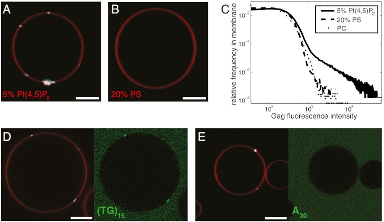
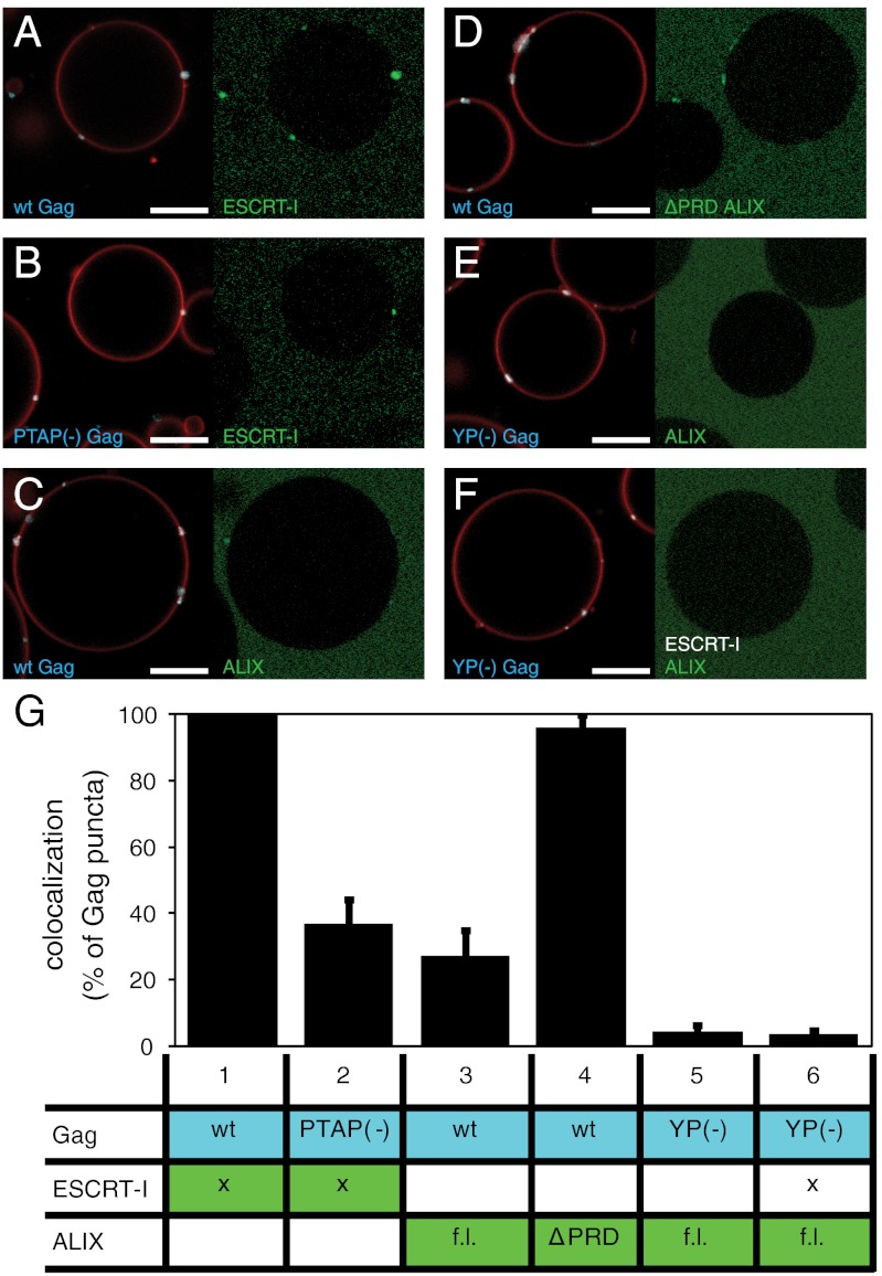
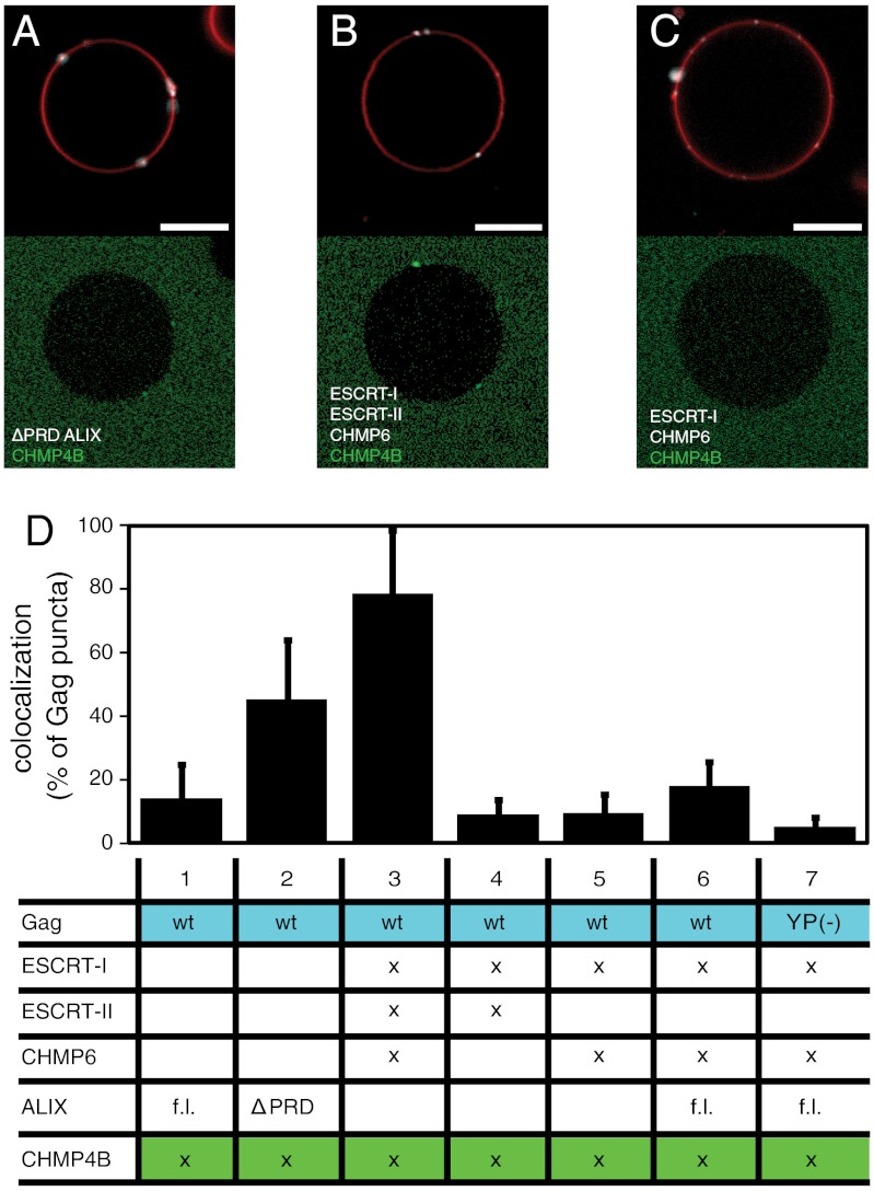
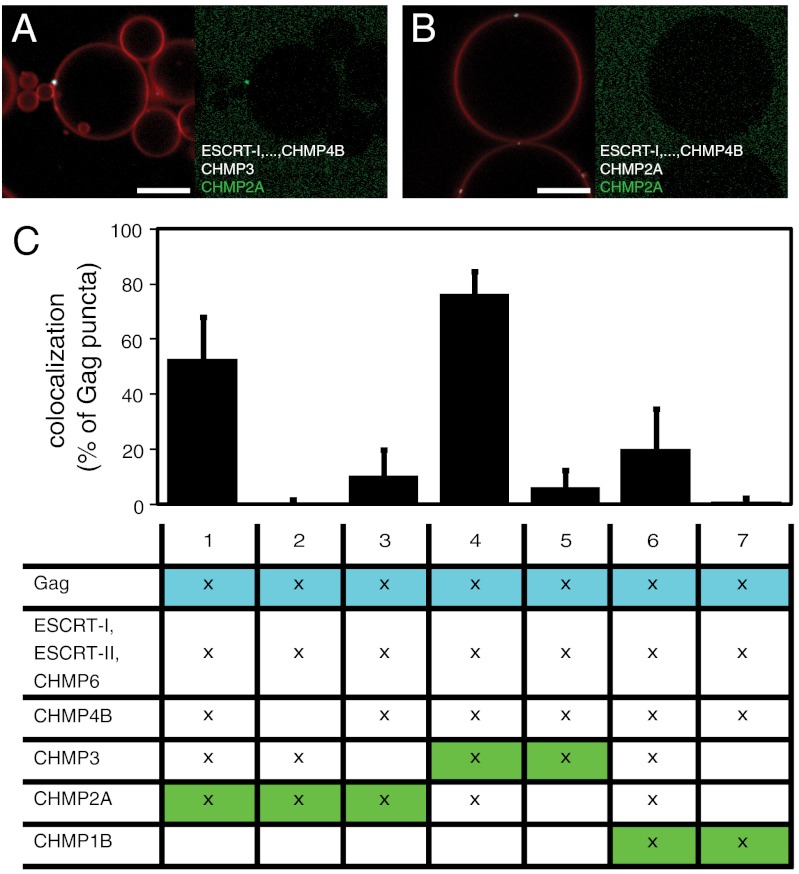
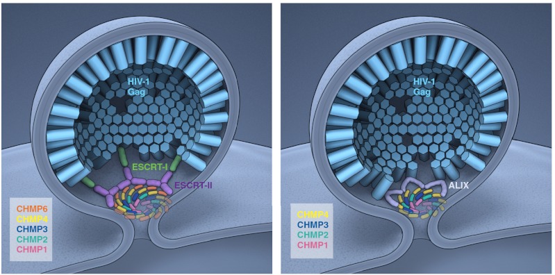
Most membrane-enveloped viruses depend on host proteins of the endosomal sorting complex required for transport (ESCRT) machinery for their release. HIV-1 is the prototypic ESCRT-dependent virus. The direct interactions between HIV-1 and the early ESCRT factors TSG101 and ALIX have been mapped in detail. However, the full pathway of ESCRT recruitment to HIV-1 budding sites, which culminates with the assembly of the late-acting CHMP4, CHMP3, CHMP2, and CHMP1 subunits, is less completely understood. Here, we report the biochemical reconstitution of ESCRT recruitment to viral assembly sites, using purified proteins and giant unilamellar vesicles. The myristylated full-length Gag protein of HIV-1 was purified to monodispersity. Myr-Gag forms clusters on giant unilamellar vesicle membranes containing the plasma membrane lipid PI(4,5)P(2). These Gag clusters package a fluorescent oligonucleotide, and recruit early ESCRT complexes ESCRT-I or ALIX with the appropriate dependence on the Gag PTAP and LYP(X)(n)L motifs. ALIX directly recruits the key ESCRT-III subunit CHMP4. ESCRT-I can only recruit CHMP4 when ESCRT-II and CHMP6 are present as intermediary factors. Downstream of CHMP4, CHMP3 and CHMP2 assemble synergistically, with the presence of both subunits required for efficient recruitment. The very late-acting factor CHMP1 is not recruited unless the pathway is completed through CHMP3 and CHMP2. These findings define the minimal sets of components needed to complete ESCRT assembly at HIV-1 budding sites, and provide a starting point for in vitro structural and biophysical dissection of the system.


Conflict of interest statement

The authors declare no conflict of interest.

Figures

Fig. 1.
Fig. 1.
HIV-1 Gag puncta assemble on PI(4,5)P2-containing GUVs. (A and B) Confocal microscopy images of GUVs containing 5% PI(4,5)P2 or 20% PS. When added to GUV exterior at 100 nM, Gag binds the outer leaflet of the PI(4,5)P2-containing GUV, forming clusters (A). Gag does not bind to GUVs with similar charge density in the form of 20% PS (B). (C) Brightness histograms of Gag fluorescence calculated in GUV membrane for GUVs containing POPC and 25% cholesterol (PC), along with 5% PI(4,5)P2 (5% PIP2), or 20% POPS (PS). Each curve is calculated from a total of approximately 50 GUVs in 10 z stacks recorded on random positions. (D) Fluorescent DNA oligo (TG)15 at 1 μM is packaged by Gag puncta. (E) Fluorescent DNA oligo A30 at 1 μM is not packaged by Gag puncta. Membrane is red, Gag is white, and fluorescent oligonucleotide is green. Scale bar, 10 μm.
Fig. 2.
Fig. 2.
Recruitment of early ESCRT proteins to Gag puncta. (AF) 30 nM ESCRT-I or 1 μM ALIX [full-length (f.l.) or ΔPRD], as indicated, were added to GUVs together with 100 nM Gag [wild-type (wt) or late-domain mutant as indicated]. Representative images are shown from experiments detailed in G, with Gag and membrane fluorescence in Left and ESCRT fluorescence in Right. ESCRT-I is strongly recruited to wt Gag puncta (column 1) and moderately to puncta formed by PTAP(-) Gag (column 2). Full-length ALIX is moderately recruited to Gag puncta (column 3), whereas the ΔPRD-ALIX is strongly recruited (column 4). Full-length ALIX is not colocalizing to YP(-) Gag puncta, in the presence or absence of ESCRT-I (columns 5 and 6). (G) Quantification of early ESCRT recruitment to Gag puncta. Each experiment is represented by one row in the table, showing what proteins were added (marked with x if wild type in all experiments). Flourescently labeled proteins have a colored background in the table. For each experiment, 10 z stacks were recorded at random positions. Gag puncta on GUV membranes were identified by an automated MATLAB script, and the percentage of Gag puncta having an ESCRT-I/ALIX fluorescence > 1.5 times the surrounding membrane was calculated and is shown in the bar graphs above each column. All six experiments were carried out on the same day with the same batch of GUVs for comparability, and the error bars represent the standard deviation of three independent repeats of the experiments. Membrane is red, Gag is white, and fluorphore-labeled ESCRT-I/ALIX is green. Scale bar, 10 μm.
Fig. 3.
Fig. 3.
Recruitment of CHMP4B by upstream ESCRT proteins. (A) Atto 488–labeled CHMP4B at 300 nM is recruited to Gag puncta by ΔPRD-ALIX at 4 μM. (B) Atto 488–labeled CHMP4B at 300 nM is recruited to Gag puncta by ESCRT-I (100 nM), ESCRT-II (200 nM), and CHMP6 (400 nM). (C) Same protein combination as in B except that ESCRT-II is omitted. (D) Quantification of CHMP4B recruitment to Gag puncta. Statistics of CHMP4B recrutiment was performed as for Fig. 2G, and the table shows added proteins analogously to Fig. 2G. Membrane is red, Gag is white, and fluorphore-labeled CHMP4B is green. Scale bar, 10 μm.
Fig. 4.
Fig. 4.
Interaction between ESCRT-III subunits. (A) Atto 488–labeled CHMP2A at 100 nM is recruited to a Gag punctum in the presence of ESCRT-I (100 nM), ESCRT-II (200 nM), CHMP6 (400 nM), CHMP4B (300 nM), and CHMP3 (100 nM). (B) Atto 488–labeled CHMP2A at 100 nM is not recruited to a Gag punctum in the presence of ESCRT-I (100 nM), ESCRT-II (200 nM), CHMP6 (400 nM), CHMP4B (300 nM), CHMP3 (100 nM), and unlabeled CHMP2A (100 nM). (C) Quantification of late CHMP recruitment to Gag puncta, analogous to Fig. 2G. The fluorescently labeled CHMP is shown on a green background. Labeled or unlabeled CHMP3, CHMP2A, and CHMP1B are present at 100 nM where marked with an x. In experiments 3, 5, and 7, omission of CHMP3, CHMP2A, and CHMP3 + CHMP2A were compensated by adding the same amount of unlabeled CHMP2A, CHMP3, or CHMP1B, respectively. Membrane is red, Gag is white, and fluorphore-labeled CHMPs are green. Scale bar, 10 μm.
Fig. 5.
Fig. 5.
Ordered assembly of ESCRTs at HIV-1 budding sites. Schematic depiction of ESCRT-I–dependent (Left) and ALIX-dependent (Right) assembly cascades. The Gag–ESCRT and ESCRT–ESCRT interactions in this model are based on the present study, whereas their three-dimensional arrangement is extrapolated from other available data. The ESCRT-III subunits are thought to drive membrane scission by forming a dome at the membrane neck (30). In the HIV-1 setting, it is not known if the putative dome forms from the inside (as depicted here) or the outside (3). The detailed arrangment of the subunits in the dome is not known. They are depicted here as forming whorls, which provides for all subunits of a given type to maintain equivalent interactions with other subunit types. Concentric circles, each consisting of a unique subunit composition, would also meet this criterion, but a single spiral would not. Because HIV-1 assembly sites in cells will, on average, contain approximately 2,400 Gag molecules at the time of release (52), even the small subset of Gag molecules which expose their p6 domains at the rim of the Gag lattice could provide enough opportunity for the ESCRT-I–mediated and the ALIX-mediated pathways to both contribute to ESCRT-III assembly at the same assembly site.

References

    1. Chen BJ, Lamb RA. Mechanisms for enveloped virus budding: Can some viruses do without an ESCRT? Virology. 2008;372:221–232. - PMC - PubMed
    1. Martin-Serrano J, Neil SJD. Host factors involved in retroviral budding and release. Nat Rev Microbiol. 2011;9:519–531. - PubMed
    1. Sundquist WI, Krausslich HG. HIV-1 assembly, budding, and maturation. Cold Spring Harb Perspect Med. 2012;2:a006924. - PMC - PubMed
    1. Gottlinger HG, Dorfman T, Sodroski JG, Haseltine WA. Effect of mutations affecting the p6 gag protein on human immunodeficiency virus particle release. Proc Natl Acad Sci USA. 1991;88:3195–3199. - PMC - PubMed
    1. Huang MJ, Orenstein JM, Martin MA, Freed EO. p6Gag is required for particle production from full-length human immunodeficiency virus type 1 molecular clones expressing protease. J Virol. 1995;69:6810–6818. - PMC - PubMed

Publication types

MeSH terms

Substances