. 2012 Oct 16;109(42):16951-6.
doi: 10.1073/pnas.1201408109. Epub 2012 Oct 1.

Analysis of human samples reveals impaired SHH-dependent cerebellar development in Joubert syndrome/Meckel syndrome

Affiliations

Analysis of human samples reveals impaired SHH-dependent cerebellar development in Joubert syndrome/Meckel syndrome

Andrea Aguilar et al. Proc Natl Acad Sci U S A. .

Abstract

Joubert syndrome (JS) and Meckel syndrome (MKS) are pleiotropic ciliopathies characterized by severe defects of the cerebellar vermis, ranging from hypoplasia to aplasia. Interestingly, ciliary conditional mutant mice have a hypoplastic cerebellum in which the proliferation of cerebellar granule cell progenitors (GCPs) in response to Sonic hedgehog (SHH) is severely reduced. This suggests that Shh signaling defects could contribute to the vermis hypoplasia observed in the human syndromes. As existing JS/MKS mutant mouse models suggest apparently contradictory hypotheses on JS/MKS etiology, we investigated Shh signaling directly on human fetal samples. First, in an examination of human cerebellar development, we linked the rates of GCP proliferation to the different levels and localizations of active Shh signaling and showed that the GCP possessed a primary cilium with CEP290 at its base. Second, we found that the proliferation of GCPs and their response to SHH were severely impaired in the cerebellum of subjects with JS/MKS and Jeune syndrome. Finally, we showed that the defect in GCP proliferation was similar in the cerebellar vermis and hemispheres in all patients with ciliopathy analyzed, suggesting that the specific cause of vermal hypo-/aplasia precedes this defect. Our results, obtained from the analysis of human samples, show that the hemispheres and the vermis are affected in JS/MKS and provide evidence of a defective cellular mechanism in these pathologic processes.


Conflict of interest statement

The authors declare no conflict of interest.

Figures

Fig. 1.
Fig. 1.
CEP290 is enriched at the base of murine and human GCPs; shRNA-mediated down-regulation of Cep290 reduces the number of ciliated cells. (A) Transmission EM images of a gw 21 sample of human EGL. Left: Three GCPs have a primary cilium (yellow arrow) that extends from the BB (black asterisk) near the centriole (blue asterisk). The primary cilium has a basal foot (black arrow) and a ciliary pocket, the base of which is coated by electron-dense material (yellow arrowhead). (B) Immunohistofluorescence imaging of CEP290 (green) and a BB rootlet component rootletin (red) on cryosections of mouse (Left) and human cerebellum (Right). In murine EGL, dots of CEP290 surround the rootlet; this pattern is observed from embryonic day 16 to postnatal day 14. In human EGL at gw 21, CEP290 is also localized in dots surrounding the BB rootlet. (C) Immunostaining of CEP290 (white), GFP (shRNA transfected cells, green), and the ciliary and BB marker polyglutamylated tubulin (GT335, red) in adherent astroglial stem cells, transfected with GFP-expressing plasmids driving the expression of scramble or CEP290 shRNA (green). Most control transfected cells express CEP290 and have a primary cilium. When CEP290 is no longer detectable in CEP290 shRNA-expressing cells, they do not possess a primary cilium. (D) Quantification of primary cilia in scramble or CEP290 shRNA-transfected cells. For CEP290 shRNA-transfected cells, only those in which CEP290 was no longer immunodetectable were quantified. CEP290 down-regulation leads to a 69% reduction in cells presenting a primary cilium. The result shown is the mean of three replicates. Error bars indicate SD. (Scale bars: A, left to right, top to bottom, 10 μm, 2.5 μm, and 0.5 μm; B, 5 μm; C, left to right, 2 μm and 1 μm.)
Fig. 2.
Fig. 2.
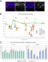
GCP proliferation is severely impaired in the cerebellar vermis and hemispheres of most JS/MKS cases. (A) Immunolabeling of cycling cells expressing the marker Ki67 (red) in the EGL in the gw 23 JS/MKS case 070151 and a gw 21 control. In all panels, a dashed line outlines the EGL. Note that there are fewer labeled cells in the EGL of the JS/MKS case (Right) compared with control (Left), although thickness of the EGL is comparable in both cases. (B) Quantification of Ki67-positive cells per 100 μm2 in the EGL of 12 cases of JS/MKS (CEP290, MKS3, CC2D2A, and mutations still under investigation), one case of JATD, one case of HLS, and 11 age-matched controls. In the controls, GCP proliferation is high at 12 gw, greatly decreases at approximately 14 to 15 gw (P < 0.05), increases again from gw 15 to gw 21 (P < 0.005), and then stabilizes at a significantly higher plateau. The number of Ki67-positive cells in the cerebellum (vermis and hemispheres) is dramatically decreased in the majority of JS/MKS cases (10 of 12) and in the JATD case, compared with controls. (C) Ki67-positive cells per 100 μm2 in the cerebellar vermis and hemispheres in the control and JS cases in B. As in all control cases (green), in five of seven of the JS/MKS cases (blue) studied, the proliferation defect was similar in the vermis (light green/blue) and the hemispheres (dark green/blue). (Scale bar: 10 μm.) ***P < 0.001, **P < 0.01, and *P < 0.05.
Fig. 3.
Fig. 3.
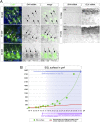
SHH signaling is active in control cases in which the exponential increase in the EGL surface begins. (A) Combined in situ hybridization of SHH or GLI1 mRNA and calbindin immunostaining at gw 12, 17, and 33. At gw 12, neither the PCs (arrows) nor the EGL express SHH, whereas the EGL expresses GLI1. At 17 gw, around the observed peak of GCP proliferation, some calbindin-positive PCs still migrating toward the pial surface express detectable levels of SHH mRNA (arrows) whereas others do not (arrowheads). At the same time, GLI1 mRNA is expressed in the EGL. At gw 33, PCs, aligned in a single-cell row beneath the EGL, all express high levels of SHH mRNA (arrows). (B) Quantification of the EGL surface on horizontal H&E-stained sections (green dots). From the onset of detectable SHH expression (i.e., gw 17, blue arrow), the exponential curve fitting all the data points starts to diverge from the linear regression calculated from the data points at gw 10 to gw 15. The divergence corresponds with the timing of the GCP proliferation peak (Fig. 2, purple arrows), suggesting that SHH-dependent increase in the rate of GCP proliferation is responsible for the exponential increase in the surface of the EGL. (Scale bars: 20 μm.)
Fig. 4.
Fig. 4.
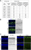
Conserved SHH expression and reduced GLI1 mRNA and PTC levels in most JS/MKS cases with defective GCP proliferation. (A) GLI1, PTC, and SHH expression in JS samples compared with controls. SHH, GLI1, and PTC levels of expression were assessed by in situ hybridization (SHH, GLI1) or immunohistochemistry (PTC). SHH mRNA expression is conserved in the vast majority of JS/MKS cases with defective GCP proliferation, but GLI1 mRNA and PTC levels are reduced or undetectable, indicating a defective response to SHH. (∼, comparable to WT; ND, not determined; NE, not expressed; −, diminished.) (B) Combined SHH in situ hybridization and calbindin immunolabeling of JS case 080144 and an age-matched control. SHH mRNA levels in JS calbindin-positive PCs (green) are comparable to control levels. The EGL and PC layer (PCL) are delimited by a dashed line. (C) GLI1 in situ hybridization or PTC immunohistochemistry and DAPI staining in two JS cases and age-matched controls. GLI1 mRNA and PTC expression are reduced in the JS EGL. (Scale bar: 20 μm.)

References

    1. Brancati F, Dallapiccola B, Valente EM. Joubert syndrome and related disorders. Orphanet J Rare Dis. 2010;5:20. - PMC - PubMed
    1. Louie CM, Gleeson JG. Genetic basis of Joubert syndrome and related disorders of cerebellar development. Hum Mol Genet. 2005;14 spec no 2:R235–R242. - PubMed
    1. Sang L, et al. Mapping the NPHP-JBTS-MKS protein network reveals ciliopathy disease genes and pathways. Cell. 2011;145:513–528. - PMC - PubMed
    1. Novarino G, Akizu N, Gleeson JG. Modeling human disease in humans: The ciliopathies. Cell. 2011;147:70–79. - PMC - PubMed
    1. Garcia-Gonzalo FR, et al. A transition zone complex regulates mammalian ciliogenesis and ciliary membrane composition. Nat Genet. 2011;43:776–784. - PMC - PubMed

Publication types

MeSH terms

Supplementary concepts