. 2012 Oct 16;109(42):16829-34.
doi: 10.1073/pnas.1210131109. Epub 2012 Oct 1.

Bacillus cereus iron uptake protein fishes out an unstable ferric citrate trimer

Affiliations

Bacillus cereus iron uptake protein fishes out an unstable ferric citrate trimer

Tatsuya Fukushima et al. Proc Natl Acad Sci U S A. .

Abstract

Citrate is a common biomolecule that chelates Fe(III). Many bacteria and plants use ferric citrate to fulfill their nutritional requirement for iron. Only the Escherichia coli ferric citrate outer-membrane transport protein FecA has been characterized; little is known about other ferric citrate-binding proteins. Here we report a unique siderophore-binding protein from the gram-positive pathogenic bacterium Bacillus cereus that binds multinuclear ferric citrate complexes. We have demonstrated that B. cereus ATCC 14579 takes up (55)Fe radiolabeled ferric citrate and that a protein, BC_3466 [renamed FctC (ferric citrate-binding protein C)], binds ferric citrate. The dissociation constant (K(d)) of FctC at pH 7.4 with ferric citrate (molar ratio 1:50) is 2.6 nM. This is the tightest binding observed of any B. cereus siderophore-binding protein. Nano electrospray ionization-mass spectrometry (nano ESI-MS) analysis of FctC and ferric citrate complexes or citrate alone show that FctC binds diferric di-citrate, and triferric tricitrate, but does not bind ferric di-citrate, ferric monocitrate, or citrate alone. Significantly, the protein selectively binds triferric tricitrate even though this species is naturally present at very low equilibrium concentrations.


Conflict of interest statement

The authors declare no conflict of interest.

Figures

Fig. 1.
Fig. 1.
Proposed structures of ferric citrate complexes in neutral aqueous solution. Citrate coordinates Fe(III) as FeCit (ML) (A), FeCit2 (ML2) (B), Fe2Cit2 (M2L2) (C), and Fe3Cit3 (M3L3) (D). Structures B and C are 2D bond-line drawings generated from the crystal structures of Gautier-Luneau et al. (17). Structure D is a 2D model proposed by Gautier-Luneau et al. (17). The Gautier-Luneau et al. model shows dangling citrate carboxylates uncoordinated to Fe(III), which we suggest does not occur (Fig. 5).
Fig. 2.
Fig. 2.
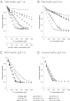
FctC FQ experiments. FQ of FctC with several ferric citrate molar ratios was measured at pH 7.4 (A), pH 8.0 (B), pH 5.5 (C), and pH 4.5 (D). The ferric:citrate molar ratios used in this study were 1:1 (closed triangles), 1:2 (open triangles), 1:5 (open diamonds), 1:10 (closed diamonds), 1:25 (closed squares), 1:50 (closed circles), 1:100 (open circles), and 1:200 (open squares). Crosses represent citrate alone and the concentrations (μM) are shown in the lower x axis in A. The calculated Kds of FctC with ferric:citrate molar ratios of 1:1, 1:2, 1:5, and 1:10 at pH 7.4 and with ferric:citrate molar ratios of 1:1, 1:10, and 1:25 at pH 8.0 were 500–900 nM and >1 μM, respectively.
Fig. 3.
Fig. 3.
Speciation diagrams of ferric citrate. A speciation diagram was generated with HySS as described in SI Materials and Methods. (A and B) The calculation was performed with ferric:citrate molar ratios of 1:50 (1.25 μM:62.5 μM, A) and 1:100 (1.25 μM:125 μM, B). The Fe(III) and citrate concentrations are derived from the final conditions of the FQ experiments (1.25 μM Fe concentration in Fig. 2 and Fig. S4 B and C). (C and D) The calculation was performed with ferric:citrate molar ratios of 1:1 (50 μM:50 μM, C) and 1:10 (50 μM:500 μM, D). The Fe(III) and citrate concentrations are the same as those in the nano ESI-MS analysis conditions in Fig. 4 and in Figs. S6 AC and S7C. Fe2L2, [Fe2(Cit)2]2−; Fe3L3, [Fe3(Cit)3]3−; FeL2, [Fe(Cit)2]5−; FeLH, Fe(HCit); FeL2H, [Fe(HCit2)]4−; FeH-2, [Fe(OH)2]+.
Fig. 4.
Fig. 4.
Nano ESI-MS analysis of FctC and ferric citrate complexes at pH 5.5 (AC) and pH 8.0 (DF). FctC and ferric citrate complexes were observed with several charge states in positive mode. (A and D) FctC only. (B and E) FctC and ferric citrate (1:1) mixture. (C and F) FctC and ferric citrate (1:10) mixture. Predicted ferric citrate compositions are shown with a speciation diagram in Fig. 3 C and D. Peak A series, FctC protein only; peak B series, FctC and Fe3Cit3 complex. Charged states of +15 and +16 were detected, and the numbers correspond to the calculated values in Table S2. The measured molecular weights of FctC and ferric citrate complexes using MassLynx software (Waters) are shown in Table S1.
Fig. 5.
Fig. 5.
Schematic model for Fe3Cit3. Each iron center (orange spheres) is pseudo-octahedral, and each has a coordinated water molecule or peptide ligand from the binding pocket of FctC [green atoms (Left) and L (Right)] to fill the sixth coordination site. Red atoms are oxygen atoms and hydrogen atoms are removed for clarity. The geometric parameters (distances and angles) around iron are shown in Table S3.

References

    1. Raymond KN, Dertz EA. Biochemical and physical properties of siderophores. In: Crosa JH, Mey AR, Payne SM, editors. Iron Transport in Bacteria. Washington, DC: ASM Press; 2004. pp. 3–17.
    1. Braun V, Hantke K, Koster W. Bacterial iron transport: Mechanisms, genetics, and regulation. In: Sigel A, Sigel H, editors. Metal Ions in Biological Systems. Vol 35. New York: Marcel Dekker, Inc.; 1998. pp. 67–145. - PubMed
    1. Raymond KN, Dertz EA, Kim SS. Enterobactin: An archetype for microbial iron transport. Proc Natl Acad Sci USA. 2003;100:3584–3588. - PMC - PubMed
    1. Koppisch AT, et al. Petrobactin is the primary siderophore synthesized by Bacillus anthracis str. Sterne under conditions of iron starvation. Biometals. 2005;18:577–585. - PubMed
    1. Müller SI, Valdebenito M, Hantke K. Salmochelin, the long-overlooked catecholate siderophore of Salmonella. Biometals. 2009;22:691–695. - PubMed

Publication types

MeSH terms

LinkOut - more resources