. 2012 Nov 1;189(9):4640-7.
doi: 10.4049/jimmunol.1201904. Epub 2012 Oct 1.

The alternative complement pathway propagates inflammation and injury in murine ischemic stroke

Affiliations

The alternative complement pathway propagates inflammation and injury in murine ischemic stroke

Andrew Elvington et al. J Immunol. .

Abstract

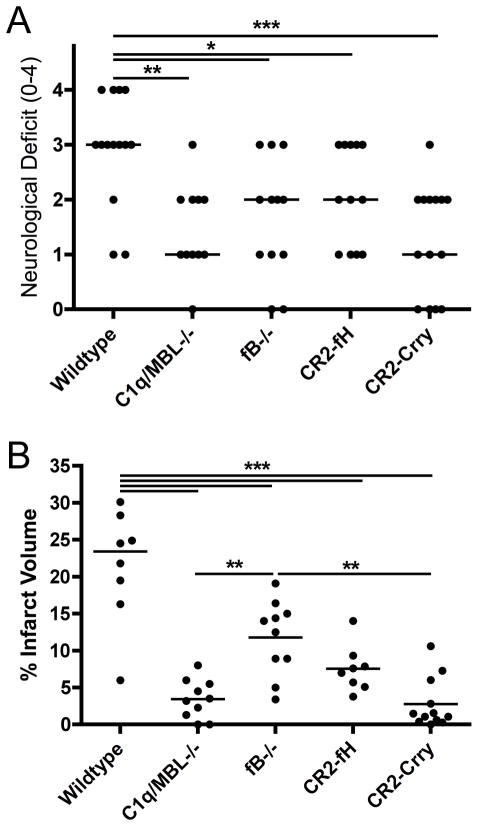
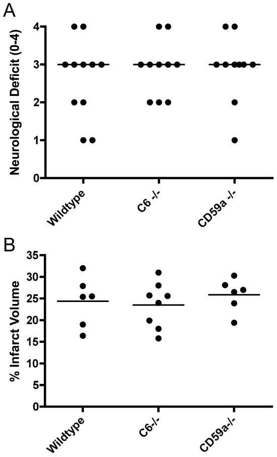
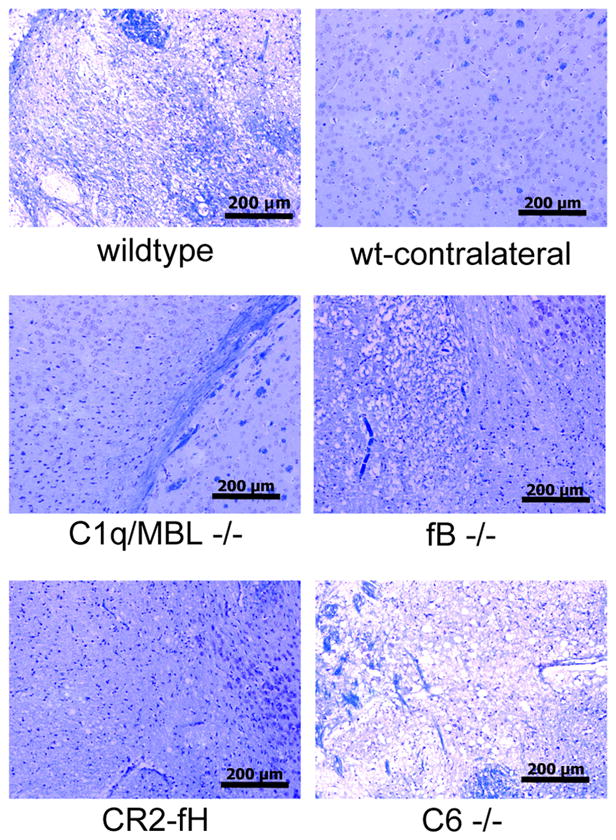
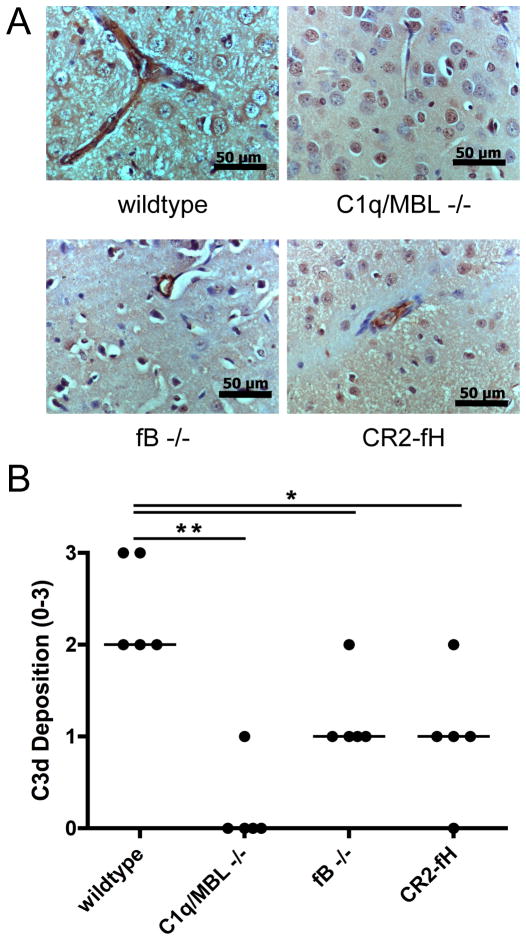
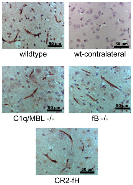
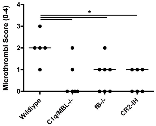
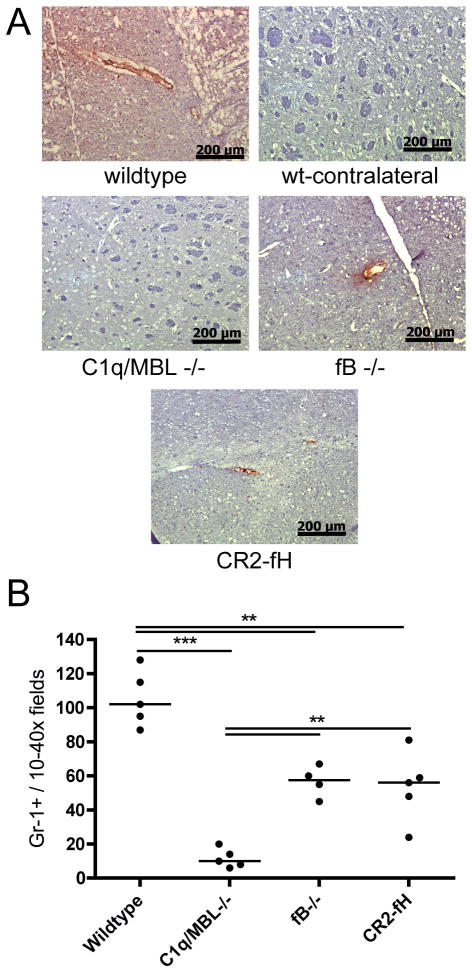
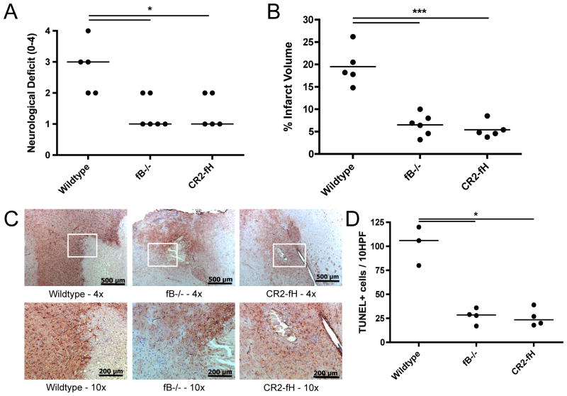
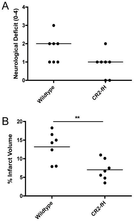
There is mounting evidence indicating an important role for complement in the pathogenesis of cerebral ischemia-reperfusion injury, or ischemic stroke. The role of the alternative complement pathway in ischemic stroke has not been investigated, and there is conflicting data on the role of the terminal pathway. In this study, we show that compared with wild-type mice, mice deficient in the alternative pathway protein factor B or mice treated with the alternative pathway inhibitor CR2-fH have improved outcomes after 60-min middle cerebral artery occlusion and 24-h reperfusion. Factor B-deficient or CR2-fH-treated mice were protected in terms of improved neurologic function and reduced cerebral infarct, demyelination, P-selectin expression, neutrophil infiltration, and microthrombi formation. Mice deficient in both the classical and lectin pathways (C1q/MBL deficient) were also protected from cerebral ischemia-reperfusion injury, and there was no detectable C3d deposition in the ipsilateral brain of these mice. These data demonstrate that the alternative pathway is not alone sufficient to initiate complement activation and indicate that the alternative pathway propagates cerebral injury via amplification of the cascade. Deficiency of C6, a component of the terminal cytolytic membrane attack complex, had no effect on outcome after ischemic stroke, indicating that the membrane attack complex is not involved in mediating injury in this model. We additionally show that the protective effect of factor B deficiency and CR2-fH treatment is sustained in the subacute stage of infarct development, adding to the clinical relevance of these findings.


Figures

Figure 1
Figure 1
Effect of complement deficiency and CR2-fH or CR2-Crry treatment on neurological deficit and infarct volume after 60 minutes MCAO and 24 hours reperfusion. (A) Neurological deficit and (B) Infarct volume in C1q/MBL −/− mice, fB−/− mice and wt mice treated with CR2-fH or CR2-Crry 30 minutes after reperfusion. Horizontal bar indicates median neurological score and mean infarct volume, n = 12–15 for neurological deficit and n = 8–11 for infarct volume; *p<0.05, **p<0.01, ***p<0.001.
Figure 2
Figure 2
Effect of terminal pathway deficiency and terminal pathway inhibitor deficiency on neurological deficit and infarct volume after 60 minutes MCAO and 24 hours reperfusion. (A) Neurological deficit in C6 −/− and CD59a−/− mice. Horizontal bar represents median; n=10–11. No statistical difference between groups. (B) Infarct volume in C6 −/− and CD59a−/− mice. Horizontal bar indicates mean, n = 6–8. No statistical difference between groups.
Figure 3
Figure 3
Neuronal tissue integrity in complement deficient and CR2-fH treated mice after 60 minutes MCAO and 24 hours reperfusion. Brain sections from the ipsilateral hemisphere were stained with luxol fast blue/Nissl. Section from contralateral hemisphere shown as undamaged control. Representative images shown, n = 4 per group; 10x magnification, scale bar = 200 μm.
Figure 4
Figure 4
C3d deposition in penumbral tissue in complement deficient and CR2-fH treated mice after 60 minutes MCAO and 24 hours reperfusion. (A) Representative immunohistochemistry images taken at 40x magnification. Scale bar = 50 μm. There was no observable C3d staining in the contralateral hemispheres in any group. (B) Quantification of C3d staining. Horizontal bar represents median, n = 5; *p<0.05, **p<0.01.
Figure 5
Figure 5
IgM deposition in penumbral tissue in complement deficient and CR2-fH treated mice after 60 minutes MCAO and 24 hours reperfusion. There were no discernable differences in the levels or distribution of IgM staining between any groups. No IgM staining was observed in the contralateral hemispheres in any group. Representative images, n = 3; 40x magnification, scale bar = 50 μm.
Figure 6
Figure 6
Microthrombi formation in penumbral tissue in complement deficient and CR2-fH treated mice after 60 minutes MCAO and 24 hours reperfusion. Microthrombi formation was assessed in H&E stained brain sections. Horizontal bars represent median microthrombi scores, n = 5–7; *p<0.05.
Figure 7
Figure 7
P-selectin expression and neutrophil infiltration in penumbral tissue in complement deficient and CR2-fH treated mice after 60 minutes MCAO and 24 hours reperfusion. (A) P-selectin expression as detected by immunohistochemistry. Expression is associated with the vasculature. Representative images, n = 3, 10x magnification, scale bar = 200 μm. No staining was detected in contralateral hemisphere of any group. (B) Neutrophil infiltration as detected by immunohistochemical staining of GR1+ cells. Horizontal bar represents median, n = 4–5; **p<0.01, ***p<0.001.
Figure 8
Figure 8
Effect of fB deficiency and CR2-fH treatment on outcomes after 60 minutes MCAO and 72 hours reperfusion. (A) Neurological deficit. Horizontal bar represents median, n = 5–6; *p<0.05. (B) Infarct volume. Horizontal bar represents mean, n = 5–6; ***p<0.001. (C) Astrocyte GFAP reactivity as assessed by GFAP immunohistochemistry. Representative images shown, n = 3. Magnifications as follows: 4x, scale bar = 500 μm; 10x, scale bar = 200 μm. Contralateral GFAP staining was not different between groups. (D) Apoptosis as evaluated by counting 10 random 40x fields from the peri-infarct region of the penumbra. Horizontal bar represents median, n=3–4; *p<0.05.
Figure 9
Figure 9
Effect of CR2-fH treatment on neurological deficit and infarct volume after 60 minutes of MCAO and 7 days reperfusion. (A) Neurological deficit. Horizontal bar represents median, n = 7; p=0.07. (B) Infarct volume. Horizontal bar indicates mean, n = 7; **p<0.01.

References

    1. Dirnagl U, Iadecola C, Moskowitz MA. Pathobiology of ischaemic stroke: an integrated view. Trends Neurosci. 1999;22:391–397. - PubMed
    1. Schaller B, Graf R. Cerebral ischemia and reperfusion: the pathophysiologic concept as a basis for clinical therapy. J Cereb Blood Flow Metab. 2004;24:351–371. - PubMed
    1. Brennan FH, Anderson AJ, Taylor SM, Woodruff TM, Ruitenberg MJ. Complement activation in the injured central nervous system: another dual-edged sword? J Neuroinflammation. 2012;9:137. - PMC - PubMed
    1. Huang J, Kim LJ, Mealey R, Marsh HC, Jr, Zhang Y, Tenner AJ, Connolly ES, Jr, Pinsky DJ. Neuronal protection in stroke by an sLex-glycosylated complement inhibitory protein. Science. 1999;285:595–599. - PubMed
    1. Atkinson C, Zhu H, Qiao F, Varela JC, Yu J, Song H, Kindy MS, Tomlinson S. Complement-dependent P-selectin expression and injury following ischemic stroke. J Immunol. 2006;177:7266–7274. - PubMed

Publication types

MeSH terms