. 2012 Oct 1;26(19):2180-91.
doi: 10.1101/gad.198085.112.

miR-26a is required for skeletal muscle differentiation and regeneration in mice

Affiliations

miR-26a is required for skeletal muscle differentiation and regeneration in mice

Bijan K Dey et al. Genes Dev. .

Abstract

Multiple microRNAs are known to be induced during the differentiation of myoblasts to myotubes. Yet, experiments in animals have not provided clear evidence for the requirement of most of these microRNAs in myogenic differentiation in vivo. miR-26a is induced during skeletal muscle differentiation and is predicted to target a well-known inhibitor of differentiation, the transforming growth factor β/bone morphogenetic protein (TGF-β/BMP) signaling pathway. Here we show that exogenous miR-26a promotes differentiation of myoblasts, while inhibition of miR-26a by antisense oligonucleotides or by Tough-Decoys delays differentiation. miR-26a targets the transcription factors Smad1 and Smad4, critical for the TGF-β/BMP pathway, and expression of microRNA-resistant forms of these transcription factors inhibits differentiation. Injection of antagomirs specific to miR-26a into neonatal mice derepressed both Smad expression and activity and consequently inhibited skeletal muscle differentiation. In addition, miR-26a is induced during skeletal muscle regeneration after injury. Inhibiting miR-26a in the tibialis anterior muscles through the injection of adeno-associated virus expressing a Tough-Decoy targeting miR-26a prevents Smad down-regulation and delays regeneration. These findings provide evidence for the requirement of miR-26a for skeletal muscle differentiation and regeneration in vivo.


Figures

Figure 1.
Figure 1.
miR-26a is abundantly expressed in skeletal muscle and up-regulated during muscle differentiation and regeneration. miR-26a is up-regulated during differentiation of C2C12 myoblast (A), mouse primary myoblast (B), and human primary myoblast (C). Quantitative RT–PCR (qRT–PCR) of miR-26a after indicated days in differentiation medium (DM). Fold change of miR-26a, normalized to U6sn RNA, is expressed relative to cells in growth medium (GM). Means ± standard deviation of three replicates. (D) miR-26a is abundantly expressed in mouse and human skeletal muscles as compared with other tissue types. qRT–PCR for miR-26a, which is normalized to U6sn RNA. Mean ± standard deviation of three replicates. (E) miR-26a is down-regulated on days 1–3 after injury and up-regulated on days 5–14 after injury by injection of CTX. The rest is as in D. (F) FISH of control adult skeletal muscles using locked nucleic acid (LNA)-based scrambled (top left two panels) or anti-miR-26a (top right two panels) probes. FISH of miR-26a and immunostaining for MyoD are shown for regenerating muscle on day 5 (middle) and on day 7 (bottom) after CTX injection. Yellow or white arrows indicate miR-26a- and MyoD-positive or -negative cells, respectively, located outside the skeletal muscle fibers. Bars, 20 μm.
Figure 2.
Figure 2.
miR-26a promotes and is required for myoblast differentiation. (A) C2C12 cells were transfected three times at 24-h intervals with GL2 or miR-26a in GM, and the cells were transferred to DM and stained for Myogenin at 32 h or for MHC at 60 h. (Green) Myogenin or MHC; (blue) nuclei stained by DAPI. (B) Fractions of Myogenin- and MHC-positive cells are presented relative to the GL2 control (100%). Mean ± standard deviation of 10 measurements (Supplemental Table 1). (C) C2C12 cells transfected with GL2 or miR-26a as above and held in GM for an extra 24 h; qRT–PCR was performed for Myogenin and MHC. The results were normalized to GAPDH in the same sample and then again to the level in GL2 transfected cells. Mean ± standard deviation of three replicates. (D) Increase in G1-phase cells after transfection of miR-26a in C2C12 cells. Propidium iodide staining for DNA content and FACS analysis (Supplemental Fig. 1). The results are expressed as the percentage change of cells in a given phase of the cell cycle in the microRNA transfected cells relative to that in the GL2 control. Mean ± standard deviation of three measurements. (E,F) 2′O-methyl antisense oligonucleotides against GL2 or miR-26a were transfected as in A, and C2C12 cells were stained for Myogenin at 32 h or for MHC at 60 h. Data are presented as in A and B. (G) qRT–PCR performed for Myogenin and MHC mRNA from cells in E, and data are presented as in C. (H) TuD for miR-26a inhibits myoblast differentiation as indicated by MCK-luciferase activity at 48 h. A Renilla (rr) luciferase construct was cotransfected with MCK-firefly (pp) luciferase as a transfection control. The result is expressed as pp/rr relative to the control TuD-infected samples. Mean ± standard deviation of three replicates.
Figure 3.
Figure 3.
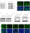
miR-26a targets Smad1 and Smad4. (A,B) Smad1 and Smad4 proteins are down-regulated during differentiation. (A) Western blot of indicated proteins in C2C12 cells in GM or on various days following transfer to DM. GAPDH served as loading control. (B) Quantification of expression of Smad1 and Smad4 protein normalized to GAPDH. (C,D) Relative Smad1 and Smad4 mRNA levels normalized to GAPDH mRNA in C2C12 cells as in A. (E,F) Luciferase assays were performed to measure the effect of transfected miR-26a on a Renilla luciferase reporter fused to Smad1 or Smad4 3′ UTRs. Mutations at target sites indicated in Supplemental Figure 4. A firefly (pp) luciferase plasmid was cotransfected with the Renilla (rr) luciferase construct as a transfection control. The rr/pp was normalized to that for a control Renilla luciferase plasmid without a Smad1 or Smad4 3′ UTR segment and is expressed relative to the normalized rr/pp in cells transfected with the GL2 control. Mean ± SD of three measurements.
Figure 4.
Figure 4.
miR-26a down-regulates Smad1 and Smad4 in differentiating myoblasts by directly targeting their 3′ UTRs. (A) Transfection of miR-26a in C2C12 cells down-regulates Smad1 and Smad4 protein levels as detected by Western blotting on day 4 of transfection. GAPDH served as a loading control. (B) 2′O-methyl antisense oligonucleotide against miR-26a (Anti-26a) causes longer persistence Smad1, Smad4, and pSmad1/5 protein levels in C2C12 cells held in DM for 2 d compared with cells transfected with the anti-GL2 oligonucleotides. GAPDH served as a loading control. (C) siRNA of Smad1 and Smad4 causes differentiation of C2C12 in GM as detected by MHC immunostaining. (Green) MHC; (blue) DAPI. (D, left) Flag Western blot of C2C12 cells stably expressing empty retroviral vector, the Flag-tagged Smad1 ORF, or the Flag-tagged Smad1 ORF+3′ UTR. (Right) Similarly, for Flag-Smad4. GAPDH represents the loading control. (E) Level of Flag-Smad1 or Smad4 (top panel) and MHC (middle panel) proteins after transfer to DM3 of C2C12 cells expressing constructs indicated at the top. GAPDH served as the loading control. (F) C2C12 cells stably expressing various constructs were induced to differentiate by transfer to DM. Overexpression of the Smad1 or Smad4 ORF inhibited differentiation in DM3, whereas overexpression of Smad1 ORF+3′ UTR or Smad4 ORF+3′ UTR permitted cells to differentiate to a degree similar to cells infected with empty vector. (Green) MHC; (blue) DAPI staining of nuclei.
Figure 5.
Figure 5.
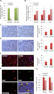
Knockdown of miR-26a increases the Pax7-positive proliferating cells in vivo. (A) Injection of Ant26a knocks down endogenous miR-26a in neonatal skeletal muscle. qRT–PCR of microRNAs normalized to U6sn RNA are expressed relative to control antagomir-injected samples. Mean ± standard deviation of three mice. (B) Ant26a increases Smad1, Smad4, and Id3 mRNA levels. qRT–PCR of Smad1, Smad4, and Id3 on hind leg skeletal muscle, normalized to GAPDH in the same sample and then again to the level in controls. Mean ± standard deviation of the sample from three mice. (*) P < 0.001. (C, left) Immunostaining images of pSmad1/5 showing that Ant26a increases pSmad1/5-positive cells. (Right) Quantitation of ∼2000 nuclei from random fields. Mean ± standard deviation of three different samples of Ant26a-injected or control-injected neonates. (*) P < 0.001. Bar, 50 μm. (D) As in C, immunostaining images of Ki67 (left) and quantitation (right). (*) P < 0.001. (E, left) Confocal microscopy images of skeletal muscle 2 h after BrdU labeling from Ant26a-injected or control antagomir-injected neonates. Cell proliferation was determined by anti-BrdU antibody (green), cell surface was marked by laminin (red), and nuclei were counterstained with DAPI (blue). (Right) The percentage of BrdU-positive nuclei was quantitated. Mean ± standard deviation of 10 different random fields from three different samples. (*) P < 0.001. Bar, 50 μm. (F) Confocal microscopy images of Ant26a-injected neonatal skeletal muscles from E immunostained for Pax7 and BrdU. (Yellow arrows) Satellite cells doubly stained for BrdU and Pax7. Bar, 25 μm. (G) Ant26a decreases differentiation marker Myogenin and MHC. qRT–PCR performed for Myogenin and MHC mRNAs. The results were normalized to GAPDH and expressed relative to control. Mean ± standard deviation of the sample from three mice. (*) P < 0.001.
Figure 6.
Figure 6.
TuD-26a delays muscle regeneration after injury. (A) Smad1 and Smad4 are up-regulated in TA muscles on days 1–3 after injury with CTX, and the opposite pattern was seen on days 5–14 after injury. qRT–PCR of Smad1 and Smad4 normalized to GAPDH. Mean ± standard deviation of the samples from three mice. (B,C) miR-26a level was decreased, and Smad1, Smad4, Id3, and Pax7 mRNA was increased after 10 d of injection of TuD-26a into the TA muscles as compared with NC-TuD. 26a-TuD qRT–PCR values were normalized to GAPDH in the same sample and then again to the level in NC-TuD. Mean ± standard deviation of the samples from three mice. (*) P < 0.001. (DG) Injection of TuD-26a before CTX injury disrupts the expression patterns of miR-26a, Smads, and their targets during regeneration (days 7, 10, and 14). qRT–PCR was performed and data are represented as in B and C. (H) H&E staining of representative images showing that the TuD-26a-treated sample delays regeneration. Bar, 100 μm. (I,J) Desmin and laminin staining and cross-section area of regenerating fibers on day 7 using ImageJ software. More than 200 fibers were counted in each group. NC-TuD or 26a-TuD and CTX were injected as in H. Bar, 100 μm.

Similar articles

Cited by

References

    1. Andres V, Walsh K 1996. Myogenin expression, cell cycle withdrawal, and phenotypic differentiation are temporally separable events that precede cell fusion upon myogenesis. J Cell Biol 132: 657–666 - PMC - PubMed
    1. Bartel DP 2004. MicroRNAs: Genomics, biogenesis, mechanism, and function. Cell 116: 281–297 - PubMed
    1. Berkes CA, Tapscott SJ 2005. MyoD and the transcriptional control of myogenesis. Semin Cell Dev Biol 16: 585–595 - PubMed
    1. Buckingham M, Bajard L, Daubas P, Esner M, Lagha M, Relaix F, Rocancourt D 2006. Myogenic progenitor cells in the mouse embryo are marked by the expression of Pax3/7 genes that regulate their survival and myogenic potential. Anat Embryol 211: 51–56 - PubMed
    1. Chen JF, Mandel EM, Thomson JM, Wu Q, Callis TE, Hammond SM, Conlon FL, Wang DZ 2006. The role of microRNA-1 and microRNA-133 in skeletal muscle proliferation and differentiation. Nat Genet 38: 228–233 - PMC - PubMed

Publication types

MeSH terms

LinkOut - more resources