. 2012 Oct 1;26(19):2206-21.
doi: 10.1101/gad.193409.112.

Synaptic vesicle clustering requires a distinct MIG-10/Lamellipodin isoform and ABI-1 downstream from Netrin

Affiliations

Synaptic vesicle clustering requires a distinct MIG-10/Lamellipodin isoform and ABI-1 downstream from Netrin

Andrea K H Stavoe et al. Genes Dev. .

Abstract

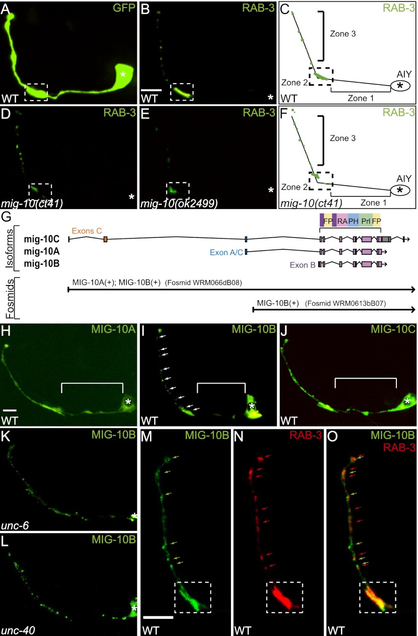
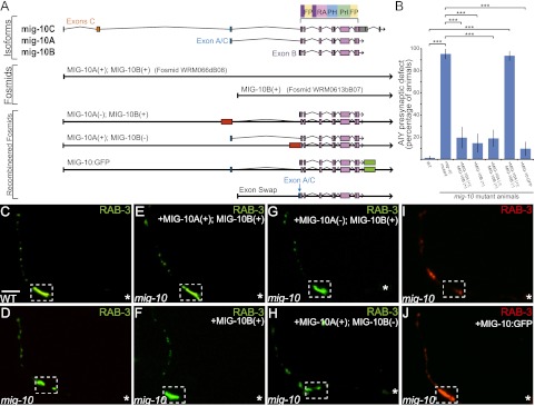
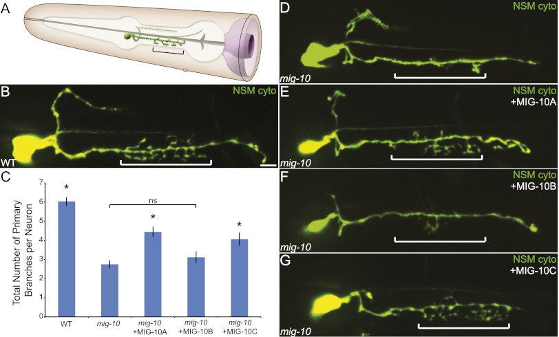
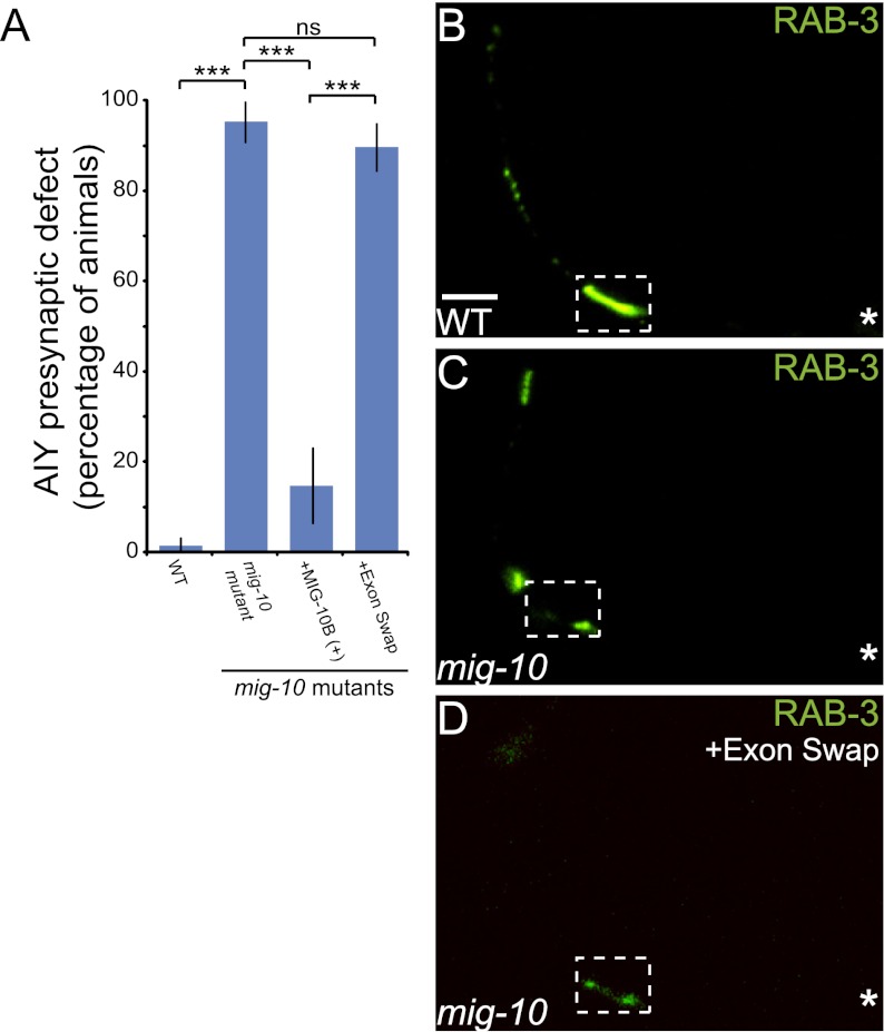
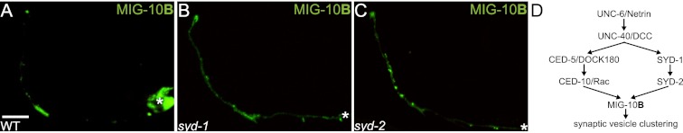
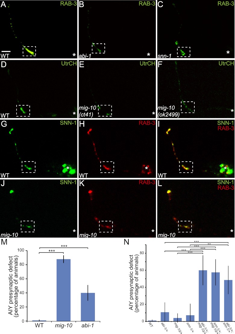
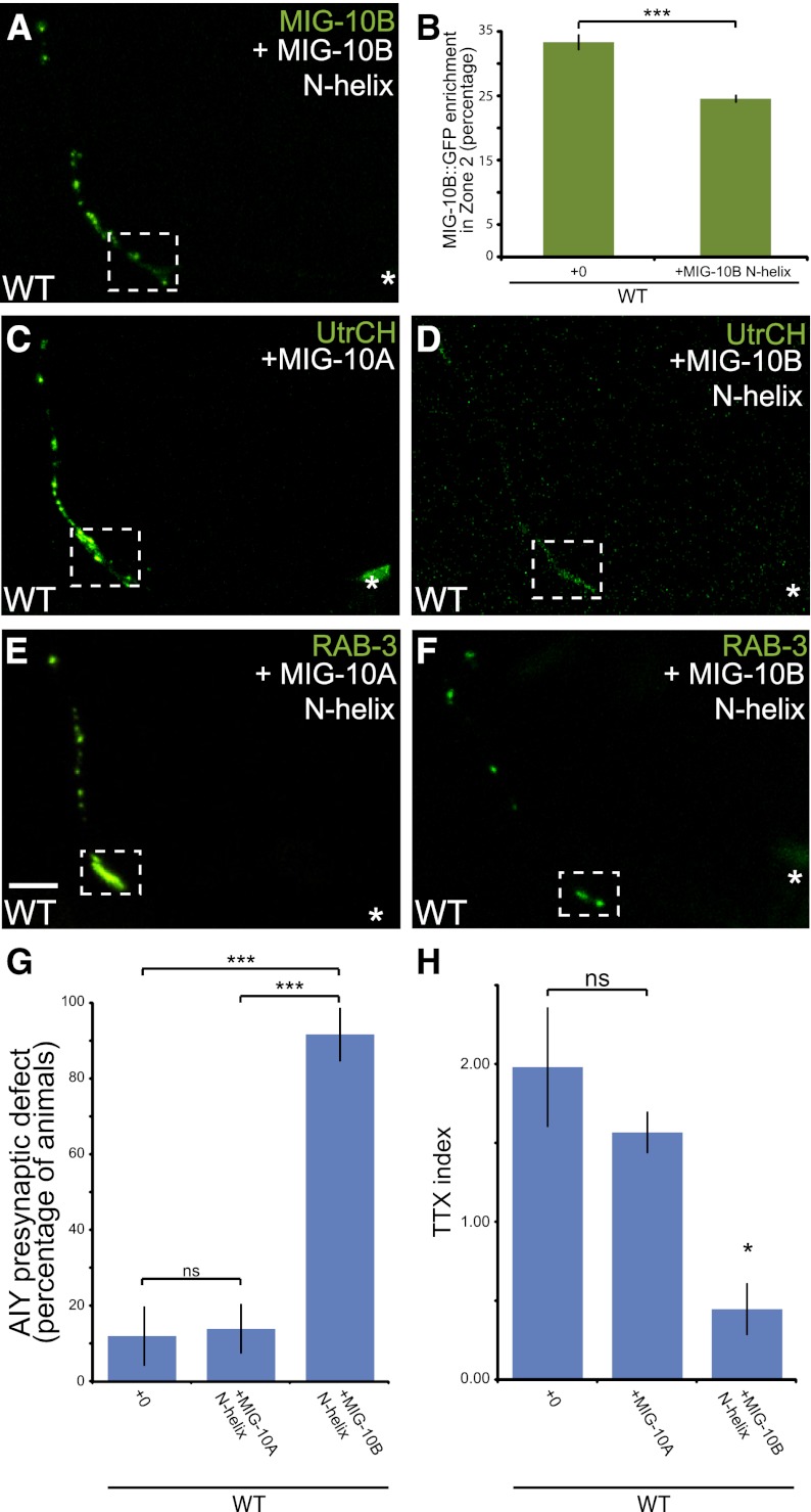
The chemotrophic factor Netrin can simultaneously instruct different neurodevelopmental programs in individual neurons in vivo. How neurons correctly interpret the Netrin signal and undergo the appropriate neurodevelopmental response is not understood. Here we identify MIG-10 isoforms as critical determinants of individual cellular responses to Netrin. We determined that distinct MIG-10 isoforms, varying only in their N-terminal motifs, can localize to specific subcellular domains and are differentially required for discrete neurodevelopmental processes in vivo. We identified MIG-10B as an isoform uniquely capable of localizing to presynaptic regions and instructing synaptic vesicle clustering in response to Netrin. MIG-10B interacts with Abl-interacting protein-1 (ABI-1)/Abi1, a component of the WAVE complex, to organize the actin cytoskeleton at presynaptic sites and instruct vesicle clustering through SNN-1/Synapsin. We identified a motif in the MIG-10B N-terminal domain that is required for its function and localization to presynaptic sites. With this motif, we engineered a dominant-negative MIG-10B construct that disrupts vesicle clustering and animal thermotaxis behavior when expressed in a single neuron in vivo. Our findings indicate that the unique N-terminal domains confer distinct MIG-10 isoforms with unique capabilities to localize to distinct subcellular compartments, organize the actin cytoskeleton at these sites, and instruct distinct Netrin-dependent neurodevelopmental programs.


Figures

Figure 1.
Figure 1.
MIG-10B localizes to AIY presynaptic regions in response to Netrin. (A) Confocal micrograph of cytoplasmic GFP expressed cell-specifically in AIY, depicting the morphology of AIY. (B–F) Synaptic vesicle pattern in maximal projection confocal micrographs of GFP∷RAB-3 in AIY in wild-type (B), mig-10(ct41) (D), and mig-10(ok2499) (E) mutant animals and schematics (C,F). The schematic diagram of wild-type AIY (shown in C) indicates neurite zones. Zone 1 is asynaptic and projects anteriorly from the cell body. AIY forms synapses onto RIA, AIZ, and RIB in zone 2. Zone 3 is within the nerve ring and is the distal portion of the neurite after the neurite turns dorsally (Colon-Ramos et al. 2007). Bar: B, 5 μm (applies to A,B,D,E). (G) Diagram of the mig-10 gene, with exons as boxes and introns as lines. The first three schematics show the relative positions of the three MIG-10 isoforms—MIG-10A, MIG-10B, and MIG-10C—drawn to scale. Shared exons code for conserved domains. Cartoon diagram of the conserved protein domains in the top right corner. Ras association (RA), plekstrin homology (PH), proline-rich (Prl), and FPPP (FP) (Holt and Daly 2005). The fourth and fifth schematics include a diagram of the rescuing fosmids, drawn to scale and aligned with the genetic regions of mig-10 and the isoforms. Modified image adapted with permission from WormBase (http://www.wormbase.org). (H–J) Confocal micrographs showing the cell-specific subcellular localization of MIG-10A∷GFP (H), MIG-10B∷GFP (I), and MIG-10C∷GFP (J). Note that MIG-10A and MIG-10C signal is diffuse throughout the neurite, including asynaptic regions (bracket) (H,J), while MIG-10B is enriched in presynaptic regions (I, arrows). (K,L) Confocal micrographs showing MIG-10B∷GFP subcellular localization in AIY in unc-6(ev400) (K) and unc-40(e271) (L) mutant animals. (I) Note that MIG-10B∷GFP is less enriched in zone 2 in the two mutant backgrounds, as compared with wild-type animals. (M–O) Confocal micrographs of zones 2 and 3 of a transgenic animal expressing MIG-10B∷GFP (M) and mCh∷RAB-3 (N) in AIY. Merge is shown in O. Synaptic regions are expanded in these images to allow evaluation of partial colocalization (arrows). In all images, the asterisk (*) represents the location of the cell body, and the dashed box encloses zone 2. Bar: H, 5 μm (applies to H–L); M, 5 μm (applies to M–O).
Figure 2.
Figure 2.
MIG-10B is sufficient and necessary to rescue synaptic vesicle clustering. (A) Diagram of the mig-10 gene, as in Figure 1G. The sixth, seventh, eighth, and ninth schematics correspond to the recombineered fosmids used to assay the necessity of MIG-10B. Red boxes represent the introduction of a cassette with a stop codon in lieu of the mig-10a unique exon [MIG-10A(−); MIG-10B(+)] or in lieu of the mig-10b unique exon [MIG-10A(+); MIG-10B(−)].Green boxes represent the introduction of GFP in-frame after the mig-10 coding region (“MIG-10:GFP”). For the “exon swap” recombineered fosmid, the shared exon A/C sequence (in blue) was introduced in place of the unique exon B. Image modified with permission from WormBase (http://www.wormbase.org). (B) Quantification of the percentage of animals displaying the AIY presynaptic defect. Rescuing fosmids (see A) were expressed in mig-10(ct41) mutants and scored for rescue of the AIY presynaptic defect. Note that only fosmid MIG-10A(+);MIG-10B(−) is not capable of rescuing the AIY presynaptic defect. (***) P < 0.0001 between indicated groups by Fisher's exact test. Error bars represent 95% confidence interval. (C,D) Distribution of synaptic vesicles in AIY in wild-type (C) and mig-10(ct41) mutants (D). (E–H) Distribution of synaptic vesicles in AIY in mig-10(ct41) mutants expressing rescuing constructs fosmid MIG-10A (+); MIG-10B (+) (E), fosmid MIG-10B (+) (F), fosmid MIG-10A(−); MIG-10B(+) (G), or fosmid MIG-10A(+); MIG-10B(−) (H). Note that the distribution of synaptic vesicles is disrupted in mig-10(ct41) mutants (shown in D) and is rescued in E–G, but not in H. (I,J) Distribution of synaptic vesicles in AIY in mig-10(ct41) mutants (I) and mig-10(ct41) mutants expressing rescuing construct MIG-10:GFP (J). Note that MIG-10:GFP (shown in J) rescues the mig-10(ct41) synaptic patterning defect. In all images, the asterisk (*) represents the location of the cell body, and the dashed box encloses zone 2. Bar: C, 5 μm (applies to C–J).
Figure 3.
Figure 3.
MIG-10A and MIG-10C rescue an axon arborization defect in the NSM neuron. (A) Schematic of the pharyngeal NSM neuron. Note the presence of axonal arbors extending from the ventral neurite in the pharyngeal isthmus. The bracket indicates the pharyngeal isthmus region in which axon arbors typically form. Modified image adapted with permission from Wormatlas (http://www.wormatlas.org). (B) Confocal micrograph of a wild-type animal expressing GFP under the control of the NSM-specific tph-1 promoter (Sze et al. 2002). (C) Quantification of the axon arborization defect in the NSM. Total primary branches extending off of the ventral NSM neurite were counted for each genotype. Cell-specific expression of MIG-10A∷GFP or MIG-10C∷GFP results in statistically significant rescue of the arborization defect with respect to mig-10(ct41) mutants, but NSM-specific expression of MIG-10B∷GFP does not result in rescue. Error bars represent SEM. (*) P < 0.05 between indicated groups and mig-10(ct41). Post-hoc analysis was performed using Tukey's HSD. (D–G) Confocal micrographs of mig-10(ct41) mutant (D) or mig-10(ct41) mutants expressing MIG-10 cDNA constructs under the control of the NSM-specific tph-1 promoter (Sze et al. 2002) (E–G). Note the presence of axon arbors in the bracketed region in wild-type (B) or rescued (E,G) animals, but note the absence in arbors in mig-10(ct41) mutant animals (D) or mig-10(ct41) mutant animals expressing MIG-10B (F). Bar: B, 5 μm (applies to B,D–G).
Figure 4.
Figure 4.
MIG-10B unique N-terminal helix is required for proper AIY synaptic vesicle clustering. (A) Quantification of the percentage of animals displaying the AIY presynaptic defect. Rescuing fosmids (see Fig. 2A) were expressed in mig-10(ct41) mutants and scored for rescue. (***) P < 0.0001 between indicated groups by Fisher's exact test. Error bars represent 95% confidence interval. (B–D) Distribution of synaptic vesicles in AIY in wild-type (B), mig-10(ct41) mutants (C), or mig-10(ct41) mutants expressing the “exon swap” construct (D). The “exon swap” construct contains the first MIG-10A exon in place of the unique MIG-10B exon, thus expressing MIG-10A under the MIG-10B endogenous promoter (see Fig. 2A). Note that the distribution of synaptic vesicles is disrupted in mig-10(ct41) mutants (shown in C) and is not rescued in D. In all images, the asterisk (*) represents the location of the cell body, and the dashed box encloses zone 2. Bar: B, 5 μm (applies to B–D).
Figure 5.
Figure 5.
Active zone proteins SYD-1 and SYD-2 are necessary for MIG-10B synaptic localization. (A–C) Confocal micrographs showing MIG-10B∷GFP subcellular localization expressed cell-specifically in AIY in wild-type (A) and syd-1(ju82) (B) and syd-2(ju37) (C) mutant animals. Note that MIG-10B∷GFP is less enriched in zone 2 and is more diffuse in zone 3 in the two mutant backgrounds as compared with wild-type animals. (D) Schematic of the Rac GTPase and active zone protein pathways converging on MIG-10B to instruct MIG-10B localization and synaptic vesicle clustering. In all images, the asterisk (*) represents the location of the cell body. Bar: A, 5 μm (applies to A–C).
Figure 6.
Figure 6.
MIG-10B and ABI-1 organize the actin cytoskeleton at presynaptic sites to instruct synaptic vesicle clustering through synapsin. (A–C) Confocal micrographs of GFP∷RAB-3 expressed in AIY in wild-type (A) and abi-1(tm494) (B) and snn-1(tm2557) (C) mutant animals. Note that the presynaptic pattern is disrupted in abi-1(tm494) and snn-1(tm2557) mutants. (D–F) Confocal micrographs of animals expressing the F-actin probe UtrCH∷GFP in AIY in wild-type (D) and mig-10(ct41) (E) and mig-10(ok2499) (F) mutant animals. Note that F-actin is no longer enriched in zone 2 in both mig-10 mutant alleles as compared with wild-type animals. (G–L) Confocal micrographs of a transgenic animal expressing SNN-1A∷GFP (G) and RAB-3∷mCh (H) in AIY. Merge is shown in I. Note that SNN-1A∷GFP and RAB-3∷mCh colocalize in AIY presynaptic regions and are enriched in zone 2 (dashed box). (J–L) In a mig-10(ct41) mutant animal, both SNN-1A∷GFP (J) and RAB-3∷mCh (K) are disrupted and no longer enriched in zone 2. Merge is shown in L. (M) Quantification of penetrance of AIY synaptic vesicle clustering defect in wild-type (n = 318), mig-10(ct41) (n = 141), and abi-1(tm494) (n = 75) mutant animals. Unlike mig-10(ct41) and snn-1(tm2557) alleles, abi-1(tm494) is not a null allele, which likely accounts for the reduced penetrance of presynaptic defect. (***) P < 0.0001 between indicated groups by Fisher's exact test. Error bars represent 95% confidence interval. (N) Transheterozygote analysis. Quantification of penetrance of AIY synaptic vesicle clustering defect in wild-type (n = 318), abi-1(tm494)/+ heterozygote (n = 28), mig-10(ct41)/+ heterozygote (n = 51), snn-1(tm2557)/+ heterozygote (n = 14), abi-1(tm494)/+;mig-10(ct41)/+ transheterozygote (n = 30), snn-1/+;mig-10(ct41)/+ transheterozygote (n = 40), and abi-1(tm494)/+;snn-1(tm2557)/+ transheterozygote (n = 35) animals. Note that transheterozygote animals display significantly higher penetrance of AIY presynaptic defects when compared with wild-type and heterozygote animals. (**) P < 0.01 between indicated groups; (***) P < 0.005 between indicated groups by Fisher's exact test. Error bars represent 95% confidence interval. In all images, the asterisk (*) represents the location of the cell body, and the dashed box encloses zone 2. Bar: A, 5 μm (applies to A–L).
Figure 7.
Figure 7.
Overexpression of the MIG-10B N terminus disrupts AIY synaptic vesicle clustering and thermotactic behavior. (A) Confocal micrograph showing the subcellular localization of MIG-10B∷GFP in wild-type animals overexpressing MIG-10B N-terminal helix. Note that overexpression of the MIG-10B N-terminal helix affects MIG-10B∷GFP localization (cf. Fig. 1I). (B) Quantification of MIG-10B∷GFP distribution in AIY presynaptic zone 2 in wild-type animals (n = 49) and wild-type animals overexpressing the MIG-10B N-terminal helix (n = 45). (***) P < 0.0001 between indicated groups by unpaired t-test. Error bars represent SEM (C,D) F-actin organization in AIY in wild-type animals overexpressing MIG-10A in AIY (C) or the MIG-10B N-terminal helix in AIY (D). Note that overexpression of the MIG-10B N-terminal helix disrupts F-actin enrichment in zone 2. (E,F) Synaptic vesicle distribution in AIY in wild-type animals overexpressing the MIG-10A N terminus in AIY (E) or the MIG-10B N-terminal helix in AIY (F). Note that overexpression of the MIG-10B N-terminal helix disrupts synaptic vesicle clustering in AIY. (G) Quantification of the percentage of animals displaying a disrupted presynaptic pattern in AIY in wild-type animals (n = 67) and wild-type animals overexpressing MIG-10A (n = 108) or the MIG-10B N-terminal helix (n = 60). (***) P < 0.0001 between indicated groups by Fisher's exact test. Error bars represent 95% confidence interval. (H) Thermotaxis (TTX) index of wild-type animals (n = 5 independent behavioral assays) and wild-type animals overexpressing MIG-10A (n = 5 independent behavioral assays) or the MIG-10B N-terminal helix (n = 6 independent behavioral assays). In each assay, ∼20 animals were tracked for 1 h on a thermotaxis gradient, and their behavior was quantified (see the Materials and Methods). Note that overexpression of the MIG-10B N-terminal helix results in abnormal thermotaxis behavior. (*) P < 0.05 between the MIG-10B N-terminal helix and controls by Fischer individual and Tukey simultaneous confidence intervals. Error bars represent SEM.

Similar articles

Cited by

References

    1. Adler CE, Fetter RD, Bargmann CI 2006. UNC-6/Netrin induces neuronal asymmetry and defines the site of axon formation. Nat Neurosci 9: 511–518 - PMC - PubMed
    1. Asakura T, Ogura K, Goshima Y 2007. UNC-6 expression by the vulval precursor cells of Caenorhabditis elegans is required for the complex axon guidance of the HSN neurons. Dev Biol 304: 800–810 - PubMed
    1. Bahler M, Greengard P 1987. Synapsin I bundles F-actin in a phosphorylation-dependent manner. Nature 326: 704–707 - PubMed
    1. Bloom O, Evergren E, Tomilin N, Kjaerulff O, Low P, Brodin L, Pieribone VA, Greengard P, Shupliakov O 2003. Colocalization of synapsin and actin during synaptic vesicle recycling. J Cell Biol 161: 737–747 - PMC - PubMed
    1. Bompard G, Caron E 2004. Regulation of WASP/WAVE proteins: Making a long story short. J Cell Biol 166: 957–962 - PMC - PubMed

Publication types

MeSH terms