Review
. 2012 Oct;192(2):289-318.
doi: 10.1534/genetics.112.140863.

Response to hyperosmotic stress

Affiliations
Review

Response to hyperosmotic stress

Haruo Saito et al. Genetics. 2012 Oct.

Abstract

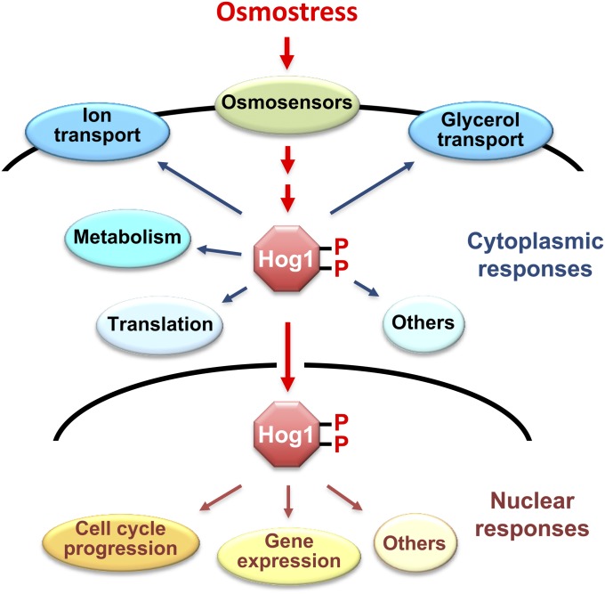
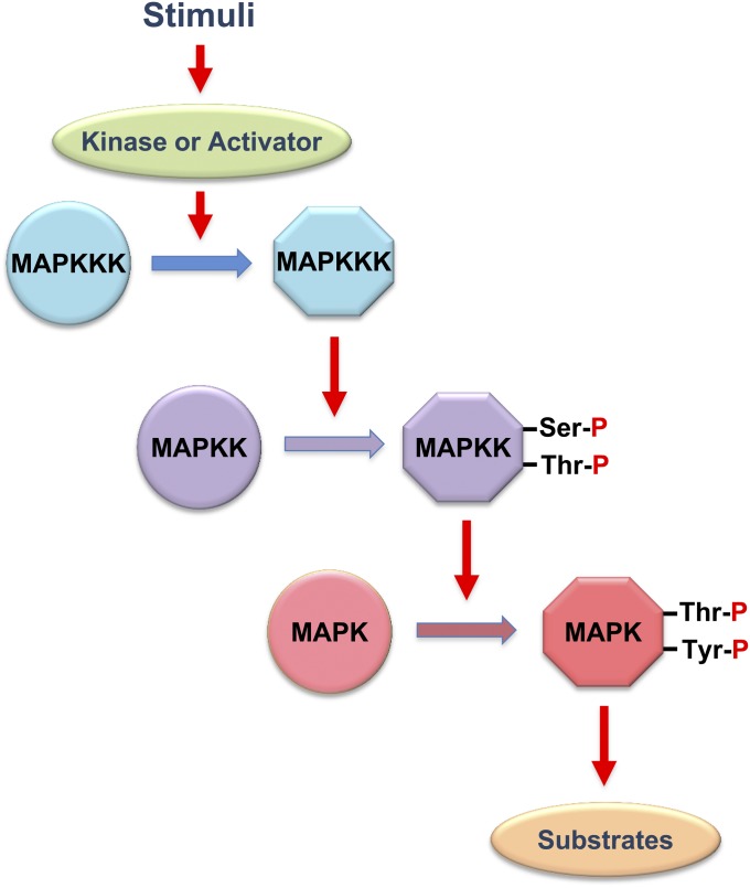
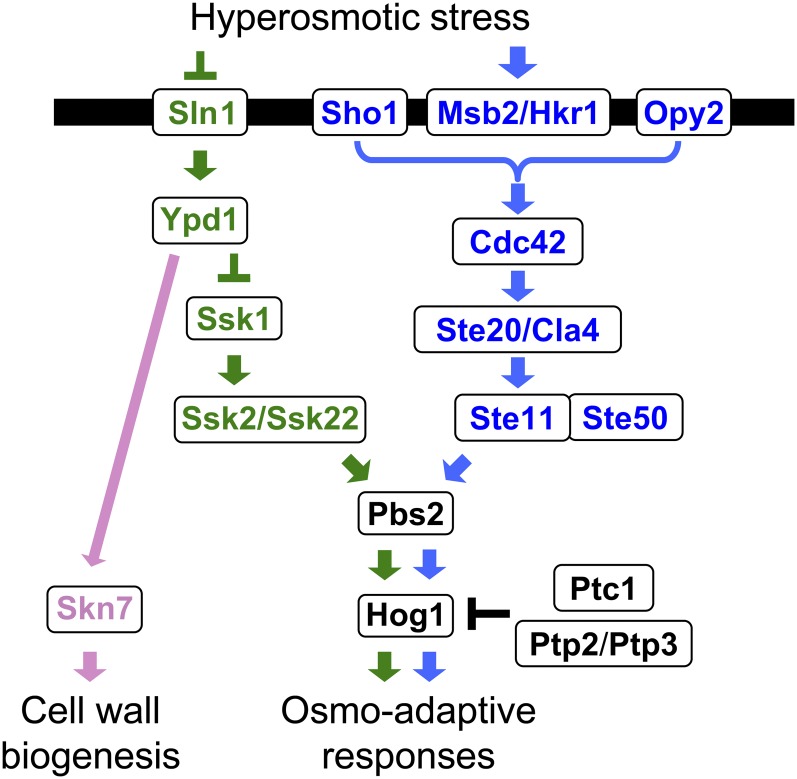
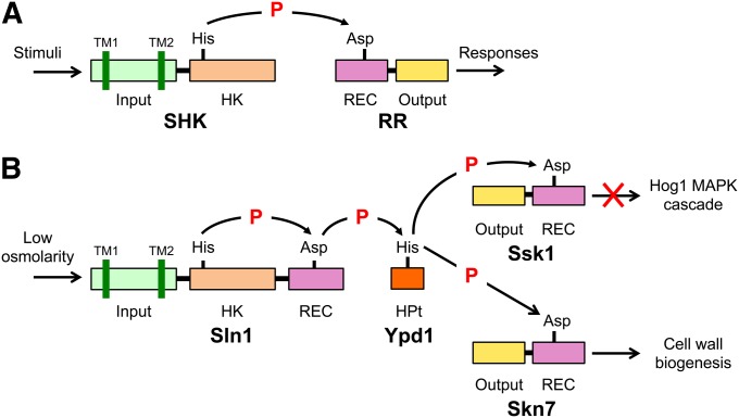
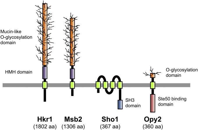
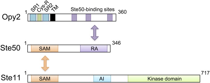
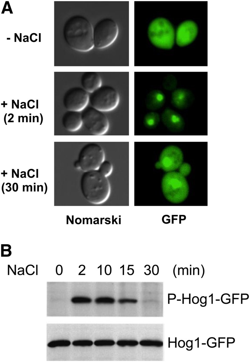
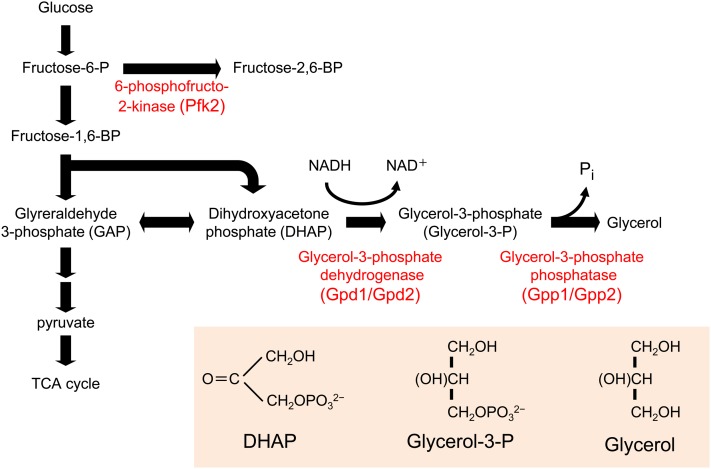
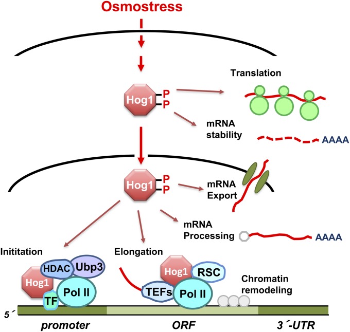
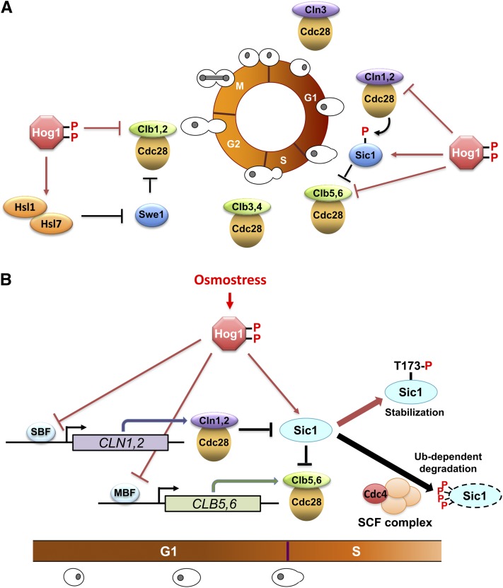
An appropriate response and adaptation to hyperosmolarity, i.e., an external osmolarity that is higher than the physiological range, can be a matter of life or death for all cells. It is especially important for free-living organisms such as the yeast Saccharomyces cerevisiae. When exposed to hyperosmotic stress, the yeast initiates a complex adaptive program that includes temporary arrest of cell-cycle progression, adjustment of transcription and translation patterns, and the synthesis and retention of the compatible osmolyte glycerol. These adaptive responses are mostly governed by the high osmolarity glycerol (HOG) pathway, which is composed of membrane-associated osmosensors, an intracellular signaling pathway whose core is the Hog1 MAP kinase (MAPK) cascade, and cytoplasmic and nuclear effector functions. The entire pathway is conserved in diverse fungal species, while the Hog1 MAPK cascade is conserved even in higher eukaryotes including humans. This conservation is illustrated by the fact that the mammalian stress-responsive p38 MAPK can rescue the osmosensitivity of hog1Δ mutations in response to hyperosmotic challenge. As the HOG pathway is one of the best-understood eukaryotic signal transduction pathways, it is useful not only as a model for analysis of osmostress responses, but also as a model for mathematical analysis of signal transduction pathways. In this review, we have summarized the current understanding of both the upstream signaling mechanism and the downstream adaptive responses to hyperosmotic stress in yeast.


Figures

Figure 1
Figure 1
Osmo-adaptive responses in yeast. In response to an increase in extracellular osmolarity, the Hog1 MAPK is activated, which leads to the induction of cytoplasmic and nuclear adaptive responses. Cytoplasmic responses include the control of ionic fluxes and glycerol transport, metabolic enzymes, and protein translation. Nuclear responses include the modulation of cell-cycle progression and the control of gene expression.
Figure 2
Figure 2
A schematic diagram of the MAP kinase module. Circles and hexagons represent, respectively, inactive and active forms of kinases. MAPK, MAP kinase; MAPKK, MAPK kinase; MAPKKK, MAPKK kinase.
Figure 3
Figure 3
A schematic diagram of the yeast HOG pathway. The protein names separated by a thrash (/) are functionally redundant. Proteins that are specific to the Sln1 branch are colored green, those that are specific to the Sho1 branch are colored blue, and those that are common are colored black. The black horizontal bar represents the plasma membrane. Arrows indicate activation, whereas the T-shaped bars represent inhibition.
Figure 4
Figure 4
Schematic diagram of two-component signaling systems. (A) The prototypical two-component system that is characterized by the conserved phosphotransfer reaction between a histidine residue and an aspartate residue. (B) The Sln1-Ypd1-Ssk1 multistep phosphorelay. SHK, sensor histidine kinase; RR, response regulator; HK histidine kinase domain; REC, receiver domain; HPt, histidine-containing phospho-transfer protein; TM, transmembrane segment; P, phosphoryl group.
Figure 5
Figure 5
Schematic representations of the four transmembrane proteins involved in the Sho1 branch of the HOG pathway. HMH, Hkr1-Msb2 homology domain. Not drawn to scale.
Figure 6
Figure 6
Schematic diagram of the Ste11/Ste50/Opy2 complex. Ste11 and Ste50 bind together through their SAM domains, whereas the RA domain of Ste50 binds to any of three binding sites in Opy2. AI, autoinhibitory domain; Cys-R, cysteine-rich domain; SR, Serine rich domain; TM, transmembrane domain.
Figure 7
Figure 7
Transient phosphorylation and nuclear localization of the Hog1 MAPK after osmostress. GFP-tagged Hog1 (Hog1-GFP) was expressed in a hog1Δ host strain, and cells were exposed to 0.4 M NaCl for the time indicated. (A) Hog1-GFP was detected by fluorescence microscopy (GFP), while the cell shape was pictured by differential interference contrast microscopy (Nomarski). (B) Total Hog1-GFP and phosphorylated Hog1-GFP were detected by immunoblotting using, respectively, anti-GFP and anti-phosphotyrosine antibody. Modified from Ferrigno et al., 1998.
Figure 8
Figure 8
Glycerol biosynthetic pathway. Glycerol is synthesized from an intermediate in the glycolysis, dihydroxyacetone phosphate (DHAP), by two-step enzymatic reactions. The first enzyme is glycrol-3-phosphate dehydrogenase (Gpd1/Gpd2), which reduces DHAP using NADH as reducing agent. The second enzyme is glycerol-3-phosphate phosphatase (Gpp1/Gpp2), which removes phosphate from glycerol-3-P to generate glycerol.
Figure 9
Figure 9
Control of mRNA biogenesis by the Hog1 MAPK. Once activated upon osmostress, Hog1 controls many aspects of mRNA biogenesis both in the nucleus and in the cytoplasm. Hog1 phosphorylates and activates transcription factors (TFs). Remarkably, Hog1 associates to loci of stress-responsive genes to modulate both initiation and elongation. Hog1 also seems to control mRNA processing, nuclear export, translation and mRNA stability.
Figure 10
Figure 10
Control of the cell-cycle progression by the Hog1 MAPK. (A) The dominant species of the cyclin/Cdc28 complex at each cell-cycle phase are shown around the circle that represents the cell cycle (G1→S→G2→M). Once activated by osmostress, Hog1 seems to modulate all phases of the cell cycle. In the G1 and G2 phases, Hog1 controls cell-cycle regulators both directly and indirectly, and Hog1 also regulates expression of cyclins. Hog1 also modulates the S and M phases, but the mechanisms remain unclear (not shown). (B) Details of the control of the G1/S transition by Hog1. The transition from G1 to S phase is mediated by the expression of cyclins Cln1,2 and Clb5,6, and their binding to the Cdc28 kinase. Initially, Clb5,6/Cdc28 is inhibited by the Sic1 (CDKi). As the activity of Cln1,2/Cdc28 increases, Sic1 is phosphorylated at multiple sites, prompting ubiquitination of Sic1 by the SCF (Cdc4) complex, and its degradation by proteasome. This degradation of Sic1 releases active Clb5,6/Cdc28, which then promotes DNA replication. Osmostress-activated Hog1 delays G1/S transition both by inhibiting transcription of cyclin genes (both CLN and CLB), and by directly phosphorylating Sic1 at Thr-173, which inhibits ubiquitination of Sic1 and stabilizes Sic1.

References

    1. Adrover M., Zi Z., Duch A., Schaber J., González-Novo A., et al. , 2011. Time-dependent quantitative multicomponent control of the G1-S network by the stress-activated protein kinase Hog1 upon osmostress. Sci. Signal. 4: ra63. - PubMed
    1. Aguilera A., 2002. The connection between transcription and genomic instability. EMBO J. 21: 195–201 - PMC - PubMed
    1. Aguilera A., 2005. mRNA processing and genomic instability. Nat. Struct. Mol. Biol. 12: 737–738 - PubMed
    1. Aguilera A., Gómez-González B., 2008. Genome instability: a mechanistic view of its causes and consequences. Nat. Rev. Genet. 9: 204–217 - PubMed
    1. Albertyn J., Hohmann S., Thevelein J. M., Prior B. A., 1994. GPD1, which encodes glycerol-3-phosphate dehydrogenase, is essential for growth under osmotic stress in Saccharomyces cerevisiae, and its expression is regulated by the high-osmolarity glycerol response pathway. Mol. Cell. Biol. 14: 4135–4144 - PMC - PubMed

Publication types

MeSH terms

Substances