Review
. 2012 Oct;192(2):319-60.
doi: 10.1534/genetics.112.140467.

The ubiquitin-proteasome system of Saccharomyces cerevisiae

Affiliations
Review

The ubiquitin-proteasome system of Saccharomyces cerevisiae

Daniel Finley et al. Genetics. 2012 Oct.

Abstract

Protein modifications provide cells with exquisite temporal and spatial control of protein function. Ubiquitin is among the most important modifiers, serving both to target hundreds of proteins for rapid degradation by the proteasome, and as a dynamic signaling agent that regulates the function of covalently bound proteins. The diverse effects of ubiquitylation reflect the assembly of structurally distinct ubiquitin chains on target proteins. The resulting ubiquitin code is interpreted by an extensive family of ubiquitin receptors. Here we review the components of this regulatory network and its effects throughout the cell.


Figures

Figure 1
Figure 1
Protein ubiquitylation. (A) Ubiquitin is activated by E1 in an ATP-dependent step, transferred to the active site cysteine in an ubiquitin-conjugating enzyme (E2), and covalently attached to substrate proteins. Substrate selection depends on ubiquitin ligases (E3). Conjugation of a single ubiquitin molecule generates monoubiquitylated proteins. Repeated rounds of ubiquitin activation and conjugation lead to multi- or polyubiquitylated proteins. (B) Different polyubiquitin chain topologies can be synthesized depending on the specific lysine residue in ubiquitin used for chain formation. Three of the eight possible unbranched chain topologies (K6, K11, K27, K29, K33, K48, K63, and linear chains), and only one type of the possible forked polyubiquitin chains are shown. (C) Structural model for synthesis of K63-linked polyubiquitin chains by Ubc13/Mms2. Mms2 positions the acceptor ubiquitin with K63 in proximity to the active site cysteine of Ubc13. Figure adapted with permission from Macmillan Publishers Ltd: Chan, N. L., and C. P. Hill, 2001 Nat. Struct. Biol. 8: 650–652.
Figure 2
Figure 2
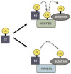
HECT and RING E3 ubiquitin ligases. Substrate ubiquitylation with HECT E3s involves an E3∼Ub thioester intermediate. Ubiquitin is transferred from the HECT E3 to the substrate. RING E3s typically do not form thioester intermediates but promote ubiquitin conjugation by bridging the interaction between E2 and substrate proteins. RING E3s also stimulate E2 activity. A subclass of RING-based ligases, the RING-in-between-RING (RBR) proteins, function like RING/HECT hybrids and form thioester intermediates. This mechanism remains to be confirmed for putative yeast RBR ligases.
Figure 3
Figure 3
Cullin RING ligases (CRLs). A large class of multisubunit RING-based ligases is nucleated around cullins. Yeast has three classes of CRLs formed with the cullins Cdc53 (cullin 1), Cul3, and Rtt101 (functionally similar to human Cul4). The C-terminal regions of cullins bind the RING protein Hrt1/Rbx1/Roc1, and the N-terminal portions interact with specific adaptor proteins (Skp1, Elc1, and Mms1), which recruit substrate receptor proteins (F-box, SOCS-box, or DCAF proteins). Putative substrate receptors are listed in Table 3.
Figure 4
Figure 4
Proteasome core particle. (A) Space-filling exterior view of the CP, with subunits differentiated by color. Note the α7β7β7α7 organization. (B) Medial cut-away view of the CP, showing the interior cavity and active sites (red) sequestered within it. The substrate transloction channel is fully closed in the crystal structure of the free CP, but brackets indicate the approximate position of the channel in its open state. (C) Detail of the CP gate. The N-terminal tails of the α subunits, particularly α2, α3, and α4, as shown, block substrate access. The bodies of the α subunits are rendered in gray. Arrow indicates the movement of the tails that constitutes gate opening, a likely upward and outward migration (Förster et al. 2003). Images modified from Groll et al. 1997 and Tian et al. 2011, with permission.
Figure 5
Figure 5
The proteasome holoenzyme. (A) Model of the Rpt ring of the proteasome in association with the yeast CP. Medial cut-away view, with the Rpt ring modeled from observations of the PAN ATPase from Archaea (adapted from Zhang et al. 2009b, with permission). The ATPase domain of the Rpt ring and the smaller OB domain above it both in blue. Coiled-coil elements (turquoise) emerge distally from the OB domain with their trajectory influenced by Pro91 (pink). The CP is in green, with proteolytic sites in red. Slice surfaces of the CP and Rpt ring are in black. The presumptive substrate translocation channel is demarcated with yellow lines: The entry port of the translocation channel is thought to be the OB ring, and substrates must migrate to the proteolytic active sites (red) to be hydrolyzed. The driving force for translocation is thought to be axial motions of the pore loops from the ATPase domain that line the translocation channel (gold rectangles). (B) Tilted view of the RP based on EM studies (Lander et al. 2012). The Rpt ring and CP are colored as in A. The DUB Rpn11 is in turquoise, with the presumptive substrate entry port directly beneath it (red-orange). The ubiquitin receptor Rpn13 is in orange. To its left is Ubp6 (approximate position), contacting Rpn1. To the right is Rpn10, with its Von Willebrand A (VWA) domain in yellow and its ubiquitin-binding UIM domain in red. All other RP subunits are in gray. Shown for comparison at upper right is free ubiquitin (pink). (C) Lateral view of the RP (derived from Lander et al. 2012). Highlighted are Rpn1 (red-orange), Rpn2 (pink), Rpn13 (orange), and Rpn10 (yellow). Lid subunits are in gray. B and C are from Tian et al. (2012), with permission.
Figure 6
Figure 6
Structure of p97/Cdc48. Left: Ribbon representations of full-length p97. Top and side views are shown. The N, D1, and D2 domains are indicated in different colors. Right: Ribbon representations of p97 N and D1 domains interacting with p47. Top and side views, as at left. These images were reproduced with permission from Dreveny et al. (2004).
Figure 7
Figure 7
HRD ubiquitin ligase. (A) HRD ubiquitin ligase consists of six core subunits: Hrd1 exposes a RING-finger domain on the cytoplasmic surface of the ER membrane and acts together with the E2 enzymes Ubc7/Cue1 and Ubc1 (both not depicted). Hrd3 together with Yos9 forms the ER luminal domain of the ligase complex. Usa1 bridges Hrd1 with Der1. Ubx2 binds Hrd1 and also, via a UBX domain, Cdc48. The transmembrane organization of the ligase complex suggests that it connects ER-luminal quality-control functions, dislocation, ubiquitylation, and the generation of pulling forces with proteolysis by the proteasome. (B) Hypothetical model of how the ER-luminal domain of the HRD ligase selects ERAD substrates. The glycans of misfolded proteins are processed by Htm1 to generate the glycan signal Man7GlcNAc2. Hrd3 first binds the misfolded protein in a “recruitment step” (left). Then Yos9 controls the identity of the glycan signal in a “commitment step” (center). Only when both interactions are productive is the client protein dislocated into the cytoplasm for proteasomal digestion.
Figure 8
Figure 8
Modifications of the replication factor PCNA. During undisturbed replication, PCNA (blue ring shape) promotes processive DNA synthesis by replicative polymerases δ and ε (Pol δ/ε), and is modified by SUMO (red lollipop shape). The modification prevents binding of Eco1, but causes the recruitment of Elg1 and Srs2. Srs2 prevents the formation of the recombinogenic Rad51 filament (51), inhibiting unscheduled recombination at replication forks. Upon damage-induced replication fork stalling, PCNA is modified by mono- and polyubiquitin (black lollipop shapes) at postreplicative daughter-strand gaps. Monoubiquitylation recruits damage-tolerant DNA polymerases (TLS) for translesion synthesis, while K63 polyubiquitylation causes recruitment of Mgs1 and initiates damage bypass by template switching in an unknown manner. Conjugating enzymes, ligases, and DUBs are highlighted in shades of purple, green, and pink, respectively.
Figure 9
Figure 9
Ubiquitylation during nucleotide excision repair. (A) For global genome repair, lesions are recognized by Rad4 in complex with Rad23. Ubiquitylation of Rad4 is important for subsequent steps of repair. Ubiquitylated Rad4 is degraded by the proteasome. (B) Lesions on the transcribed strand of actively expressed genes are repaired by transcription-coupled repair, where RNA polymerase II (RNA Pol II) contributes to lesion recognition. Following removal of the enzyme by the action of Rad26, strand unwinding, excision of the lesion and resynthesis proceed as in global genome repair. (C) An irreversibly stalled RNA polymerase II is targeted for ubiquitylation and proteasomal degradation in a Def1-dependent manner. This frees the lesion and allows global genome repair. Conjugating enzymes, ligases, and DUBs are highlighted in shades of purple, green, and pink, respectively. Distinct polyubiquitin chain linkages are indicated as K48 or K63.

References

    1. Albert T. K., Hanzawa H., Legtenberg Y. I., De Ruwe M. J., Van Den Heuvel F. A., et al. , 2002. Identification of a ubiquitin-protein ligase subunit within the CCR4-NOT transcription repressor complex. EMBO J. 21: 355–364 - PMC - PubMed
    1. Amerik A. Y., Swaminathan S., Krantz B. A., Wilkinson K. D., Hochstrasser M., 1997. In vivo disassembly of free polyubiquitin chain by yeast Ubp14 modulates rates of protein degradation by the proteasome. EMBO J. 16: 4826–4838 - PMC - PubMed
    1. Amerik A. Y., Nowak J., Swaminathan S., Hochstrasser M., 2000a The Doa4 deubiquitinating enzyme is functionally linked to the vacuolar protein-sorting and endocytic pathways. Mol. Biol. Cell 11: 3365–3380 - PMC - PubMed
    1. Amerik A. Y., Li S. J., Hochstrasser M., 2000b Analysis of the deubiquitinating enzymes of the yeast Saccharomyces cerevisiae. Biol. Chem. 381: 981–992 - PubMed
    1. Amerik A., Sindhi N., Hochstrasser M., 2006. A conserved late endosome-targeting signal required for Doa4 deubiquitylating enzyme function. J. Cell Biol. 175: 825–835 - PMC - PubMed

Publication types

MeSH terms