

Direct patterning of coplanar polyethylene glycol alkylsilane monolayers by deep-ultraviolet photolithography as a general method for high fidelity, long-term cell patterning and culture
- PMID: 23028211
- PMCID: PMC3427986
- DOI: 10.1116/1.3549127
Direct patterning of coplanar polyethylene glycol alkylsilane monolayers by deep-ultraviolet photolithography as a general method for high fidelity, long-term cell patterning and culture
Abstract
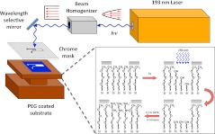
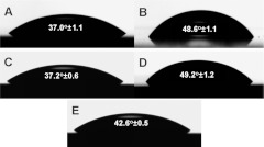
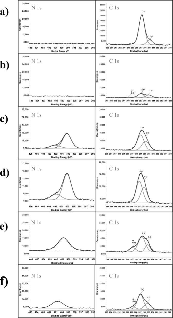
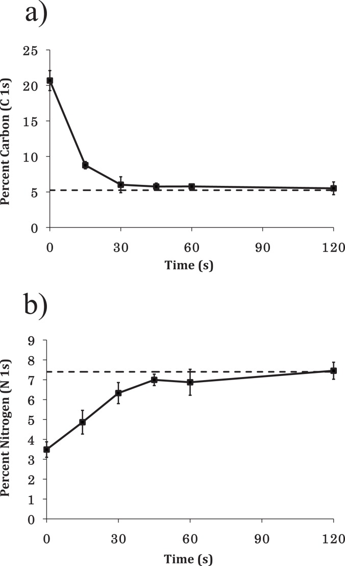
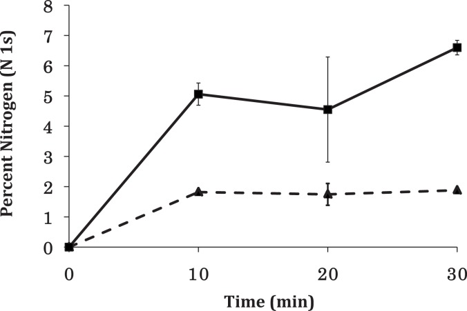
This manuscript details a general method for patterning coplanar alkylsilane monolayers using deep-ultraviolet photolithography that has broad application for high fidelity patterning of cells of varying phenotype in long-term cultures. A polyethylene glycol monolayer was formed on a silica substrate and then patterned using 193 nm light from an ArF excimer laser. The regions of photoablation were then rederivatized with (3-trimethoxysilyl propyl) diethyltriamine (DETA), yielding high contrast cytophilic islands that promoted cell adhesion and growth. Rat hippocampal neurons, motoneurons, and myoblasts were then cultured in a defined, serum-free medium on the patterned surfaces for periods in excess of 40 days. This approach has been shown to be useful as a general method for the long-term culture of multiple cell types in highly defined spatial patterns and can be used for supporting complex cocultures for creating in vitro models for biological systems.
Figures

Similar articles
-
Two cell circuits of oriented adult hippocampal neurons on self-assembled monolayers for use in the study of neuronal communication in a defined system.ACS Chem Neurosci. 2013 Aug 21;4(8):1174-82. doi: 10.1021/cn300206k. Epub 2013 May 20. ACS Chem Neurosci. 2013. PMID: 23611164 Free PMC article.
-
Precise cell patterning using cytophobic self-assembled monolayer deposited on top of semi-transparent gold.Biomed Microdevices. 2010 Oct;12(5):935-48. doi: 10.1007/s10544-010-9448-8. Biomed Microdevices. 2010. PMID: 20571865
-
Electrophysiological and immunocytochemical characterization of DRG neurons on an organosilane surface in serum-free medium.In Vitro Cell Dev Biol Anim. 2008 May-Jun;44(5-6):162-8. doi: 10.1007/s11626-008-9097-x. Epub 2008 May 14. In Vitro Cell Dev Biol Anim. 2008. PMID: 18478304
-
Patterning Quantum Dots via Photolithography: A Review.Adv Mater. 2023 Oct;35(41):e2300546. doi: 10.1002/adma.202300546. Epub 2023 Aug 2. Adv Mater. 2023. PMID: 36892995 Review.
-
Self-Assembled Monolayers as Patterning Tool for Organic Electronic Devices.Adv Mater. 2017 May;29(18). doi: 10.1002/adma.201605286. Epub 2017 Feb 3. Adv Mater. 2017. PMID: 28160336 Review.
Cited by
-
Myelination and node of Ranvier formation on sensory neurons in a defined in vitro system.In Vitro Cell Dev Biol Anim. 2013 Sep;49(8):608-618. doi: 10.1007/s11626-013-9647-8. Epub 2013 Aug 16. In Vitro Cell Dev Biol Anim. 2013. PMID: 23949775 Free PMC article.
-
Validation of a functional human AD model with four AD therapeutics utilizing patterned iPSC-derived cortical neurons integrated with microelectrode arrays.Res Sq [Preprint]. 2024 May 20:rs.3.rs-4313679. doi: 10.21203/rs.3.rs-4313679/v1. Res Sq. 2024. Update in: Sci Rep. 2024 Oct 22;14(1):24875. doi: 10.1038/s41598-024-73869-9. PMID: 38826367 Free PMC article. Updated. Preprint.
-
Classical Complement Pathway Inhibition in a "Human-On-A-Chip" Model of Autoimmune Demyelinating Neuropathies.Adv Ther (Weinh). 2022 Jun;5(6):2200030. doi: 10.1002/adtp.202200030. Epub 2022 Apr 5. Adv Ther (Weinh). 2022. PMID: 36211621 Free PMC article.
-
Morphological and functional characterization of human induced pluripotent stem cell-derived neurons (iCell Neurons) in defined culture systems.Biotechnol Prog. 2015 Nov-Dec;31(6):1613-22. doi: 10.1002/btpr.2160. Epub 2015 Sep 11. Biotechnol Prog. 2015. PMID: 26317319 Free PMC article.
-
Self-contained, low-cost Body-on-a-Chip systems for drug development.Exp Biol Med (Maywood). 2017 Nov;242(17):1701-1713. doi: 10.1177/1535370217694101. Epub 2017 Feb 17. Exp Biol Med (Maywood). 2017. PMID: 29065797 Free PMC article. Review.
References
-
- Ratner B., Hoffman A., Schoen F., and Lemons J., Biomaterials Science: An Introduction to Materials in Medicine (Elsevier Academic, Amsterdam, 2004).
-
- Capadona J. R., Collard D. M., and Garcia A. J., Langmuir LANGD5 19, 1847 (2003).10.1021/la026244+ - DOI
-
- Michael K. E., Vernekar V. N., Keselowsky B. G., Meredith J. C., Latour R. A., and Garcia A. J., Langmuir LANGD5 19, 8033 (2003).10.1021/la034810a - DOI
Grants and funding
LinkOut - more resources
Full Text Sources
Other Literature Sources