. 2012 Sep;8(9):e1002948.
doi: 10.1371/journal.ppat.1002948. Epub 2012 Sep 20.

A unique protein phosphatase with kelch-like domains (PPKL) in Plasmodium modulates ookinete differentiation, motility and invasion

Affiliations

A unique protein phosphatase with kelch-like domains (PPKL) in Plasmodium modulates ookinete differentiation, motility and invasion

David S Guttery et al. PLoS Pathog. 2012 Sep.

Abstract

Protein phosphorylation and dephosphorylation (catalysed by kinases and phosphatases, respectively) are post-translational modifications that play key roles in many eukaryotic signalling pathways, and are often deregulated in a number of pathological conditions in humans. In the malaria parasite Plasmodium, functional insights into its kinome have only recently been achieved, with over half being essential for blood stage development and another 14 kinases being essential for sexual development and mosquito transmission. However, functions for any of the plasmodial protein phosphatases are unknown. Here, we use reverse genetics in the rodent malaria model, Plasmodium berghei, to examine the role of a unique protein phosphatase containing kelch-like domains (termed PPKL) from a family related to Arabidopsis BSU1. Phylogenetic analysis confirmed that the family of BSU1-like proteins including PPKL is encoded in the genomes of land plants, green algae and alveolates, but not in other eukaryotic lineages. Furthermore, PPKL was observed in a distinct family, separate to the most closely-related phosphatase family, PP1. In our genetic approach, C-terminal GFP fusion with PPKL showed an active protein phosphatase preferentially expressed in female gametocytes and ookinetes. Deletion of the endogenous ppkl gene caused abnormal ookinete development and differentiation, and dissociated apical microtubules from the inner-membrane complex, generating an immotile phenotype and failure to invade the mosquito mid-gut epithelium. These observations were substantiated by changes in localisation of cytoskeletal tubulin and actin, and the micronemal protein CTRP in the knockout mutant as assessed by indirect immunofluorescence. Finally, increased mRNA expression of dozi, a RNA helicase vital to zygote development was observed in ppkl(-) mutants, with global phosphorylation studies of ookinete differentiation from 1.5-24 h post-fertilisation indicating major changes in the first hours of zygote development. Our work demonstrates a stage-specific essentiality of the unique PPKL enzyme, which modulates parasite differentiation, motility and transmission.


Conflict of interest statement

The authors have declared that no competing interests exist.

Figures

Figure 1
Figure 1. Phylogenetic analysis of Plasmodium PPKL.
A. Schematic representations of the predicted protein architectures (Pfam domains) of the PPKL orthologues in A. thaliana and P. falciparum. B. Bayesian phylogeny of kelch-like phosphatases. Tree shown is the consensus of trees from four independent inferences based on a trimmed alignment of the phosphatase domain. Numbers beside nodes represent support from Bayesian posterior probabilities.
Figure 2
Figure 2. Expression of PPKL-GFP during the Plasmodium life-cycle in the mosquito and PPKL-GFP phosphatase activity.
A. Wild-type mRNA expression of ppkl relative to hsp70 and arginyl-tRNA synthetase as endogenous controls (ΔΔCt method). Error bar = ±SEM, n = 3 from three independent experiments. ASB = asexual stages in blood; NAG = non-activated gametocytes; AG = activated gametocytes. B. PPKL-GFP expression in transgenic parasites during the sexual phase of the life-cycle and sporogony. Bar = 5 µm. Note that in gametocytes (the male gametocyte contains the enlarged nucleus) only the female expresses PPKL-GFP. C. Upper panel: phosphatase activity in immunoprecipitated lysates of PPKL-GFP and WT parasite lines in the presence or absence of MFP substrate. Error bar = ±SEM, n = 6. Lower panel: anti-GFP Western blot showing amounts of PPKL-GFP retained from the corresponding lysate.
Figure 3
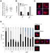
Figure 3. Gametocyte activation and ookinete differentiation of ppkl mutants.
A. Exflagellation of activated male gametocytes of ppkl mutants compared to wild-type (bar = arithmetic mean, n = 20, ×40 magnification). B. Ookinete conversion in wild-type and ppkl parasites. Conversion rate is the percentage of P28 (ookinete surface protein)-positive parasites that had differentiated into ‘banana-shaped’ ookinetes (error bar = ±SD; n = 3). C. Different morphological shapes of ppkl abnormal retorts as assessed by P28 staining. Bar = 1 µm. D. Time-course analysis of ookinete differentiation in wild-type and ppkl parasites. Morphologies were grouped into four categories: unfertilized macrogametes/zygotes (round – black bars); stages I–III (gray bars); abnormal/retorts (light blue bars); and stages IV–VI (white bars). Error bar = ±SD, n = 3. Panels to the right of the graph show ookinetes at different stages of maturity immunolabelled with the anti-P28 Cy3-conjugated 13.1 antibody used for scoring. Bar = 5 µm.
Figure 4
Figure 4. Genetic crossing, in vivo transmission and gliding motility of ppkl parasites.
A. Ookinete conversion after crossing ppkl mutants with female-defective (nek4) or male-defective (map2) mutants. Wild-type parasites were used as a control. Bar graph shows the percentage of round P28-positive parasites that had converted into elongated ookinetes and retorts (error bar = ±SD; n = 3). B. Average number of oocysts per mosquito gut (day 14 post-infection; bar = arithmetic mean; n = 60 of wild-type or ppkl infected mosquitoes from three independent experiments). Overall infection prevalence was 85% for wild-type and 0% for ppkl. C. Representative frames from time-lapse videos of a wild-type ookinete (upper panels) and ppkl abnormal retort (lower panels) in Matrigel. Black arrow indicates the apical end of the ookinete/abnormal retort. Bar = 10 µm. Speed of individual wild-type ookinetes or ppkl abnormal retorts from 24 h ookinete cultures measured over a 10 min period is shown in the dot plot. Bar = arithmetic mean; n = 17 for wild-type and 28 for ppkl lines.
Figure 5
Figure 5. Ultrastructure analysis and indirect immunofluorescence of ookinetes.
A. (i) Transmission electron micrograph (TEM) of a longitudinal section though a wild-type ookinete showing the conical apical end. The cytoplasm contains a number of apically located micronemes (Mn), a more posteriorly located nucleus (N) and a central crystalloid body (Cr). Bar = 1 µm. (ii) TEM of a longitudinal section though an ookinete of the ppkl mutant showing partial collapse and elongation of the apical end. Within the cytoplasm, the micronemes (Mn) are more randomly distributed. Bar = 1 µm. (iii) Enlargement of the anterior of a wild-type ookinete illustrating the complex nature of the apical end consisting of a conical shaped electron dense collar (C) with a central aperture. Underlying the collar and in contact with it is a less electron dense ring (R) that is also in contact with longitudinally running sub-pellicular microtubules (Mt). Micronemes (Mn) are located within the cytoplasm with small ducts (D) running though the apical aperture to the plasmlemma. Bar = 100 nm. (iv) Detail of the anterior of a ppkl ookinete showing a less conical shape associated with a reduction in the length of the electron dense collar (C) and some separation (arrow) from the electron lucent ring (R) with associated microtubules (Mt). The few micronemes (Mn) present showed ducts (D) running to the anterior. Bar = 100 nm. (v) Part of a cross section though the anterior end of a wild-type ookinete showing the specific inter-relationship between the microtubules (arrowheads) and the inner membrane complex (IMC). Bar = 100 nm. (vi) Cross-section though a ppkl ookinete illustrating microtubules (arrowheads) within the cytoplasm that have lost contact with the IMC. Bar = 100 nm. (vii) Longitudinal section though the apical end of a severely affected ppkl ookinete showing the collapsed and elongate neck-like structure containing longitudinally running microtubules (Mt). Bar = 100 nm. (viii) Cross section though the collapsed neck region showing it to consist of microtubules. Bar = 100 nm. B. Indirect immunofluorescence detection of a number of cytoskeletal, glideosomal and micronemal proteins in wild-type ookinetes and ppkl abnormal retorts (24 h) post-gametocyte activation. Abnormally intense tubulin staining was observed at the ookinetes apical end (red). Motor protein GAP45 and MTIP distributions (green) did not show any difference in the mutant compared to wild-type. The apical localisation of actin and micronemal CTRP was more diffuse throughout the cell body (red) in the ppkl mutant. However, the usual diffuse intracellular staining of SOAP showed no obvious abnormal pattern in the mutant. The nucleus was counter-stained using DAPI (blue). Arrows indicate the apical end of the ookinete. Bar = 5 µm.
Figure 6
Figure 6. Differential transcript and phosphorylation levels in ppkl and mutants of genes essential for zygote development.
A. Relative expression of ppkl, nek4 and dozi in ppkl, nek4 and dozi mutant parasites compared to wild-type controls (Pfaffl method). Error bar = ±SEM, n = 3 from three independent experiments. ***p≤0.001; **p≤0.01. ASB = asexual blood stages; NAG = non-activated gametocytes; AG = activated gametocytes. B. Upper panel: autoradiograph showing phosphorylation in lysates of schizonts and activated gametocytes from WT-GFP and PPKL-GFP parasite lines. Lower panel: corresponding Western blot using anti-GFP antibody. Sch = schizonts; AG = activated gametocytes. C. Autoradiograph from three fractions showing alterations in global phosphorylation 1.5, 6 and 24 h post-activation of wild-type and ppkl gametocytes. Differential phosphorylation in ppkl compared to wild-type 1.5 h after gametocyte activation is represented by arrows, 6 h after gametocyte activation by an arrowhead and 24 h after gametocyte activation by an asterisk. Representative radiographs of three independent experiments are shown.

References

    1. Graves JD, Krebs EG (1999) Protein phosphorylation and signal transduction. Pharmacol Ther 82: 111–121. - PubMed
    1. Blume-Jensen P, Hunter T (2001) Oncogenic kinase signalling. Nature 411: 355–365. - PubMed
    1. Tan CS, Bodenmiller B, Pasculescu A, Jovanovic M, Hengartner MO, et al. (2009) Comparative analysis reveals conserved protein phosphorylation networks implicated in multiple diseases. Sci Signal 2: ra39. - PubMed
    1. Barford D, Das AK, Egloff MP (1998) The structure and mechanism of protein phosphatases: insights into catalysis and regulation. Annu Rev Biophys Biomol Struct 27: 133–164. - PubMed
    1. Gallego M, Virshup DM (2005) Protein serine/threonine phosphatases: life, death, and sleeping. Curr Opin Cell Biol 17: 197–202. - PubMed

Publication types

MeSH terms

LinkOut - more resources