. 2012 Sep;8(9):e1002926.
doi: 10.1371/journal.pgen.1002926. Epub 2012 Sep 20.

Cadm1 is a metastasis susceptibility gene that suppresses metastasis by modifying tumor interaction with the cell-mediated immunity

Affiliations

Cadm1 is a metastasis susceptibility gene that suppresses metastasis by modifying tumor interaction with the cell-mediated immunity

Farhoud Faraji et al. PLoS Genet. 2012 Sep.

Abstract

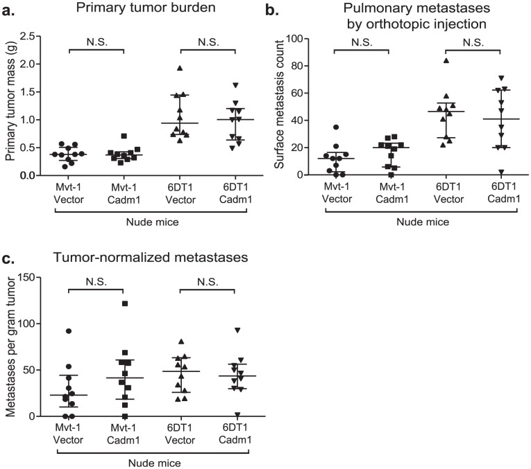
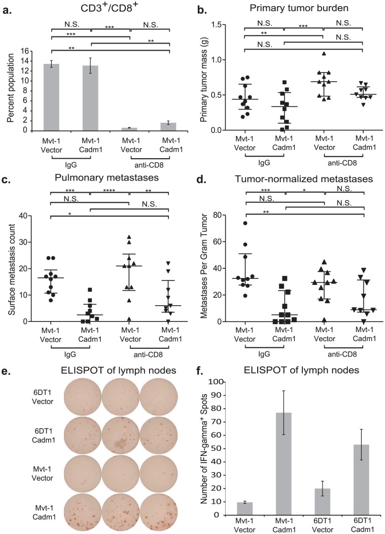
Metastasis is a complex process utilizing both tumor-cell-autonomous properties and host-derived factors, including cellular immunity. We have previously shown that germline polymorphisms can modify tumor cell metastatic capabilities through cell-autonomous mechanisms. However, how metastasis susceptibility genes interact with the tumor stroma is incompletely understood. Here, we employ a complex genetic screen to identify Cadm1 as a novel modifier of metastasis. We demonstrate that Cadm1 can specifically suppress metastasis without affecting primary tumor growth. Unexpectedly, Cadm1 did not alter tumor-cell-autonomous properties such as proliferation or invasion, but required the host's adaptive immune system to affect metastasis. The metastasis-suppressing effect of Cadm1 was lost in mice lacking T cell-mediated immunity, which was partially phenocopied by depleting CD8(+) T cells in immune-competent mice. Our data show a novel function for Cadm1 in suppressing metastasis by sensitizing tumor cells to immune surveillance mechanisms, and this is the first report of a heritable metastasis susceptibility gene engaging tumor non-autonomous factors.


Conflict of interest statement

The authors have declared that no competing interests exist.

Figures

Figure 1
Figure 1. A genetic screen in mice identifies Cadm1 as a candidate germline modifier of metastasis.
(a) NZB and C58 mice were crossed to FVB mice then re-crossed with MMTV-PyMT-transgenic FVB mice. (b) Primary tumor, pulmonary metastasis, and genotype data was obtained from transgene-positive mice developed tumors and was used to generate QTL maps for pulmonary metastasis. Subcongenic lines containing fragments of NZB chromosome 9 (c) were crossed to MMTV-PyMT transgenic FVB mice and tumor size and pulmonary surface metastases were measured in the progeny (d). No significant change in tumor mass was observed, however a significant increase in metastasis was observed in the N9(49–60) subcongenic line (p = 0.022), resolving the locus of susceptibility was resolved to 49–60 Mb of chromosome 9. (e) Real time quantitative PCR measurement of cDNA from mammary tumor and lung tissue demonstrated a 1.5 fold higher expression of Cadm1 in NZB and NF9 relative to FVB.
Figure 2
Figure 2. The effect of Cadm1 over-expression on tumor growth and metastasis in vivo.
(a) Expression of endogenous and exogenous V5-epitope tagged Cadm1 protein (IB, immunoblot) and total protein by ECL densitometry on immunoblot. (d) Total RNA levels of Cadm1 measured by qRT-PCR (mean +/− standard error of the mean [SEM]). Mvt-1 and 6DT1 mouse mammary tumor cells stably expressing Cadm1 were implanted into mammary fat pads of syngeneic FVB mice. Data on primary tumor burden (Mvt-1: p = 0.0031; 6DT1: p = 0.2261) (b), pulmonary surface metastases (c) was collected on day 30 (Mvt-1: p = 0.0026; 6DT1: p = 0.0089; median +/− interquartile range; n = 10 mice per group). (e) Pulmonary surface metastases were normalized by primary tumor mass to determine if Cadm1 overexpression showed an effect on metastasis independent of its potential tumor suppressive activity (Mvt-1: p = 0.0048; 6DT1: p = 0.0185; median +/− interquartile range; n = 10 mice per group). Arrows indicate several but not all visible metastatic nodules present on lung surfaces. (f) Images of whole lungs of two mice implanted with 6DT1 control cells and two mice implanted with 6DT1 cells expressing Cadm1. (g) Results of tail vein injection study (Mvt-1: p = 0.0361; 6DT1: p = 0.0475; median +/− interquartile range; n = 10 mice per group). (N.S., not significant; * defined as p<0.05; ** defined as p<0.01.)
Figure 3
Figure 3. The effect of Cadm1 knockdown on tumor growth and metastasis in vivo.
6DT1 cells expressing shRNA constructs targeting Cadm1 showed (a) protein level knockdown of Cadm1 by immunoblot and ECL densitometry quantification and (d) total RNA level knockdown (mean +/− SEM). Orthotopic implant data showing the effects of Cadm1 knockdown on primary tumor burden (shScr vs. sh14: p = 0.0090; shScr vs. sh15: p = 0.0943; sh14 vs. sh15: p = 0.0001) (b), pulmonary surface metastases (shScr vs. sh14: p = 0.0085; shScr vs. sh15: p = 0.0709; sh14 vs. sh15: p = 0.3485) (c), and tumor-normalized metastases (e) ((shScr vs. sh14: p = 0.00005; shScr vs. sh15: p = 0.0444; sh14 vs. sh15: p = 0.0152; median +/− interquartile range; n = 10 mice per group).
Figure 4
Figure 4. Pulmonary metastases of Mvt-1/Cadm1 cells express attenuated levels of Cadm1.
Matched primary tumor and lung sections were immunohistochemically (IHC) stained with antibody against the V5 epitope. Immunohistochemical staining of the primary tumors produced by control (a) and Cadm1-V5 (b) expressing cells. IHC stained lung sections of mice implanted with control (c) and Cadm1-V5 (d). Hematoxylin and eosin (H&E) stained lung sections of mice implanted with control (e) and Cadm1-V5 (f) expressing cells. Insets show 20× magnification of boxed area (L.N., lymph node; black bar indicates 1 um). Level of (g) exogenous and (h) total Cadm1 mRNA from grossly dissected pulmonary metastatic nodules of mice injected with Mvt-1/Vector versus Mvt-1/Cadm1 relative to the original cell lines prior to injection.
Figure 5
Figure 5. The effect of Cadm1 expression on tumor and metastasis in athymic mice.
Orthotopic injections of control and Cadm1-expressing cells into athymic nude mice resulted in no significant difference in (a) primary tumor burden, (b) pulmonary metastasis, or (c) tumor-normalized metastasis. (median +/− interquartile range; n = 10 mice per group).
Figure 6
Figure 6. The effect of Cadm1 expression on tumor and metastasis in CD8+ T cell depleted mice.
Orthotopic injections of control and Cadm1-expressing Mvt-1 cells into immune-competent FVB mice that were treated with IgG or anti-CD8 resulted in (a) >90% depletion of CD8+ T cells. (b) No significant differences were observed in tumor burden between control and Cadm1 expressing within the same antibody treatment group, though a significant increase was observed in control Mvt-1 cells upon anti-CD8 treatment. (c) Total pulmonary metastases showed significant suppression in Mvt-1/Cadm1 in both control and anti-CD8 treatment groups. (d) Metastases normalized by matched primary tumor mass showed highly significant Cadm1-mediated metastasis suppression in the control IgG treatment group but was not significant in the anti-CD8 group (median +/− interquartile range; n = 10 mice per group). ELISPOT plate (e) and quantitative representation (f) of interferon-γ secreting T cells (mean +/− SEM). (N.S., not significant; * defined as p<0.05; ** defined as p<0.01; *** defined as p<0.001, **** defined as p<0.0001).
Figure 7
Figure 7. The effect of Cadm1 expression levels on survival in patient datasets.
(a) Compiled results of -Log10 transformed p-values for all survival analyses. Kaplan-Meier plots of (b) estrogen receptor (ER) positive invasive ductal carcinoma (IDC) tumors from the Oncomine TCGA set, (c) ER positive tumors from the Desmedt set, (d) ER negative tumors from the Boersma set, (e) ER positive tumors from GOBO, (f) all tumors from the Boersma set.

References

    1. Steeg PS, Theodorescu D (2008) Metastasis: a therapeutic target for cancer. Nat Clin Pract Oncol 5: 206–219. - PMC - PubMed
    1. Chaffer CL, Weinberg RA (2011) A perspective on cancer cell metastasis. Science 331: 1559–1564. - PubMed
    1. Valastyan S, Weinberg RA (2011) Tumor metastasis: molecular insights and evolving paradigms. Cell 147: 275–292. - PMC - PubMed
    1. Park YG, Zhao X, Lesueur F, Lowy DR, Lancaster M, et al. (2005) Sipa1 is a candidate for underlying the metastasis efficiency modifier locus Mtes1. Nat Genet 37: 1055–1062. - PMC - PubMed
    1. Crawford NP, Alsarraj J, Lukes L, Walker RC, Officewala JS, et al. (2008) Bromodomain 4 activation predicts breast cancer survival. Proc Natl Acad Sci U S A 105: 6380–6385. - PMC - PubMed

Publication types

MeSH terms