. 2012 Sep;8(9):e1002943.
doi: 10.1371/journal.pgen.1002943. Epub 2012 Sep 20.

Characterization of inducible models of Tay-Sachs and related disease

Affiliations

Characterization of inducible models of Tay-Sachs and related disease

Timothy J Sargeant et al. PLoS Genet. 2012 Sep.

Abstract

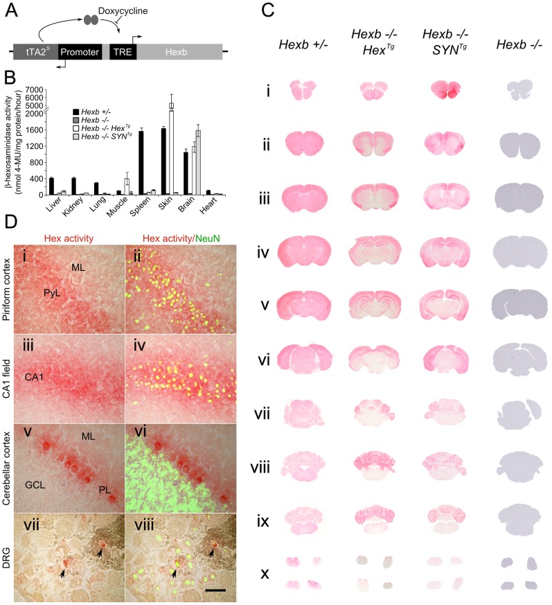
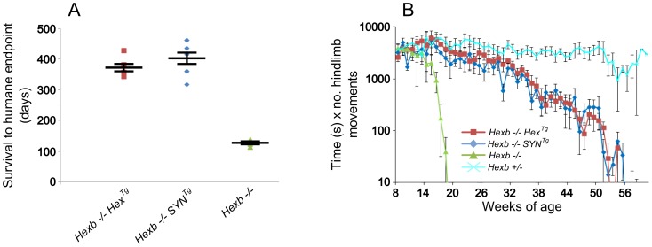
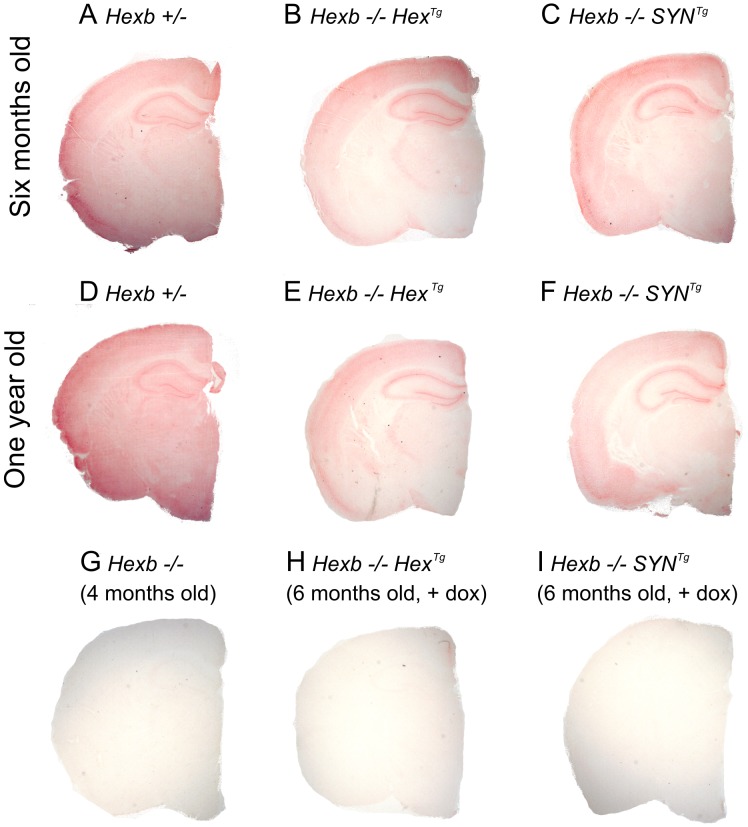
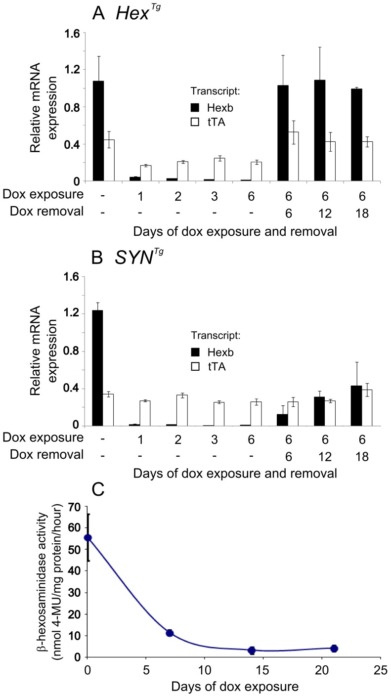
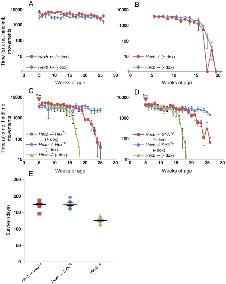
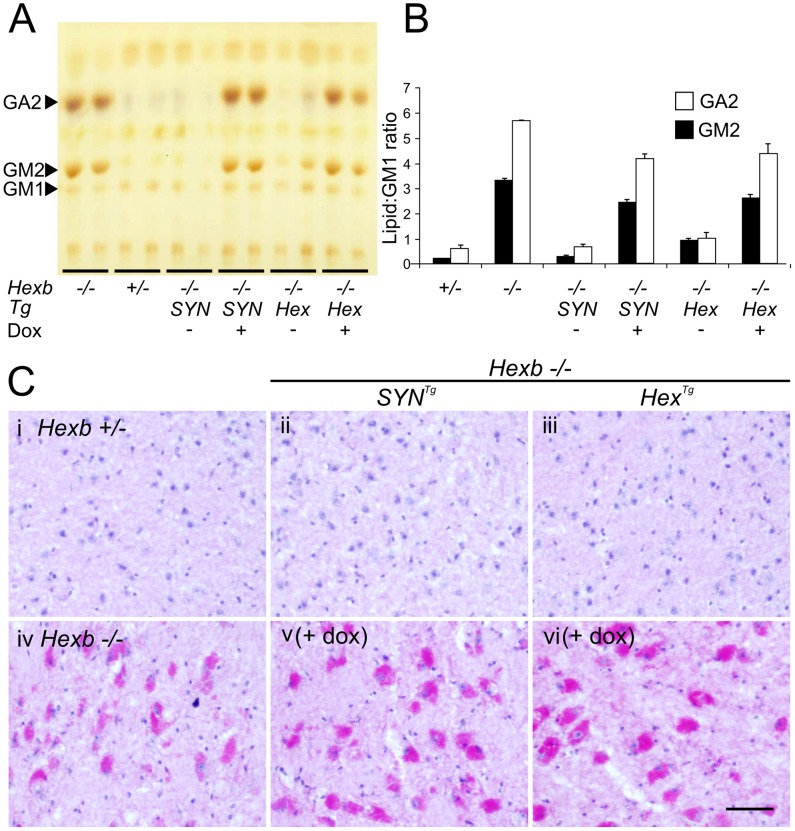
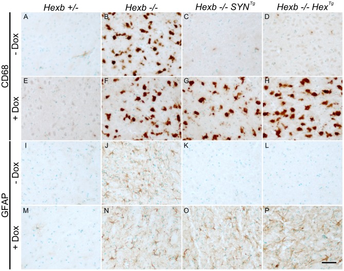
Tay-Sachs and Sandhoff diseases are lethal inborn errors of acid β-N-acetylhexosaminidase activity, characterized by lysosomal storage of GM2 ganglioside and related glycoconjugates in the nervous system. The molecular events that lead to irreversible neuronal injury accompanied by gliosis are unknown; but gene transfer, when undertaken before neurological signs are manifest, effectively rescues the acute neurodegenerative illness in Hexb-/- (Sandhoff) mice that lack β-hexosaminidases A and B. To define determinants of therapeutic efficacy and establish a dynamic experimental platform to systematically investigate cellular pathogenesis of GM2 gangliosidosis, we generated two inducible experimental models. Reversible transgenic expression of β-hexosaminidase directed by two promoters, mouse Hexb and human Synapsin 1 promoters, permitted progression of GM2 gangliosidosis in Sandhoff mice to be modified at pre-defined ages. A single auto-regulatory tetracycline-sensitive expression cassette controlled expression of transgenic Hexb in the brain of Hexb-/- mice and provided long-term rescue from the acute neuronopathic disorder, as well as the accompanying pathological storage of glycoconjugates and gliosis in most parts of the brain. Ultimately, late-onset brainstem and ventral spinal cord pathology occurred and was associated with increased tone in the limbs. Silencing transgenic Hexb expression in five-week-old mice induced stereotypic signs and progression of Sandhoff disease, including tremor, bradykinesia, and hind-limb paralysis. As in germline Hexb-/- mice, these neurodegenerative manifestations advanced rapidly, indicating that the pathogenesis and progression of GM2 gangliosidosis is not influenced by developmental events in the maturing nervous system.


Conflict of interest statement

The authors have declared that no competing interests exist.

Figures

Figure 1
Figure 1. Expression of Hexb from two inducible constructs throughout the Sandhoff mouse neuraxis.
(A) Expression of transgenic Hexb from a single autoregulatory cassette was driven by either the mouse Hexb promoter (Hex line) or the human SYN1 promoter (SYN line). Tet-transactivator expressed from tTA2s coding sequence promoted expression of Hexb cDNA from tet-response elements (TRE). This was inhibited by doxycycline. (B) Expression of transgenic Hexb in different tissues was assessed with the MUG assay. β-hexosaminidase activity was found in the brain for both Hex and SYN lines, but also in the skin and skeletal muscle for the transgenic Hex mouse line. Bars = mean ± SEM. n = 3. Panel C shows expression of β-hexosaminidase activity in Hexb−/−HexTg and Hexb−/−SYNTg mice assessed by staining using the enzyme substrate naphthol AS-BI N-acetyl-β-glucosaminide (red staining). Strong expression of β-hexosaminidase activity is seen in the cortex (C i–vi) and the cerebellum (C vii–ix) of both lines. Weaker expression is seen in the diencephalon in the SYN line (C iv–v) while activity is very weak to absent in the mid (C vi–vii) and hindbrain (C vii–ix). (D) β-hexosaminidase activity staining was associated with neurons, shown by co-labelling with NeuN (green fluorescence) in the piriform cortex (i and ii), CA1 field of Ammon's horn (iii and iv), the cerebellar cortex (v and vi) and the dorsal root ganglia (vii and viii), where only some neurons expressed transgenic β-hexosaminidase activity. Images represent staining from Hexb−/−HexTg mice (DRG and cerebellar cortex) and Hexb−/−SYNTg mice (CA1 field and piriform cortex). ML = molecular layer, PyL = pyramidal layer, CA1 = CA1 field, PL = Purkinje neuron layer, GCL = granule cell layer. Scale bar = 50 µm.
Figure 2
Figure 2. Inducible transgenic constructs rescue mice from Sandhoff disease.
(A) Hexb−/− mice that carry the Hex or SYN transgenic cassettes show an average survival of 373 or 404 days, respectively. This is a three-fold increase on Hexb−/− mice that do not carry a transgenic expression construct and only survive to an average of 127 days. Plots show data points overlaid with the mean ± SEM. (B) Motor performance of transgenic animals was assessed using the inverted screen test and performance measured by multiplying latency to fall (seconds) by number of hindlimb movements. Hexb−/− Sandhoff mice rapidly deteriorated after 14 weeks of age (green triangles). Hexb−/−HexTg and Hexb−/−SYNTg mice showed motor performance comparable with Hexb+/− mice up until six months of age, by which point transgenic mice began progressive deterioration that culminated in humane endpoint at approximately one year of age. n = 6, 8, 11 and 9 mice for Hexb−/−HexTg, Hexb−/−SYNTg, Hexb−/− and Hexb+/− respectively. Data points represent mean ± SEM.
Figure 3
Figure 3. Pattern of β-hexosaminidase activity staining in transgenic mouse brain.
Staining for β-hexosaminidase activity (red) was performed on 30 µm cryo-sections of mouse cerebrum. Controls are Hexb+/− (A) and Hexb−/− (G). Both Hexb−/−HexTg and Hexb−/−SYNTg brains show staining for β-hexosaminidase activity in the absence of doxycycline (B and C). At one year of age, both Hex and SYN transgenic lines still show stable transgene expression (E and F). Expression of activity is completely repressed in the presence of doxycycline (H and I).
Figure 4
Figure 4. Localized glycolipid storage and microgliosis in Hexb−/−HexTg mice at humane endpoint.
PAS stained brain sections show regions of the cerebrum such as the primary motor cortex (A) and the striatum (B) are devoid of glycolipid storage that stains magenta. However, storage is a prominent feature in the hindbrain of the same animals. C and D show glycolipid storage in neurons of the brainstem (gigantocellular reticular nucleus) and in the spinal cord grey matter respectively (C, arrowheads; D, dashed line). (E–H) Staining for activated microglia is revealed by brown DAB staining for CD68 and coincides with storage (G, arrowheads show CD68 staining microglia; H, dashed line shows spinal grey matter). (I) PAX2-positive ventral horn interneurons were quantified for Hexb−/−HexTg, Hexb−/− (both humane endpoint) and Hexb+/− (one year old) animals (n = 6, 8 and 6. Bars = mean ± SEM. *, P<0.05; **, P<0.01; ***, P<0.001 – Bonferroni post hoc test). Both Hexb−/−HexTg and Hexb−/− animals showed loss of PAX2-positive neuron density in multiple regions of the ventral spinal cord compared with Hexb+/− animals. J and K show PAX2 stained lumbar spinal cord used for quantification. The dashed line encompasses the region quantified. Scale bars: A–C and E–G = 50 µm; D, H, J and K = 100 µm.
Figure 5
Figure 5. Inducible expression of transgenic constructs in the brain.
Relative mRNA expression was analysed in mouse forebrain using primers specific for transgenic Hexb (black bars) and tet-transactivator (white bars), standardized to β-actin transcript. Animals used for analysis of transgene expression were Hexb+/−HexTg or Hexb+/−SYNTg. Panel A shows expression analysis of mice bearing the Hex construct. When no doxycycline is present, transgenic Hexb exceeds tet-transactivator expression. Within one day of doxycycline exposure, Hexb expression is almost completely repressed. When doxycycline is removed, Hexb expression returns within six days and is stable thereafter. In SYN transgenic animals (B), suppression of transgenic Hexb with doxycycline resembled the Hex line. In contrast, when doxycycline was removed, transgenic Hexb recovered more slowly. Bars represent mean ± SEM. n = 3 per time point except the first timepoint of each A and B where n = 4. (C) Total β-hexosaminidase activity in brain lysates was measured by MUG cleavage at timepoints post doxycycline exposure to determine how long transgenic Hexb protein lasted in the Sandhoff mouse brain. When Hexb−/−SYNTg animals were exposed to doxycycline, low levels of β-hexosaminidase activity could still be seen one week later, and reached its minimum by two weeks of doxycycline exposure. Data points = mean ± SEM. n = 3 animals per timepoint.
Figure 6
Figure 6. Suppression of Hexb expression results in development of stereotypic Sandhoff disease.
A to D show motor performance measured by the inverted screen test. (A) No difference exists between two groups of healthy control mice (Hexb+/−); with (red, n = 5) and without (blue, n = 6) doxycycline treatment starting at five weeks of age. To determine if doxycycline itself modified Sandhoff disease (B), Hexb−/− mice were also maintained with (red, n = 6) and without (blue, n = 10) doxycycline. (C) Hexb−/−HexTg mice maintained steady performance on the inverted screen test (blue, n = 6). In contrast, when Hexb−/−HexTg mice were exposed to doxycycline from five weeks of age, their performance began to deteriorate from about 20 weeks of age onward (red, n = 8). This rapid deterioration in performance mimics that of Hexb−/− mice (green, n = 10). (D) Similar results were obtained for Hexb−/−SYNTg mice with (red, n = 8) and without (blue, n = 8) doxycycline. Data points represent mean ± SEM. E shows survival of Hexb−/−HexTg and Hexb−/−SYNTg mice exposed to doxycycline from five weeks of age (mean = 172.5 days, n = 8, and 175 days, n = 8, respectively). Survival of germline Hexb−/− mice is on average 127 days of age (n = 11), similar to the length of time inducible mice survive under doxycycline mediated suppression of transgenic Hexb. Plots show data points overlaid with the mean ± SEM.
Figure 7
Figure 7. Doxycycline mediated silencing of transgenic Hexb expression induces storage of glycolipids.
(A and B) Thin layer chromatography shows increase in the amount of GA2 and GM2 lipid in extracts of Sandhoff mouse cerebrum that were taken at the humane endpoint. Only trace amounts of the same lipids exist in age-matched heterozygous controls. Both SYN and Hex transgenic constructs prevented the accumulation of GM2 and GA2 in the Sandhoff mouse at approximately six months of age. When Hexb−/−HexTg or Hexb−/−SYNTg mice were fed doxycycline from five weeks of age until their humane endpoint, these lipids accumulated to amounts seen in the Sandhoff mouse at humane endpoint (n = 4, 2, 3, 3, 4, 4, for Hexb+/−, Hexb−/−, Hexb−/−SYNTg (−Dox) and (+Dox), Hexb−/−HexTg (−Dox) and (+Dox), respectively). (C) PAS staining in the thalamus shows weak staining in the Hexb+/− animal (i) and strong staining in neurons of the Sandhoff animal at humane endpoint (iv, magenta staining). Hexb−/−HexTg and Hexb−/−SYNTg animals were protected from accumulation of lipids in the thalamus, shown by a lack of PAS staining (ii and iii). In animals that were fed doxycycline, PAS staining revealed significant accumulation of glycoconjugates (v and vi). Sections were counterstained with haematoxylin. Scale bar = 50 µm.
Figure 8
Figure 8. Induction of neuroinflammation by doxycycline-mediated suppression of transgenic Hexb.
CD68 staining (brown DAB staining) shows activated microglia in the thalamus (A–H). In animals heterozygous for Hexb (A and E), limited CD68 staining was present. Hexb−/− animals (B and F) had large amoeboid microglia that stained for CD68 in the presence or absence of doxycycline (Dox). In Sandhoff animals with either the SYN or the Hex cassette, no neuroinflammation is present in the absence of doxycycline (C and D). However, in the presence of doxycycline, animals developed marked microgliosis (G and H) similar to Sandhoff animals at their humane endpoint. Comparable results were observed with GFAP staining for astrocytes (I–P). Scale bar = 50 µm.

References

    1. Tay W (1881) Symmetrical changes in the region of the yellow spot in each eye of an infant. Trans Opthalmol Soc 1: 55–57.
    1. Sachs B (1887) On arrested cerebral development with special reference to cortical pathology. J Nerv Ment Dis 14: 541–554.
    1. Sandhoff K, Andreae U, Jatzkewitz H (1968) Deficient hexosaminidase activity in an exceptional case of Tay-Sachs disease with additional storage of kidney globoside in visceral organs. Life Sci 7: 283–288. - PubMed
    1. Svennerholm L (1962) The chemical structure of normal human brain and Tay-Sachs gangliosides. Biochem Biophys Res Commun 9: 436–441. - PubMed
    1. Makita A, Yamakawa T (1963) The glycolipids of the brain of Tay-Sachs' disease. The chemical structures of globoside and main ganglioside. Jpn J Exp Med 33: 361–368. - PubMed

Publication types

MeSH terms