. 2012 Sep;8(9):e1002960.
doi: 10.1371/journal.pgen.1002960. Epub 2012 Sep 20.

Rif2 promotes a telomere fold-back structure through Rpd3L recruitment in budding yeast

Affiliations

Rif2 promotes a telomere fold-back structure through Rpd3L recruitment in budding yeast

Heiko Poschke et al. PLoS Genet. 2012 Sep.

Abstract

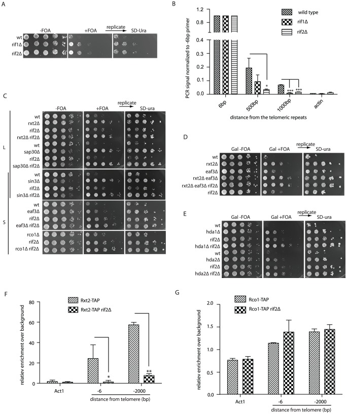
Using a genome-wide screening approach, we have established the genetic requirements for proper telomere structure in Saccharomyces cerevisiae. We uncovered 112 genes, many of which have not previously been implicated in telomere function, that are required to form a fold-back structure at chromosome ends. Among other biological processes, lysine deacetylation, through the Rpd3L, Rpd3S, and Hda1 complexes, emerged as being a critical regulator of telomere structure. The telomeric-bound protein, Rif2, was also found to promote a telomere fold-back through the recruitment of Rpd3L to telomeres. In the absence of Rpd3 function, telomeres have an increased susceptibility to nucleolytic degradation, telomere loss, and the initiation of premature senescence, suggesting that an Rpd3-mediated structure may have protective functions. Together these data reveal that multiple genetic pathways may directly or indirectly impinge on telomere structure, thus broadening the potential targets available to manipulate telomere function.


Conflict of interest statement

The authors have declared that no competing interests exist.

Figures

Figure 1
Figure 1. Histone deacetylation is required for proper telomere structure.
(A) Construct 2 consists of the URA3 gene followed by a downstream Gal UAS with a mutated TATA box (UAS*). When integrated at telomere 7L, the UAS* folds back and drives URA3 transcription in the presence of galactose. (B) Using the SGA (synthetic genetic array) technology, construct 2 was introduced into the ∼4800 strains of the viable haploid yeast gene deletion collection. Subsequently cells were robotically pinned onto −FOA and +FOA galactose-containing media in quadruplicate and scored for growth. A positively scoring hit is highlighted (box in bottom panel) as an example of a non-looping mutant. (C) Validated hits were analyzed using the cytoscape plugin, BinGO, which created a tree of significantly enriched GO (gene ontology) processes over background. (D) All indicated deletion mutants within the Rpd3L, Rpd3S and Hda1 complexes were re-constructed and spotted onto the indicated media in 10-fold serial dilutions following an overnight culture in YPD to confirm the looping defects identified in the high-throughput screen. Plates were incubated 2–3 days before being imaged. +FOA plates were subsequently replica plated onto SD-URA media to ensure that construct was not lost or mutated. (E) For construct 4, the UAS* was placed in front of the URA3 gene and subsequently integrated at telomere 7L (top). Cell spottings were performed exactly as described in (D) with the indicated genotypes.
Figure 2
Figure 2. Chromatin immunoprecipitation confirms structural defects.
(A) The immunoprecipitation of Rap1 following cross-linking should be associated with subtelomeric sequences at natural telomere 6R if the fold-back structure is intact (upper diagram); however the subtelomeric ChIP will be lost upon loop opening (lower diagram). (B) Upon Rap1 ChIP from exponentially growing cells, a subtelomeric signal was detected up to 1 kb away from the base of the telomeric repeats in wild type cells, whereas the signal was largely diminished in hda1Δ, sin3Δ and sir4Δ mutants. DNA stemming from the actin locus (ACT1) was not detected following Rap1 ChIP and was used as a background control. Error bars represent SD from three independent experiments. (C) Cdc13-TAP (13) was also able to precipitate subtelomeric DNA up to 1 kb away from the start of the telomeric sequence at telomere 6R following cross-linking (n = 3, error as SD) in comparison to wild type (non-tagged controls). This ChIP signal at -1000 was reduced to that of non-tagged controls in the sin3Δ strain. The difference in ChIP signal distribution between Rap1 (B) and Cdc13-TAP (C) is likely due to the different positioning of the two proteins on the telomere (compare diagrams in A and C for explanation). For all experiments above error bars represent SD of the mean from at least 3 independent experiments and * indicates statistically significant differences as determined through unpaired student's t-tests whereby * = p<0.05, ** = p<0.01, *** = p<0.001.
Figure 3
Figure 3. The Rif proteins promote Rpd3L recruitment to telomeres.
(A) Cells with the indicated genotypes and harboring construct 2 were spotted onto the galactose media (+/− FOA) (B) Rap1 ChIP was performed as described for Figure 2B. The defect in looping of the rif1Δ and rif2Δ strains is reflected in the loss of Rap1 association to subtelomeric DNA following cross-linking (n = 3, error as SD). (C) Looping defects of the indicated mutants were assayed as in Figure 1D in order to assess genetic interactions between rif2Δ and the Rpd3L and Rpd3S complexes. All colonies were replicated from +FOA onto SD-URA plates to ensure that construct 2 was intact. (D) The looping defect in Rpd3L (rxt2Δ) mutants is additive when combined with Rpd3S mutations (eaf3Δ), and not further exacerbated by deletion of RIF2. (E) Both hda1Δ rif2Δ and hda2Δ rif2Δ double mutants have more severe looping defects than either of the single mutants as seen by their increased resistance to 5-FOA. (F and G) Cells expressing a TAP (tandem affinity purification) tagged version of either Rxt2-TAP (Rpd3L) or Rco1-TAP (Rpd3S) were cross-linked and DNA was precipitated with IgG beads. -6 bp and -2000 bp refer to the position of the reverse primer with respect to the beginning of the telomeric tract on telomere 6R, amplicons being on average approximately 100 bp. The Rpd3L complex is lost at telomeres in a rif2Δ mutant (F) whereas Rpd3S association is not affected (G). For all experiments above error bars represent SD of the mean from at least 3 independent experiments and * indicates statistically significant differences as determined through unpaired student's t-tests whereby * = p<0.05, ** = p<0.01, *** = p<0.001.
Figure 4
Figure 4. Rpd3 promotes telomere end protection.
(A) Strains with the indicated genotypes were spotted onto YPD media following an overnight culture at 23°C and incubated at various temperatures for 3–4 days before being imaged. (B) cdc13-1, cdc13-1 sin3Δ and cdc13-1 sin3Δ exo1Δ cells were arrested in nocodazole at 23°C for three hours before being shifted to 26°C, after which DNA was extracted at 30 minute intervals. Non-denatured and denatured DNA was dot blotted onto a membrane and incubated with a DIG-labeled probe (oBL 207) to recognize the telomeric 3′ ssDNA overhang. The amount of ssDNA is represented as non-denatured DNA as a fraction of the total (as determined by the amount of denatured telomeric DNA). Error bars represent SD from three independent experiments. Pre = before the 26°C temperature shift. (C) The indicated genotypes were derived via tetrad dissection of the heterozygous diploid strain (yMD 1146) and diluted to an OD600 0.01. Cells were grown for 24-hour intervals before being measured and re-diluted. The rate of senescence was increased in est2Δ rad52Δ cells when SIN3 was subsequently deleted, n = 8 for each genotype. The growth rates of rad52Δ (n = 3) and rad52Δ sin3Δ (n = 3) were similar. Population doubling refers to the number of doublings post spore germination. (D) Genomic DNA was isolated in both non-denaturing and denaturing conditions from the indicated genotypes (each n = 3) using samples generated in (C) at the specified population doubling (PD). Single-stranded telomeric DNA was detected upon hybridization with DIG labeled oBL 207 and normalized to total telomeric DNA following a denaturation step.
Figure 5
Figure 5. The fold-back may contribute to telomere protection.
The maintenance of telomere structure requires the telomere-bound Rif2 protein to ensure that the Rpd3L complex gets properly loaded/maintained at chromosome ends. The presence of the Rpd3L KDAC (as well as Rpd3S, Sir2 and Hda1) promotes a protective structure at telomeres, which likely eminates in a fold-back of the telomeric DNA onto the subtelomeric region (1.). In the absence of this structure, telomeres remain protected due to a combination of telomerase-mediated elongation and capping via the CST complex (2.). When both capping and the fold-back structure are simultaneously compromised (3.) chromosome ends undergo accelerated nucleolytic degradation, and experience an accelerated rate of senescence in cells lacking a telomere maintenance mechanism due to the fact that rapidly resected uncapped telomeres do not get re-elongated.

References

    1. de Lange T (2009) How telomeres solve the end-protection problem. Science 326: 948–952. - PMC - PubMed
    1. Palm W, de Lange T (2008) How shelterin protects mammalian telomeres. Annu Rev Genet 42: 301–334. - PubMed
    1. Giraud-Panis MJ, Teixeira MT, Geli V, Gilson E (2010) CST meets shelterin to keep telomeres in check. Mol Cell 39: 665–676. - PubMed
    1. Lydall D, Weinert T (1995) Yeast checkpoint genes in DNA damage processing: implications for repair and arrest. Science 270: 1488–1491. - PubMed
    1. Grandin N, Damon C, Charbonneau M (2001) Ten1 functions in telomere end protection and length regulation in association with Stn1 and Cdc13. EMBO J 20: 1173–1183. - PMC - PubMed

Publication types

MeSH terms