. 2012 Sep;8(9):e1002981.
doi: 10.1371/journal.pgen.1002981. Epub 2012 Sep 20.

De novo CNV formation in mouse embryonic stem cells occurs in the absence of Xrcc4-dependent nonhomologous end joining

Affiliations

De novo CNV formation in mouse embryonic stem cells occurs in the absence of Xrcc4-dependent nonhomologous end joining

Martin F Arlt et al. PLoS Genet. 2012 Sep.

Abstract

Spontaneous copy number variant (CNV) mutations are an important factor in genomic structural variation, genomic disorders, and cancer. A major class of CNVs, termed nonrecurrent CNVs, is thought to arise by nonhomologous DNA repair mechanisms due to the presence of short microhomologies, blunt ends, or short insertions at junctions of normal and de novo pathogenic CNVs, features recapitulated in experimental systems in which CNVs are induced by exogenous replication stress. To test whether the canonical nonhomologous end joining (NHEJ) pathway of double-strand break (DSB) repair is involved in the formation of this class of CNVs, chromosome integrity was monitored in NHEJ-deficient Xrcc4(-/-) mouse embryonic stem (ES) cells following treatment with low doses of aphidicolin, a DNA replicative polymerase inhibitor. Mouse ES cells exhibited replication stress-induced CNV formation in the same manner as human fibroblasts, including the existence of syntenic hotspot regions, such as in the Auts2 and Wwox loci. The frequency and location of spontaneous and aphidicolin-induced CNV formation were not altered by loss of Xrcc4, as would be expected if canonical NHEJ were the predominant pathway of CNV formation. Moreover, de novo CNV junctions displayed a typical pattern of microhomology and blunt end use that did not change in the absence of Xrcc4. A number of complex CNVs were detected in both wild-type and Xrcc4(-/-) cells, including an example of a catastrophic, chromothripsis event. These results establish that nonrecurrent CNVs can be, and frequently are, formed by mechanisms other than Xrcc4-dependent NHEJ.


Conflict of interest statement

The authors have declared that no competing interests exist.

Figures
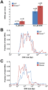
Figure 1
Figure 1. Replication stress induces CNVs in mouse ES cells.
(A) Incidence of spontaneous and induced CNVs in wild-type and Xrcc4−/− cells treated with 0.0–0.6 µM APH for 72 hours. A total of 85 independent clones of untreated and treated, wild-type and Xrcc4−/− cells were analyzed. Error bars indicate standard error. (B) Size distribution of de novo CNVs in wild-type (blue) and Xrcc4−/− cells (red). (C) Size distribution of de novo CNVs in human fibroblasts (blue) and mouse ES cells (red).
Figure 2
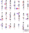
Figure 2. Locations of de novo CNVs in the mouse genome.
CNVs are mapped onto a mouse chromosome ideogram. Blue squares indicate de novo CNVs in wild-type cells. Red circles indicate de novo CNVs in Xrcc4−/− cells. Symbols to the left of a chromosome represent deletions and symbols to the right represent duplications. Ideograms adapted from www.pathology.washington.edu/research/cytopages/idiograms/mouse (Dept. of Pathology, University of Washington, with permission). Precise coordinates for all de novo CNVs are listed in Table S1.
Figure 3
Figure 3. A conserved CNV hotspot in mouse and human cells.
A mouse CNV hotspot at 5G2 in Auts2 corresponds to a previously-described human CNV hotspot at human 7q11.2 in the AUTS2 gene . The x-axis shows the position along the chromosome, while the y-axis indicates that fraction of hotspot CNVs that crossed a particular 10 kb genomic window. CNVs detected in mouse ES cells are depicted as bars. Gray areas indicate regions of inserted sequences in the human relative to mouse genomes. Although overlapping CNVs were found in these regions, all had distinct breakpoints.
Figure 4
Figure 4. Comparison of observed de novo CNV breakpoint junction sequence homology in wild-type and Xrcc4−/− cells.
Histogram showing CNV breakpoint homology in wild-type (blue) and Xrcc4−/− cells (red), compared to the expected distribution if microhomology usage was random (gray).
Figure 5
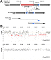
Figure 5. Example of complex APH-induced de novo CNVs in mouse ES cells.
(A) A complex CNV with two junctions at 5G2 in APH-treated Xrcc4−/− clone X6-11. Based on aCGH data, this CNV was called as a deletion, but sequencing of the breakpoint junctions revealed that this CNV was complex, containing a 219.9 kb deletion (red), as well as a duplication-insertion of 84 bp (blue) at the deletion boundary. (B) aCGH data demonstrating a region of complex CNV in APH-treated Xrcc4−/− clone X6-40 at XE3 containing 10 or more discrete deletions across a ∼2.5 Mb region. Data from the same genomic interval in a control clone (X6-38) is shown for comparison.
Figure 6
Figure 6. Models for replication-dependent, Xrcc4-independent CNV formation.
The induction of CNVs by replication stress strongly implicates stalled replication as a key intermediate (top). Template switching without fork collapse might directly create CNVs without DSB formation (left). Alternatively, fork collapse and end processing might lead to iterative template copying prior to final stable resolution of single-ended DSBs by either maturation of a one DSB end into a replication fork (MMBIR, middle) or joining of two distant DSBs by alt-EJ (right). In neither case are the single-ended DSBs good substrates for NHEJ. Results here establish that Xrcc4-dependent NHEJ is neither required for, nor suppresses, CNV formation via these inferred intermediates.

References

    1. Mills RE, Walter K, Stewart C, Handsaker RE, Chen K, et al. (2011) Mapping copy number variation by population-scale genome sequencing. Nature 470: 59–65. - PMC - PubMed
    1. Iafrate AJ, Feuk L, Rivera MN, Listewnik ML, Donahoe PK, et al. (2004) Detection of large-scale variation in the human genome. Nature Genetics 36: 949–951. - PubMed
    1. Redon R, Ishikawa S, Fitch KR, Feuk L, Perry GH, et al. (2006) Global variation in copy number in the human genome. Nature 444: 428–249. - PMC - PubMed
    1. Sebat J, Lakshmi B, Troge J, Alexander J, Young J, et al. (2004) Large-scale copy number polymorphism in the human genome. Science 305: 525–528. - PubMed
    1. Sharp AJ, Locke DP, McGrath SD, Cheng Z, Bailey JA, et al. (2005) Segmental duplications and copy-number variation in the human genome. American Journal of Human Genetics 77: 78–88. - PMC - PubMed

Publication types

LinkOut - more resources