. 2012;7(9):e44567.
doi: 10.1371/journal.pone.0044567. Epub 2012 Sep 28.

Drosophila Ninjurin A induces nonapoptotic cell death

Affiliations

Drosophila Ninjurin A induces nonapoptotic cell death

Sarah Broderick et al. PLoS One. 2012.

Abstract

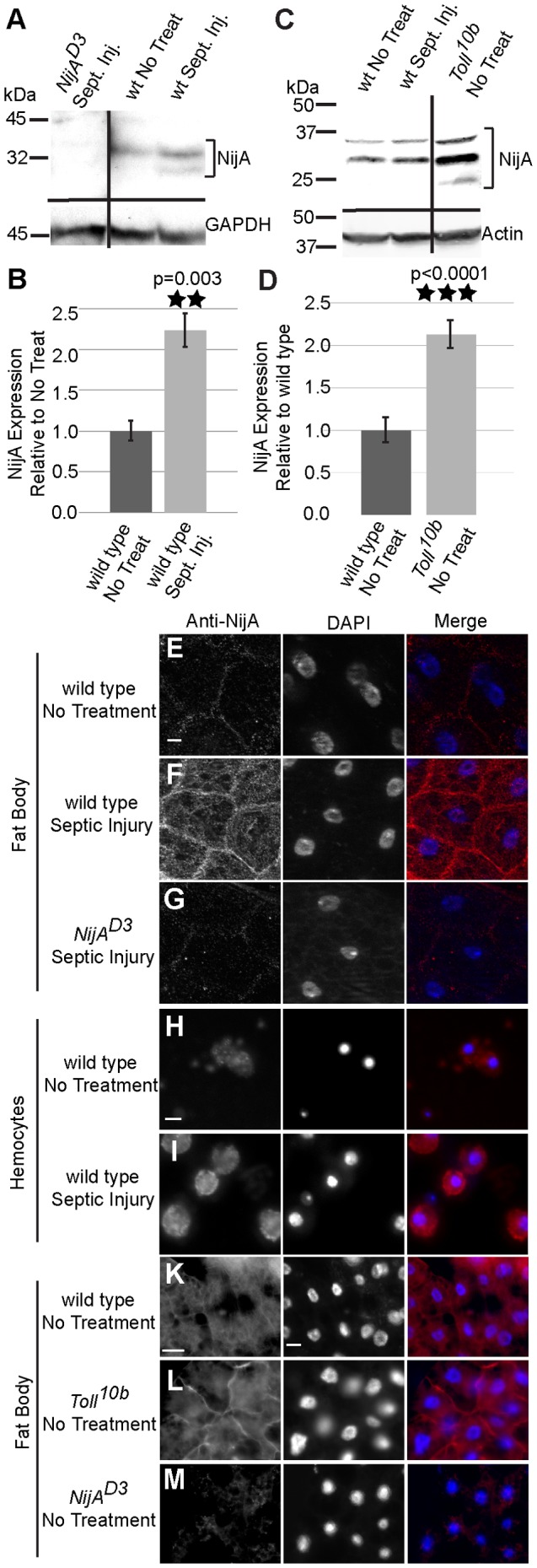
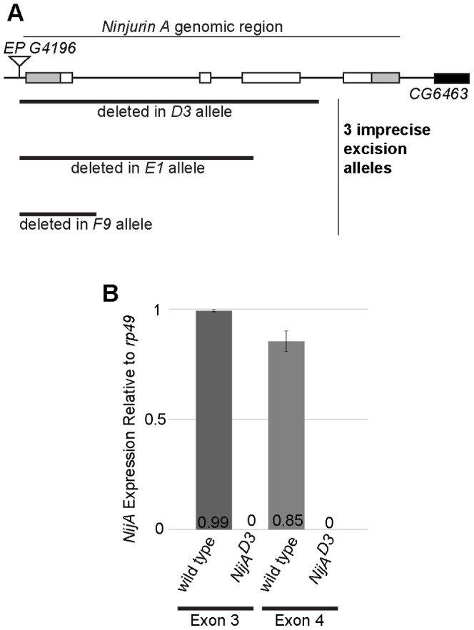
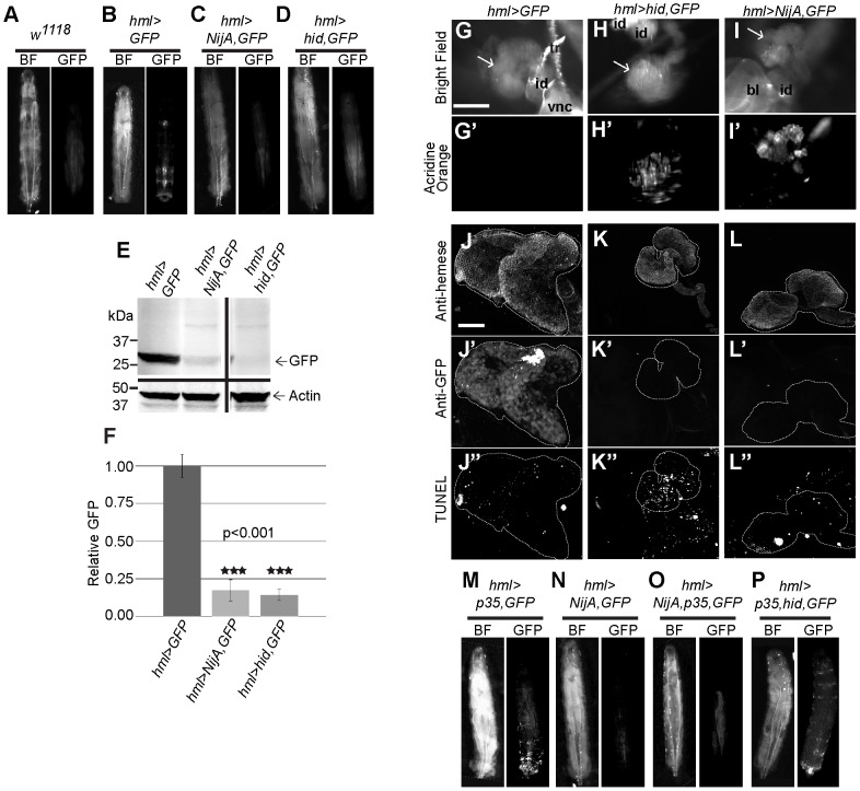
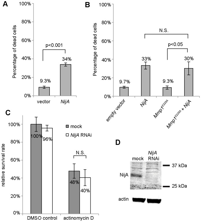
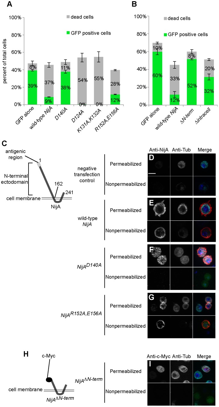
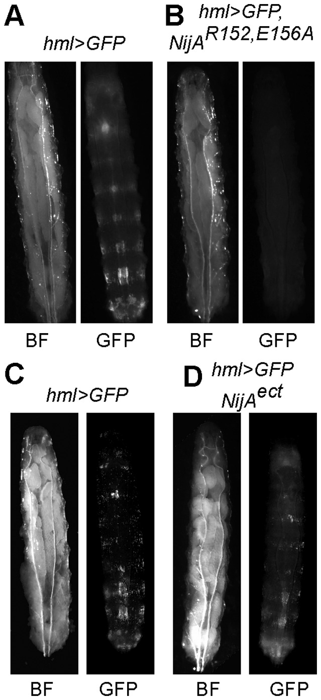
Ninjurins are conserved transmembrane proteins that are upregulated across species in response to injury and stress. Their biological functions are not understood, in part because there have been few in vivo studies of their function. We analyzed the expression and function of one of three Drosophila Ninjurins, NijA. We found that NijA protein is redistributed to the cell surface in larval immune tissues after septic injury and is upregulated by the Toll pathway. We generated a null mutant of NijA, which displayed no detectable phenotype. In ectopic expression studies, NijA induced cell death, as evidenced by cell loss and acridine orange staining. These dying cells did not display hallmarks of apoptotic cells including TUNEL staining and inhibition by p35, indicating that NijA induced nonapoptotic cell death. In cell culture, NijA also induced cell death, which appeared to be cell autonomous. These in vivo studies identify a new role for the Ninjurin family in inducing nonapoptotic cell death.


Conflict of interest statement

Competing Interests: The authors have declared that no competing interests exist.

Figures

Figure 1
Figure 1. Ninjurin A protein response to septic wounding.
(A) Western blot of whole adult male lysates probed with anti-NijA. NijA increases expression two hours after infection in adults. NijAD3 null lysates demonstrate antibody specificity. Black lines indicate regions of the blot that were omitted for clarity. (B) Graph representing three replicates of the western blot pictured in (A). NijA levels increase significantly in adults after septic injury (p = 0.003). (C) Western blot of whole male larval lysates probed with anti-NijA. NijA levels do not change 2 h after septic injury in third instar larvae; in contrast larval Toll10b gain-of-function mutant larvae have increased levels of NijA protein. (D) Graph representing five replicates of the western blot pictured in (C). NijA levels increase significantly in constitutively activate Toll10b mutant larvae (p<0.0001). (E–M) Anti-NijA (red) and DAPI (blue) labeling nuclei. All scale bars are 10 µm. (E–G) Anti-NijA stained non-permeabilized fat bodies of male third instar larvae show an increase in NijA at the cell surface 2 h after septic injury (compare E and F). (G) NijAD3 larvae demonstrate the NijA antibody specificity. (H,I) Anti-NijA stained non-permeabilized hemocytes of third instar larvae ex-vivo show an increase in NijA at the cell surface 2 h after septic injury. (K–M) Anti-NijA stained permeabilized fat bodies of male third instar larvae show increased NijA expression in gain-of-function Toll10b mutants. Error bars in (B,D) represent standard error of the mean.
Figure 2
Figure 2. NijAD3 mutants do not express mRNA from the NijA genomic locus.
(A) Schematic of the NijA locus showing all four exons. Gray indicates untranslated regions and white indicates open reading frame. Three excision alleles (D3, E1, and F9) were generated from imprecise excisions of EP G4196. (B) qPCR data from primers specific to exon 3 (a negative control, as it is deleted in the D3 allele) or exon 4 of NijA. The NijAD3 mutant did not produce any detectable mRNA from exon 4 of the NijA locus, even though exon 4 remains in the genome, indicating that the D3 allele is a null. Error bars represent standard error of the mean.
Figure 3
Figure 3. Ninjurin A over-expression in the lymph gland causes nonapoptotic cell death.
(A) Wild-type w1118 larva demonstrating background autofluorescence. (B) hml>GFP larva with GFP-positive differentiated hemocytes along posterior body wall and in lymph gland. (C) hml>NijA,GFP larva lacked GFP-positive cells. (D) hml>hid,GFP larva, a cell-death positive control, lacked GFP-positive cells. (E) Western blot of whole larval lysates probed with anti-GFP. hml>NijA,GFP and hml>hid,GFP larvae were devoid of GFP. (F) Quantification of three western blots probed for anti-GFP as in (E). GFP is virtually absent from hml>NijA,GFP and hml>hid,GFP larvae. Error bars represent standard error of the mean. (G–I′) Live partially dissected 3rd instar larval lymph glands (arrows) were stained with acridine orange to detect cell death. Scale bars are 200 µM. tr: trachea; id: imaginal disc; vnc: ventral nerve cord; bl: brain lobe. (G′) hml>GFP larval glands did not stain with acridine orange. (H′) hml>hid,GFP glands, a cell-death positive control, stained with acridine orange. (I′) hml>NijA,GFP glands stained with acridine orange, demonstrating that NijA induced cell death. (J–L″) Larval lymph glands were fixed, TUNEL labeled, and antibody stained. Scale bars are 50 µm. (J–L) Anti-Hemese staining labeled the lymph glands. (J′–L′) Anti-GFP staining shows no GFP-positive (hml+) hemocytes in hml>hid,GFP (K′) or hml>NijA,GFP (L′) larval glands. (J″–L″) TUNEL-labeled glands. (J″) Few TUNEL positive cells in hml>GFP negative control glands. (K″) Many TUNEL positive cells in hml>hid,GFP positive control glands. (L″) Few TUNEL positive-cells in the hml>NijA,GFP glands, indicating that NijA does not induce apoptosis. (M) Larvae expressing the apoptotic inhibitor p35 (hml>p35,GFP) displayed GFP-positive hemocytes similar to hml>GFP in (B). (N–O) p35 did not inhibit the NijA-induced loss of the GFP-positive cells in hml>NijA,p35,GFP larvae. (P) p35 inhibited the hid-induced loss of the GFP-positive cells in hml>hid,p35,GFP larvae, a positive control for p35 inhibition. In (A–D, M–P), anterior is up.
Figure 4
Figure 4. NijA induces cell death in Drosophila S2 cell culture.
(A) NijA expression kills S2 cells. Cells were transiently transfected with pRmHa3 empty vector or pRmHa3-NijA and induced with copper for 48 h. The percentage of dead cells was determined by dividing the number of trypan blue positive cells by that of total cells counted for each sample. Data from 8 experiments are shown. Error bars indicate S.E.M, and Student's T test was used to calculate p value. (B) Mmp1 activity is not required for NijA-induced cell death. Cells were transiently transfected and induced for 48 h. Mmp1E255A is a dominant-negative catalytically inactive mutant of Mmp1. Data from 4 experiments are shown. (C) NijA is not required for actinomycin D-induced apoptosis. Cells were treated with NijA dsRNA or no dsRNA (mock) for 48 h, then incubated with 100 nM actinomycin D for 6 h. Trypan blue staining was used to determine cell survival, which was normalized to the untreated (DMSO), wild-type (mock) sample. Data from 4 experiments are shown. (D) Western blot showing the NijA protein levels in mock and NijA dsRNA-treated cells. Actin was used as the loading control.
Figure 5
Figure 5. NijA appears to kill in a cell-autonomous manner.
(A–B) Cells were transiently co-transfected with pRmHa3-GFP and various mutants of pRmHa3-NijA as indicated under each column; mock is empty pRmHa3 vector. 48 h after induction, viability was assessed by trypan blue staining, and transfection status was assessed as GFP fluorescence. Wild-type NijA and most NijA mutants killed cells, whereas the mock control, the D140A mutant, and the N-terminal deletion (B) showed low levels of cell death. The sum of transfected live cells (GFP+) plus dead cells was relatively constant across samples despite the augmented or compromised capacity to kill cells, indicating that NijA kills the cell it transfects but not others. Data from 3 replicates are shown. Error bars indicate S.E.M. (C) Schematic showing topology of NijA (form A) protein and the extracellular region recognized by our polyclonal antibody. Amino acid residue numbers are indicated. (D–G,I) Immunofluorescence localization of wild-type NijA or NijA mutant forms expressed in S2 cells and stained with anti-NijA (D–G) or anti-c-Myc (I), both extracellular epitopes. For each construct, staining was performed on permeabilized cells to show NijA protein levels, and on unpermeabilized cells to show NijA cell-surface localization. Permeabilization status was verified by anti-tubulin staining. The merge image combines images for NijA (red), tubulin (cyan), DAPI (blue) and GFP fluorescence as a transfection control (green). Bar: 10 µm. (H) Diagram showing placement of the myc epitope for (I), necessary because the NijA antigenic region was deleted in this mutant.
Figure 6
Figure 6. NijA mutants behave in vivo as they do in cell culture.
(A) Third instar hml>GFP,NijA152156A larvae were devoid of GFP-positive hemocytes, indicating that these mutants were capable of inducing cell death and that polar amino acids 152 and 156 were not required for cell death. (B) hml>NijAect,GFP larvae displayed visible GFP-positive hemocytes on the body wall, suggesting that NijAect was not sufficient to induce cell death.

Similar articles

Cited by

References

    1. Araki T, Milbrandt J (1996) Ninjurin, a novel adhesion molecule, is induced by nerve injury and promotes axonal growth. Neuron 17: 353–361. - PubMed
    1. Araki T, Milbrandt J (2000) Ninjurin2, a novel homophilic adhesion molecule, is expressed in mature sensory and enteric neurons and promotes neurite outgrowth. J Neurosci 20: 187–195. - PMC - PubMed
    1. Zhang S, Dailey GM, Kwan E, Glasheen BM, Sroga GE, et al. (2006) An MMP liberates the Ninjurin A ectodomain to signal a loss of cell adhesion. Genes Dev 20: 1899–1910. - PMC - PubMed
    1. Koike M, Ninomiya Y, Koike A (2008) Characterization of Ninjurin and TSC22 induction after X-irradiation of normal human skin cells. The Journal of dermatology 35: 6–17. - PubMed
    1. Boutros M, Agaisse H, Perrimon N (2002) Sequential activation of signaling pathways during innate immune responses in Drosophila. Dev Cell 3: 711–722. - PubMed