. 2012;7(9):e44719.
doi: 10.1371/journal.pone.0044719. Epub 2012 Sep 13.

Expressional analysis of immunoglobulin D in cattle (Bos taurus), a large domesticated ungulate

Affiliations

Expressional analysis of immunoglobulin D in cattle (Bos taurus), a large domesticated ungulate

Beilei Xu et al. PLoS One. 2012.

Abstract

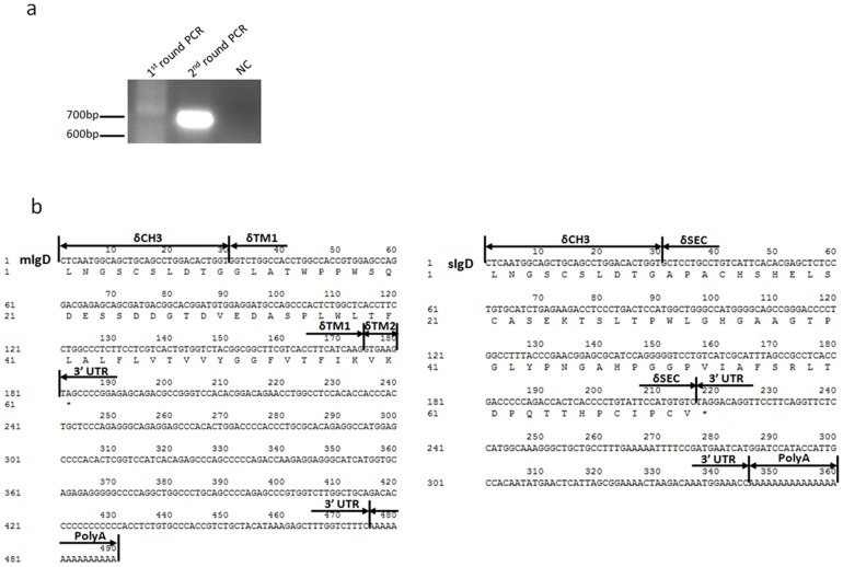
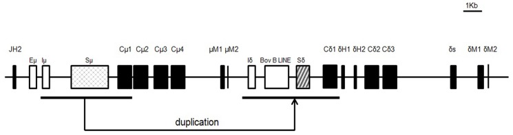
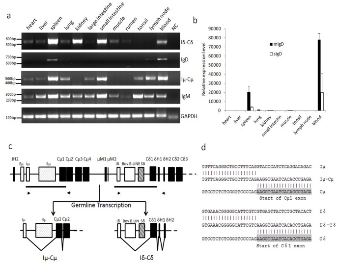
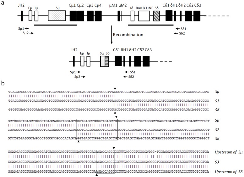
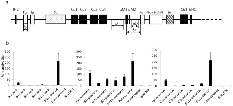
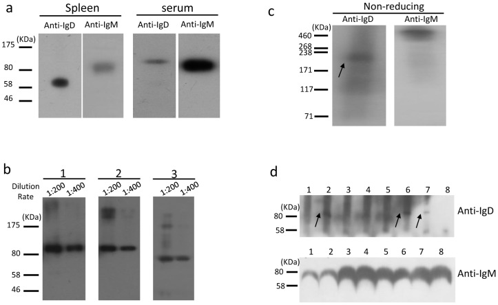
For decades, it has remained unknown whether artiodactyls, such as cattle, pigs, and sheep, express immunoglobulin D (IgD), although the δ gene was identified in these species nearly 10 years ago. By developing a mouse anti-bovine IgD heavy chain monoclonal antibody (13C2), we show that secreted bovine IgD was present mainly as a monomer in serum and was heavily glycosylated by N-linked saccharides. Nonetheless, IgD was detectable in some but not all of the Holstein cattle examined. Membrane-bound IgD was detected in the spleen by western blotting. Flow cytometric analysis demonstrated that IgD-positive B cells constituted a much lower percentage of B cells in the bovine spleen (∼6.8% of total B cells), jejunal Peyer's patches (∼0.8%), and peripheral blood leukocytes (∼1.2%) than in humans and mice. Furthermore, IgD-positive B cells were almost undetectable in bovine bone marrow and ileal Peyer's patches. We also demonstrated that the bovine δ gene can be expressed via class switch recombination. Accordingly, bovine δ germline transcription, which involves an Iδ exon and is highly homologous to Iμ, was confirmed. However, we could not identify an Iδ promoter, despite bovine Eμ demonstrating both enhancer and promoter activity. This study has answered a long-standing question in cattle B cell biology and significantly contributes to our understanding of B cell development in this species.


Conflict of interest statement

Competing Interests: The authors have declared that no competing interests exist.

Figures

Figure 1
Figure 1. Analysis of the 3′ ends of bovine IgD heavy chain cDNA.
(a)PCR product of 3′RACE of the bovine IgD heavy chain cDNA. NC, negative control. (b) The sequence of the 3′ ends of mIgD.
Figure 2
Figure 2. Genomic organization of the bovine Cδ gene.
Coding regions are indicated by black boxes. Lower thick lines indicate homologous DNA fragments involved in the duplication. Eμ, 5′ intronic enhancer; Iδ, the putative Iδ exon; Sμ, switch μ; Sδ, switch δ.
Figure 3
Figure 3. Transcription of the bovine μ and δ genes in different tissues.
(a) RT-PCR detection of the IgD, Iδ germline, IgM and Iμ germline transcripts in different tissues. GAPDH was used as an internal control. (b) Q-PCR analysis of mIgD and sIgD in different tissues. Data were analyzed using the ΔΔCT method. GAPDH was used as an internal control. (c) Germline Iμ-Cμ and Iδ-Cδ transcripts. Lower thick lines indicate homologous DNA fragments, whereas arrows indicate primers for Iμ and Iδ germline transcripts. (d) Sequence of Iμ-Cμ and Iδ-Cδ transcripts from the spleen.
Figure 4
Figure 4. Switch recombination between Sμ and Sδ.
(a) The schematic map of the CSR of the Cδ gene. Arrows indicate the positions of the Sμ1, Sμ2, Sδ1 and Sδ2 primers. (b) DNA sequence of the recombined Sμ-Sδ junctions. Upper sequence, bovine germline Sμ region (accession no. AY158087); lower sequence, bovine germline Sδ region (accession no. AF411241). The middle sequences are the cloned PCR products. Identical nucleotides are shown by vertical lines. The Sμ and Sδ breakpoints are represented by an inverted triangle and forward triangle, respectively. Overlaps are indicated by boxes.
Figure 5
Figure 5. Promoter activity analysis of the sequences upstream of the Iδ exon.
(a) Schematic positions of the cloned fragments tested for promoter activity. Eμ has been previously described . Eμ, Iδ1, Iδ2 and Iδ3 indicate the cloned fragments. (b) Detection of the promoter or enhancer activity in X16C8.5 B cell lines. Eμ, Iδ1, Iδ2 and Iδ3 were separately cloned into the pGL3-basic, pGL3-promoter and pGL3-enhancer vectors and named Eμ+basic, Eμ+promoter and Eμ+enhancer, respectively. pGL3-control was a positive transfection control. pGL3-basic, pGL3-promoter, pGL3-enhancer and untransfected cells were used as the controls. The average values of three replicates and standard deviations are shown.
Figure 6
Figure 6. Immunofluorescence staining using the 13C2 mAb.
(a) Immunofluorescence staining of full-length mIgD heavy chain or λ light chain expressed on HEK293T cells with mAb 13C2(anti-IgD) plus goat anti-mouse IgG (red) and DAPI (blue). mIgD, cells transfected with full-length mIgD plasmid only; mIgD+λ, cells transfected with full-length mIgD heavy chain and λ light chain plasmids together. NC, negative control. 1, Cells transfected with an mIgD plasmid, stained by the secondary antibody goat anti-mouse IgG. 2, Cells transfected with mIgD and λ plasmids, stained by the goat anti-mouse IgG. 3, Cells transfected with the empty vector pcDNA3.1(+), stained by 13C2 plus goat anti-mouse IgG. Original magnification, ×20. (b) Immunoblot analysis of bovine mIgD heavy and light chains in cell lysates. mAb 13C2 was used to detect mIgD, and anti-FLAG antibody was used to detect the λ light chain. 1, Cell lysates from HEK293T cells transfected with mIgD heavy chain and λ light chain plasmids separately. 2, Cell membrane proteins extracted from HEK293T cells transfected with mIgD and λ plasmids together. 3, Negative control of membrane proteins from HEK293T cells transfected with empty vector pcDNA3.1(+).
Figure 7
Figure 7. Immunoblot analysis of endogenous bovine IgD.
(a) Western blot of splenic membrane and serum proteins. (b) Western blot analysis of the IgD glycosylation in bovine serum. 1, Untreated serum proteins; 2, endo-α-N-acetylgalactosaminidase-treated serum proteins; 3, PNGase-treated serum proteins. The primary antibody 13C2 was diluted at 1∶200 or 1∶400. (c) Serum immunoblot analysis with mAb 13C2 and anti-bovine IgM polyclonal antibody under nonreducing conditions. The arrow indicates the monomer of IgD. (d) Immunoblot detection of serum IgD and IgM in differently aged cows. 1, 60 days old; 2, 180 days; 3, 1 year; 4, 2 years; 5, 3 years; 6, 4 years; 7, 5 years; 8, 6 years. The arrows indicate the IgD heavy chain.
Figure 8
Figure 8. Flow cytometric analysis of B cell populations in the PBLs, ileum PP, jejunum PP, spleen and bone marrow from a 1- year-old cow.
(a) Cells were stained for B220, and the proportion of cell populations are indicated for each gate. (b) FACS analysis of lymphocytes from the PBLs, IPP, JPP, spleen and bone marrow using FITC-conjugated anti-bovine B220, PE-conjugated anti-bovine IgD (13C2) and biotin-labeled anti-bovine IgM plus PE/Cy5-labeled streptavidin. Numbers adjacent to the outlined areas indicate the percent of IgM+ cells or IgD cells in the B220 gate. (c) Western blotting of membrane proteins extracted from the ileal PP and spleen of the same cow using anti-bovine IgD and anti-bovine IgM separately. Note: three animals were used in this experiment, but only representative data from one animal are shown.

References

    1. Rowe DS, Fahey JL (1965) A New Class of Human Immunoglobulins .I. A Unique Myeloma Protein. Journal of Experimental Medicine 121: 171–184. - PMC - PubMed
    1. Wilson M, Bengten E, Miller NW, Clem LW, Du Pasquier L, et al. (1997) A novel chimeric Ig heavy chain from a teleost fish shares similarities to IgD. Proc Natl Acad Sci U S A 94: 4593–4597. - PMC - PubMed
    1. Ohta Y, Flajnik M (2006) IgD, like IgM, is a primordial immunoglobulin class perpetuated in most jawed vertebrates. Proceedings of the National Academy of Sciences of the United States of America 103: 10723–10728. - PMC - PubMed
    1. Zhao Y, Pan-Hammarstrom Q, Yu S, Wertz N, Zhang X, et al. (2006) Identification of IgF, a hinge-region-containing Ig class, and IgD in Xenopus tropicalis. Proc Natl Acad Sci U S A 103: 12087–12092. - PMC - PubMed
    1. Ota T, Rast JP, Litman GW, Amemiya CT (2003) Lineage-restricted retention of a primitive immunoglobulin heavy chain isotype within the Dipnoi reveals an evolutionary paradox. Proceedings of the National Academy of Sciences of the United States of America 100: 2501–2506. - PMC - PubMed

Publication types

Substances