. 2012;7(9):e44899.
doi: 10.1371/journal.pone.0044899. Epub 2012 Sep 17.

Triterpenoid dihydro-CDDO-trifluoroethyl amide protects against maladaptive cardiac remodeling and dysfunction in mice: a critical role of Nrf2

Affiliations

Triterpenoid dihydro-CDDO-trifluoroethyl amide protects against maladaptive cardiac remodeling and dysfunction in mice: a critical role of Nrf2

Yifan Xing et al. PLoS One. 2012.

Erratum in

Abstract

Background and aims: Nuclear factor E2-related factor 2 (Nrf2) appears to be an attractive therapeutic target for the treatment of cardiac disease. We investigated whether a synthetic triterpenoid derivative of dihydro-CDDO-trifluoroethylamide (dh404), a novel Nrf2 activator, protects against pathological cardiac responses to hemodynamic stress in mice.

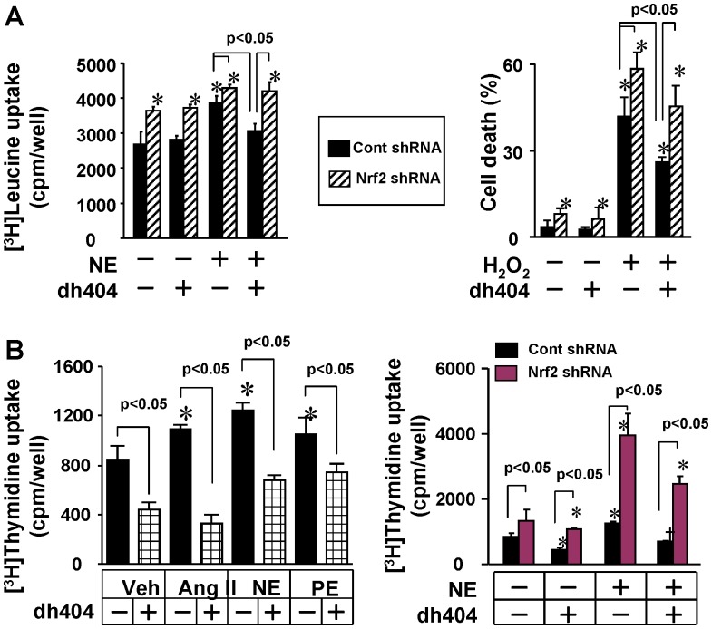
Methods: Cardiac maladaptive remodeling and dysfunction were established by transverse aortic constriction (TAC) in mice. Hypertrophic growth of rat neonatal cardiomyocytes was induced by angiotensin II (Ang II). Cell death of rat neonatal cardiomyocytes was induced with hydrogen peroxide (H₂O₂). Cellular proliferation of rat neonatal cardiac fibroblasts was induced by Ang II, norepinephrine (NE) and phenylephrine (PE). Protein expression was assessed by immunochemical staining and Western blots. Gene expression was determined by real time reverse transcription-polymerase chain reaction (Q-PCR).

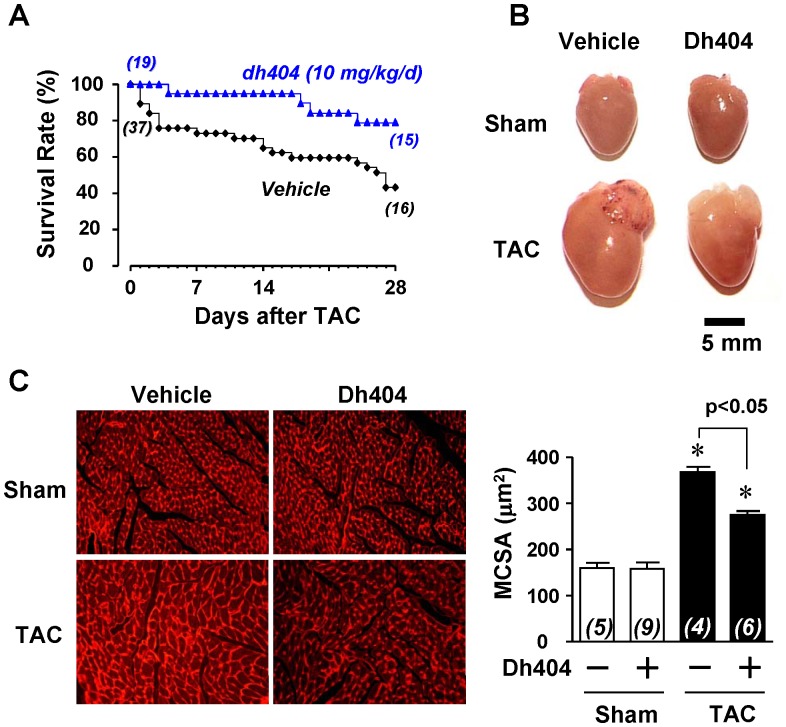
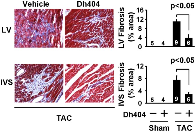
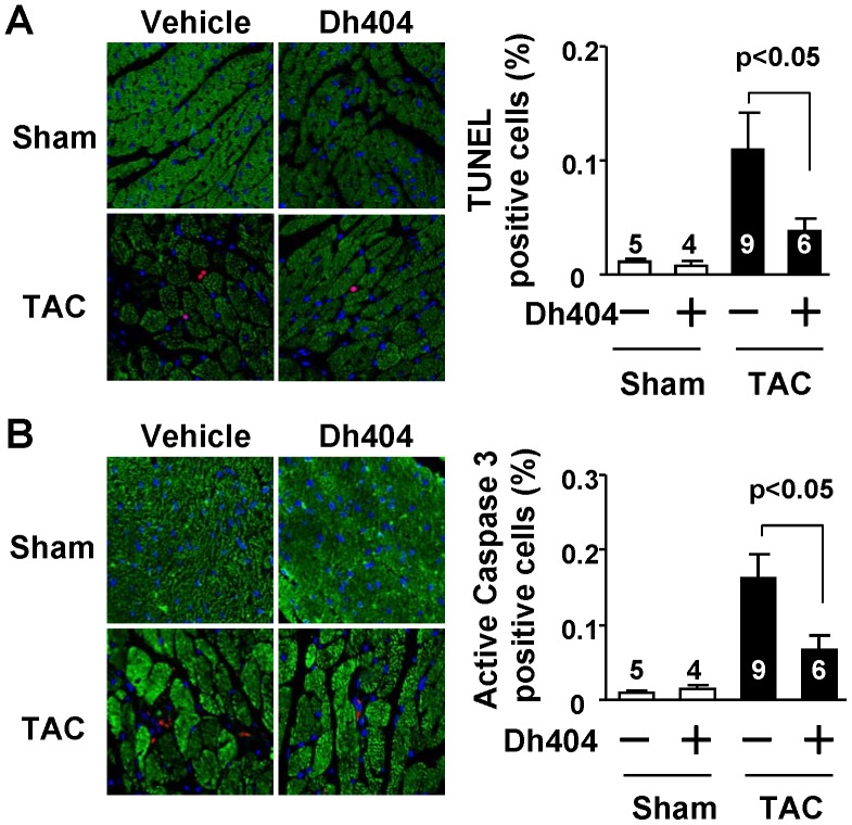
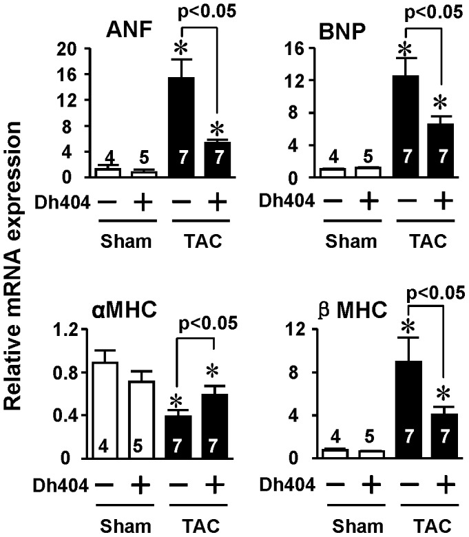
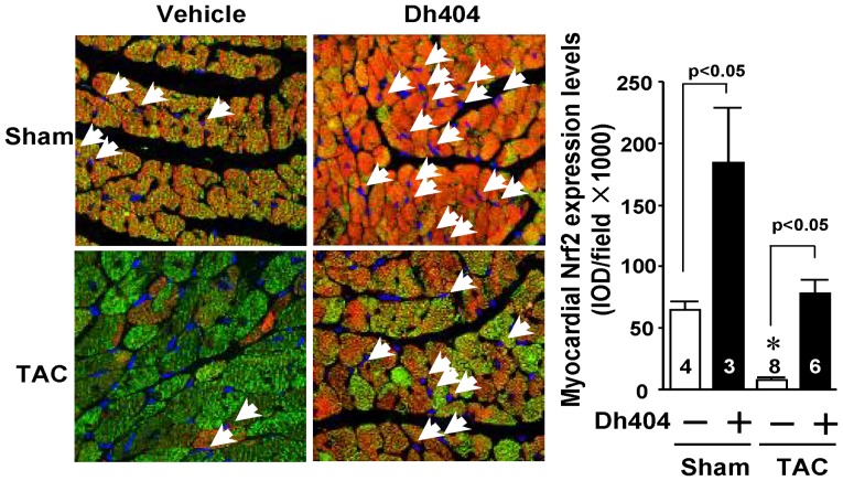
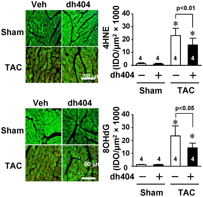
Results: TAC suppressed myocardial Nrf2 expression, increased myocardial 4-hydroxy-2-nonenal and 8-hydroxydeoxyguanosine levels, and induced cardiac hypertrophy, fibrosis and apoptosis, and overt heart failure and death in mice. Administration of dh404 inhibited the pathological cardiac remodeling and dysfunction, and reduced the mortality. Moreover, dhd404 elevated myocardial levels of Nrf2 and Nrf2 nuclear translocation with a dramatic suppression of the oxidative stress in the heart. Dh404 inhibited hypertrophic growth and death in primary culture of rat neonatal cardiomyocytes and suppressed proliferation in primary culture of rat neonatal cardiac fibroblasts. However, these effects of dh404 were blunted by knocking down of Nrf2.

Conclusion: These findings demonstrate that dh404 prevents pathological cardiac remodeling and dysfunction by activating Nrf2, indicating a therapeutic potential of dh404 for cardiac disease.


Conflict of interest statement

Competing Interests: Stacey Ruiz and Colin J. Meyer are employed by Reata Pharmaceuticals, Inc. Reata Pharmaceuticals, Inc, synthesized, purified, and provided dihydro-CDDOtrifluoroethyl amide (dh404), which Reata Pharmaceuticals has a patent on. Reata Pharmaceuticals has numerous compounds in development from this same class of compounds. There are no marketed products to declare. This does not alter the authors’ adherence to all the PLOS ONE policies on sharing data and materials, as detailed online in the guide for authors.

Figures

Figure 1
Figure 1. Effect of dh404 on survival rate and cardiac hypertrophy of mice after TAC.
(A) Effect of dh404 (10 mg/kg/d) on TAC-induced death in C57BL/6J mice. (B) Representative pictures of hearts from sham and TAC operated mice treated with vehicle or dh404 (10 mg/kg/d). (C) Left panel - representative photomicrographs of left ventricular cross-sections depicting myocyte membranes stained with Texas Red-X conjugated wheat germ agglutinin (WGA). Right panel - quantitative left ventricular cardiac myocyte cross-sectional area (MCSA) from 2000 to 5000 myocytes per heart. The number of hearts (n) analyzed is indicated.
Figure 2
Figure 2. Effect of dh404 on myocardial fibrosis in mice after TAC.
Myocardial fibrosis was assessed by staining of collagen with a Masson’s Trichrome Kit. In the left panels representative photomicrographs of a left ventricular (LV) and interventricular septum (IVS) sections from TAC hearts with and without dh404 treatment are presented. In the right panel the fibrotic areas of LV and IVS interstitial fibrosis as a percentage of total microscopic area per heart are presented. The numbers represent the number of sham and TAC hearts analyzed.
Figure 3
Figure 3. Effect of dh404 on myocardial apoptosis in mice after TAC.
(A) In the left panel, the representative TUNEL staining of apoptotic cells in left ventricle are shown. TUNEL-positive, i.e., apoptotic nuclei in TAC hearts are stained red, nuclei blue and the myocardium is green (Alexa Fluor 488 Phalloidin to mark F-actin). Magnification, X 630. In the right panel, the apoptotic index results are given. TUNEL-positive cells were quantified as a percent of all nuclei in the section of LV. The number of hearts analyzed for each group is indicated. (B) In the left panel, the representative staining of cleaved caspase-3 positive cells in the left ventricle are shown. Cleaved caspase-3-positive, (i.e., cells with activated caspase-3) in TAC hearts are stained red and nuclei are blue. Cardiomyocytes were green utilizing an antibody of anti-cardiac myosin heavy chain. Magnification, X 630. In the right panel, the relative caspase-3 activity is summarized. Cleaved caspase-3-positive cells were quantified as a percent of all in a section of LV. The number of hearts analyzed for each group is indicated.
Figure 4
Figure 4. Effect of dh404 on myocardial fetal gene expression of mice after TAC.
Hearts of mice were harvested, and left ventricles were dissected for RNA purification 4 weeks after the operation. Expression of ANF, BNP, αMHC, and βMHC was quantified by Q-PCR. *p<0.05 vs sham.
Figure 5
Figure 5. Effect of dh404 on myocardial Nrf2 expression of mice after TAC.
C57BL/6J mice at age of 8 weeks were treated with vehicle or dh404 (10 mg/kg/d) and subjected to sham or TAC operations. Their hearts were harvested 4 weeks after the operation and analyzed for Nrf2 protein expression. Left panel - representative immunohistochemical staining of Nrf2 protein expression in left ventricles from TAC mice with or without dh404 treatment. Nrf2 nuclear translocation is indicated by arrows. Nrf2 staining was performed in 4 or more sections per heart. Nrf2 is stained red, nuclei blue and cardiomyocytes green utilizing an antibody of anti-cardiac myosin heavy chain. Right panel - a semi-quantitative analysis of Nrf2 protein levels in the heart. The number of hearts analyzed is indicated.
Figure 6
Figure 6. Effect of dh404 on myocardial oxidative stress of mice after TAC.
Eight wk old C57BL/6J mice were grouped and treated as described in Figure 5. Left panel - representative confocal microscopic images of left ventricular 4-HNE staining and 8-OHdG staining. Areas positive for 4-HNE and 8-OHdG are shown in red, nuclei in blue were labelled with DAPI, and. cardiomyocytes in green were marked using anti-Tropomyosin I antibody against cardiac myocyte tropomyosin. Right panel - levels of 4-HNE and 8-OHdG were semi-quantified by measuring IOD of eight randomly chosen fields in each myocardial tissue section. The number of hearts (n) analyzed is indicated.
Figure 7
Figure 7. Role of Nrf2 in dh404-induced suppression of cardiomyocyte hypertrophy and death, and cardiac fibroblast proliferation in vitro.
(A) Effect of dh404 (200 nmol/L) on NE (20 µmol/L)-induced [3H]leucine uptake (left pannel) and H2O2 (100 µmol/L)-induced cell death of the cardiomyocytes infected with adenovirus of control (cont) shRNA and Nrf2 shRNA. *p<0.05 vs cont shRNA (-), n = 4. (B) Left pannel - Effect of dh404 (200 nmol/L) on Ang II (0.1 µmol/L)-, NE (20 µmol/L), and PE (20 µmol/L)-induced [3H]thymidine uptake in the cardiac fibroblasts. *p<0.05 vs control dh404 (-), n = 6. Right pannel – Effect of dh404 (200 nmol/L) on NE (20 µmol/L)-induced [3H]thymidine uptake in the cardiac fibroblasts infected with adenovirus of control (cont) shRNA and Nrf2 shRNA. *p<0.05 vs control dh404 (-), n = 4.

References

    1. Rosamond W, Flegal K, Furie K, Go A, Greenlund K, et al. (2008) Heart disease and stroke statistics–2008 update: a report from the American Heart Association Statistics Committee and Stroke Statistics Subcommittee. Circulation 117: e25–146. - PubMed
    1. Swynghedauw B (1999) Molecular mechanisms of myocardial remodeling. Physiol Rev 79: 215–262. - PubMed
    1. Cohn JN, Ferrari R, Sharpe N (2000) Cardiac remodeling–concepts and clinical implications: a consensus paper from an international forum on cardiac remodeling. Behalf of an International Forum on Cardiac Remodeling. J Am Coll Cardiol 35: 569–582. - PubMed
    1. Mudd JO, Kass DA (2008) Tackling heart failure in the twenty-first century. Nature 451: 919–928. - PubMed
    1. Landmesser U, Wollert KC, Drexler H (2009) Potential novel pharmacological therapies for myocardial remodelling. Cardiovasc Res 81: 519–527. - PubMed

Publication types

MeSH terms