. 2012;7(9):e44947.
doi: 10.1371/journal.pone.0044947. Epub 2012 Sep 18.

Probing the dynamics of doxorubicin-DNA intercalation during the initial activation of apoptosis by fluorescence lifetime imaging microscopy (FLIM)

Affiliations

Probing the dynamics of doxorubicin-DNA intercalation during the initial activation of apoptosis by fluorescence lifetime imaging microscopy (FLIM)

Nai-Tzu Chen et al. PLoS One. 2012.

Erratum in

  • PLoS One. 2012;7(11). doi:10.1371/annotation/4c43c8c8-0a4e-425b-a72f-74e84f6f3c28

Abstract

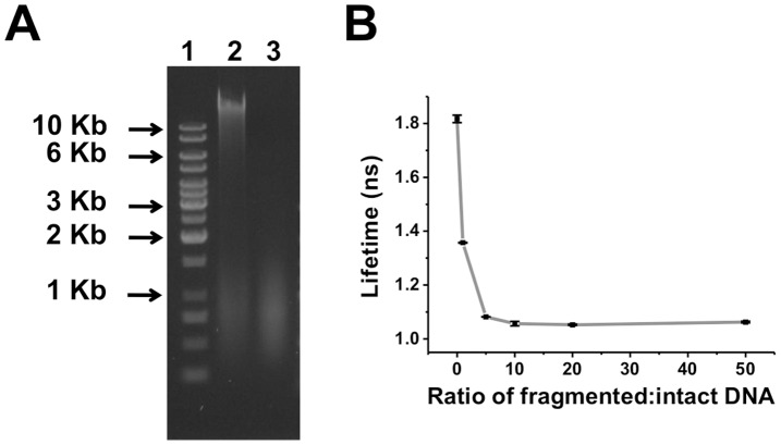
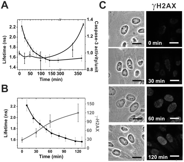
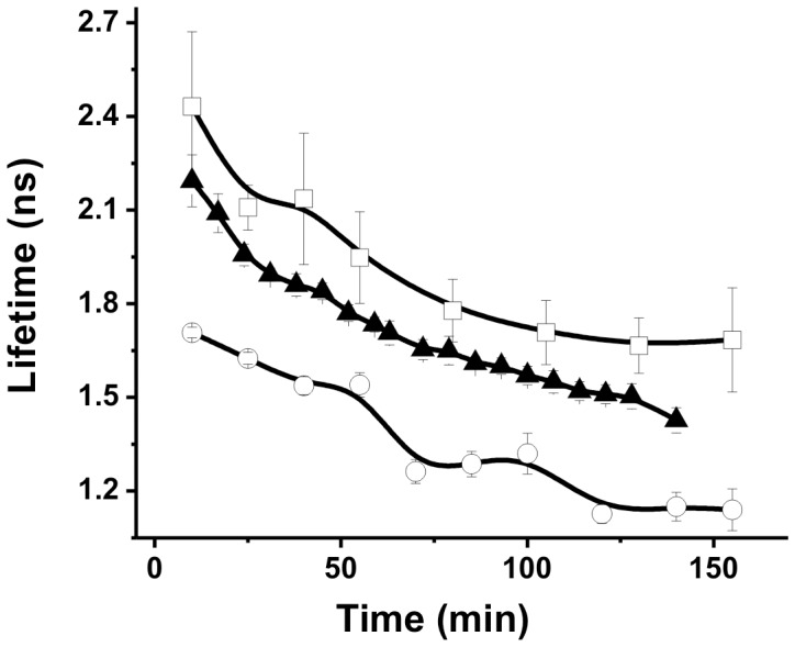
Doxorubicin is a potent anthracycline antibiotic, commonly used to treat a wide range of cancers. Although postulated to intercalate between DNA bases, many of the details of doxorubicin's mechanism of action remain unclear. In this work, we demonstrate the ability of fluorescence lifetime imaging microscopy (FLIM) to dynamically monitor doxorubicin-DNA intercalation during the earliest stages of apoptosis. The fluorescence lifetime of doxorubicin in nuclei is found to decrease rapidly during the first 2 hours following drug administration, suggesting significant changes in the doxorubicin-DNA binding site's microenvironment upon apoptosis initiation. Decreases in doxorubicin fluorescence lifetimes were found to be concurrent with increases in phosphorylation of H2AX (an immediate signal of DNA double-strand breakage), but preceded activation of caspase-3 (a late signature of apoptosis) by more than 150 minutes. Time-dependent doxorubicin FLIM analyses of the effects of pretreating cells with either Cyclopentylidene-[4-(4-chlorophenyl)thiazol-2-yl)-hydrazine (a histone acetyltransferase inhibitor) or Trichostatin A (a histone deacetylase inhibitor) revealed significant correlation of fluorescence lifetime with the stage of chromatin decondensation. Taken together, our findings suggest that monitoring the dynamics of doxorubicin fluorescence lifetimes can provide valuable information during the earliest phases of doxorubicin-induced apoptosis; and implicate that FLIM can serve as a sensitive, high-resolution tool for the elucidation of intercellular mechanisms and kinetics of anti-cancer drugs that bear fluorescent moieties.


Conflict of interest statement

Competing Interests: The authors have declared that no competing interests exist.

Figures

Figure 1
Figure 1. Diagram illustrating the principal components of the fluorescence lifetime imaging microscope.
(Abbreviattion: DM, dichoroic mirrow; BP, bandpass filter; LP, longpass filter. Please refer the details in the FLIM section of Materials and Methods.
Figure 2
Figure 2. Fluorescence intensity, lifetime mapping, and corresponding lifetime histogram of doxorubicin in HeLa cells.
Cells were treated 5 µg/ml doxorubicin for 24 hrs. (A) Fluorescence intensity image; the contours of nuclear envelop and cell membrane are delineated with yellow and red dotted lines respectively, based on the phase contrast image. (B) Fluorescence lifetime image. Scale bar, 10 µm. (C) Corresponding lifetime histogram of nucleus and cytoplasm.
Figure 3
Figure 3. Dynamic monitoring of fluorescence lifetimes within nuclei.
(A) Dynamic images of doxorubicin fluorescence lifetime in HeLa cells. (B) Changes in average fluorescence lifetime of doxorubicin in nuclei as a function of time. (C) Changes in average fluorescence lifetime of (i) (solid circle) an nucleus targeting dye, SYTO 59 in nucleus; (ii) (open triangle) an plasma membrance dye, WGA; (iii) (solid square) an tubulin labeling dye, Tubulintracker.
Figure 4
Figure 4. Fluorescence lifetime measuments at various concentrations of doxorubicin.
Fluorescence lifetimes of doxorubicin were measured at various drug concentrations. Doxorubicin samples were prepared with MEM medium for 5 µg/ml to 1 mg/ml, while fluorescence lifetimes were measured by FLIM system.
Figure 5
Figure 5. Fluorescence lifetime measurements of doxorubicin for various ratios of fragmented/intact DNA.
(A) Gel electrophoresis analysis of intact and fragmented DNA; Line1: 1 Kb DNA ladder, Line2: Genomic DNA extracted from HeLa cells, Line3: Genomic DNA digested by DNase I (1 unit/µl for 1 hr). (B) Fluorescence lifetime of doxorubicin at increasing ratios of fragmented/intact DNA at a fixed ratio of DNA/doxorubicin by weight.
Figure 6
Figure 6. Doxorubicin induces caspase-3 activation and H2AX phosphorylation.
HeLa cells were exposed to 5 µg/ml doxorubicin for different periods, after which cell lysates of each time point were extracted for either measurement of caspase-3 activity or fixed for the γH2AX staining. Doxorubicin fluorescence lifetimes following treatment (black) vs. (A) caspase-3 activity (blue); and (B) γH2AX activation (red). γH2AX activation was quantified from confocal microscopic images, as shown in (C), using MetaMorph imaging processing software. As illustrated, caspase-3 activation is preceded by the H2AX phosphorylation. (Scale bars in C: 20 µm).
Figure 7
Figure 7. Temporal profiles of doxorubicin fluorescence lifetime in nuclei after CPTH2 and TSA treatments.
CPTH2 (open square) and TSA (open circle) were added to culture medium 24 hrs before cells were exposed to 5 µg/ml doxorubicin. 5 µg/ml doxorubicin (solid triangle) served as the control.

References

    1. Blum RH, Carter SK (1974) Adriamycin, A new anticancer drug with significant clinical activity. Ann. Intern. Med. 80(1): 249–259. - PubMed
    1. Carter SK (1975) Adriamycin – thoughts for the future. Cancer Chemother. Rep. 63: 1877–1883.
    1. Hortobagyi GN (1997) Anthracyclines in the treatment of cancer an overview. Drug 54: 1–7. - PubMed
    1. Li ZX, Wang TT, Wu YT, Xu CM, Dong MY, et al. (2008) Adriamycin induces H2AX phosphorylation in human spermatozoa. Asian J Androl 10(5): 749–757. - PubMed
    1. Hurley LH (2002) DNA and its associated processes as targets for cancer therapy. Nature Reviews Cancer 2: 188–200. - PubMed

Publication types

MeSH terms