. 2012;7(9):e44985.
doi: 10.1371/journal.pone.0044985. Epub 2012 Sep 18.

The cranial osteology and feeding ecology of the metriorhynchid crocodylomorph genera Dakosaurus and Plesiosuchus from the late Jurassic of Europe

Affiliations

The cranial osteology and feeding ecology of the metriorhynchid crocodylomorph genera Dakosaurus and Plesiosuchus from the late Jurassic of Europe

Mark T Young et al. PLoS One. 2012.

Abstract

Background: Dakosaurus and Plesiosuchus are characteristic genera of aquatic, large-bodied, macrophagous metriorhynchid crocodylomorphs. Recent studies show that these genera were apex predators in marine ecosystems during the latter part of the Late Jurassic, with robust skulls and strong bite forces optimized for feeding on large prey.

Methodology/principal findings: Here we present comprehensive osteological descriptions and systematic revisions of the type species of both genera, and in doing so we resurrect the genus Plesiosuchus for the species Dakosaurus manselii. Both species are diagnosed with numerous autapomorphies. Dakosaurus maximus has premaxillary 'lateral plates'; strongly ornamented maxillae; macroziphodont dentition; tightly fitting tooth-to-tooth occlusion; and extensive macrowear on the mesial and distal margins. Plesiosuchus manselii is distinct in having: non-amblygnathous rostrum; long mandibular symphysis; microziphodont teeth; tooth-crown apices that lack spalled surfaces or breaks; and no evidence for occlusal wear facets. Our phylogenetic analysis finds Dakosaurus maximus to be the sister taxon of the South American Dakosaurus andiniensis, and Plesiosuchus manselii in a polytomy at the base of Geosaurini (the subclade of macrophagous metriorhynchids that includes Dakosaurus, Geosaurus and Torvoneustes).

Conclusions/significance: The sympatry of Dakosaurus and Plesiosuchus is curiously similar to North Atlantic killer whales, which have one larger 'type' that lacks tooth-crown breakage being sympatric with a smaller 'type' that has extensive crown breakage. Assuming this morphofunctional complex is indicative of diet, then Plesiosuchus would be a specialist feeding on other marine reptiles while Dakosaurus would be a generalist and possible suction-feeder. This hypothesis is supported by Plesiosuchus manselii having a very large optimum gape (gape at which multiple teeth come into contact with a prey-item), while Dakosaurus maximus possesses craniomandibular characteristics observed in extant suction-feeding odontocetes: shortened tooth-row, amblygnathous rostrum and a very short mandibular symphysis. We hypothesise that trophic specialisation enabled these two large-bodied species to coexist in the same ecosystem.


Conflict of interest statement

Competing Interests: The authors have declared that no competing interests exist.

Figures

Figure 1
Figure 1. Dakosaurus andiniensis, referred specimen MOZ 6146P.
Skull and mandible in, (A) lateral (right) view of the snout and (B) close-up on the posterior teeth, showing the interlocking dentition. Note the robust teeth and snout. Abbreviations: max, maxilla; mnf, maxillary neurovascular foramen; na, nasal; orb, orbit; pof, preorbital fenestra; prf, prefrontal.
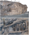
Figure 2
Figure 2. Dakosaurus maximus, neotype SMNS 8203.
(A) General view of the skull and mandible, (B) close-up on the dentary alveoli and raised lateral and medial margins, and (C) oblique forward view of the dentary tooth row. Abbreviations: den, dentary; dlp, dentary lateral plate; dmp, dentary medial plate; max, maxilla; na, nasal; pre, premaxilla; prf, prefrontal; tc, tooth crowns.
Figure 3
Figure 3. Dakosaurus maximus, neotype SMNS 8203.
(A) Anterior view of the skull and mandible, note that level to the fourth dentary tooth the mandibular ramus deflects laterally (i.e. short symphysis), and (B) oblique dorsal view of the skull, emphasising the blunt “bullet-shaped” snout (i.e. amblygnathy). Abbreviations: den, dentary; max, maxilla; na, nasal; pre, premaxilla; prf, prefrontal.
Figure 4
Figure 4. Dakosaurus maximus, neotype SMNS 8203.
(A) Close-up on the premaxillary ‘lateral plates’, (B) close-up on the maxillary alveoli in oblique ventral view and (C) close-up on the maxillary teeth showing tooth crown wear. Abbreviations: plp, premaxillary lateral plate; rp, reception pit.
Figure 5
Figure 5. Dakosaurus maximus, referred specimens SMNS 10819a and SMNS 10819b.
Snout (SMNS 10819a) in: (A) dorsal view, (B) ventral (palatal) view, and (C) close-up on the maxillary tooth-row in ventral view. Note that several teeth exhibit carinal wear, broken apices and spalling of the enamel at the apex. (D) Left-half of the posterior region of the skull (SMNS 10819b) in dorsal view. Abbreviations: en, external nares; fr, frontal; max, maxilla; pa, parietal; po, postorbital; pre, premaxilla; prf, prefrontal; rp, reception pit; sq, squamosal; stf, supratemporal fenetra.
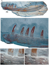
Figure 6
Figure 6. Dakosaurus maximus, referred specimen SMNS 82043.
Left mandibular ramus in lithographic limestone in: (A) general view, (B) close-up on the anterior teeth, and (C) close-up on the dentary dorsal margin at the first preserved tooth crown, showing the various foramina and a reception pit and (D) close-up on the dentary dorsal margin, slightly further back along the tooth row. Abbreviations: an, angular; art, articular; cp, coronoid process on the surangular; den, dentary; rp, reception pit; sdg, surangulodentary groove; sur, surangular.
Figure 7
Figure 7. Dakosaurus maximus, referred specimen SMNS 56999.
Left isolated maxilla in: (A) lateral view, (B) dorsal view and (C) ventral view (showing the tooth row). Abbreviations: al, alveolus; mnf, maxillary neurovascular foramen; pof, preorbital fenestra; tc, tooth crown.
Figure 8
Figure 8. Dakosaurus maximus, referred specimens SMNS 91425.
Numerous isolated teeth of D. maximus showing the occlusion wear patterns and apical breakage. Top left scale bar for (A), top right scale bar for (B), and bottom scale bar for images (C)–(D). Abbreviations: bf, basal facet; btc, broken tip; cw, carinal wear.
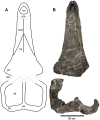
Figure 9
Figure 9. Plesiosuchus manselii, holotype NHMUK PV OR40103.
Dorsal view of the skull, (A) hypothetical skull reconstruction (grey lines represent elements that are missing) and (B) photograph of what is preserved. Abbreviations: en, external nares; max, maxilla; na, nasal; orb, orbit; pa, parietal; pre, premaxilla; prf, prefrontal; sq, squamosal; stf, supratemporal fenestra.
Figure 10
Figure 10. Plesiosuchus manselii, holotype NHMUK PV OR40103.
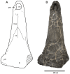
Snout in dorsal view, (A) line drawing (grey squares represent the premaxilla-maxilla suture, the exact nature of which we are unsure) and (B) photograph. Abbreviations: en, external nares; fr, frontal; max, maxilla; na, nasal; pre, premaxilla; prf, prefrontal.
Figure 11
Figure 11. Plesiosuchus manselii, holotype NHMUK PV OR40103.
Snout in lateral (right) view, (A) photograph and (B) line drawing (grey shaded area represents the orbital cavity, grey lines represents sutures we are unsure of). Abbreviations: al, alveolus; max, maxilla; na, nasal; orb, orbit; pal, palatine; pre, premaxilla; prf, prefrontal; sut?, suture?; tc, tooth crown.
Figure 12
Figure 12. Plesiosuchus manselii, holotype NHMUK PV OR40103.
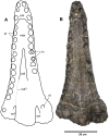
Snout in ventral (palatal) view, (A) line drawing (grey lines represent the sutures we are unsure of) and (B) photograph. Abbreviations: al, alveolus; max, maxilla; pal, palatine; pre, premaxilla; sof, suborbital fenestra; sut?, suture?; tc, tooth crown.
Figure 13
Figure 13. Plesiosuchus manselii, holotype NHMUK PV OR40103.
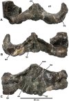
Braincase: in (A) dorsal view, (B) ventral view and (C) occipital view. Abbreviations: bt, basal tubera; eo, exocciptial; fm, foramen magnum; pa, parietal; qu, quadrate; soc, supraoccipital; sq, squamosal; stf, supratemporal fenestra.
Figure 14
Figure 14. Plesiosuchus manselii, referred specimen NHMUK PV R1089.
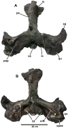
Braincase and intertemporal bar in: (A) dorsal view and (B) ventral view. Abbreviations: bt, basal tubera; crB, crest B; fr, frontal; ic, foramen for the internal carotid artery; oc, occipital condyle; pa, parietal; pop, paroccipital process; qu, quadrate; quas, quadrate articular surface; sq, squamosal.
Figure 15
Figure 15. Plesiosuchus manselii, referred specimen NHMUK PV R1089.
Close-up on the articular surface of the right quadrate. Abbreviations: bt, basal tubera; crB, crest B; oc, occipital condyle; qu, quadrate; quas, quadrate articular surface; sq, squamosal.
Figure 16
Figure 16. Plesiosuchus manselii, referred specimen NHMUK PV R1089.
Braincase in occipital view, (A) photograph and (B) line drawing (filled grey areas represent foramina). Abbreviations: XI?, foramen for cranial nerve XI?; art, articular; bo, basioccipital; bt, basal tubera; dso, depression for the supraoccipital; eo, exoccipital; fm, foramen magnum; ic, foramen for the internal carotid artery; oc, occipital condyle; pa, parietal; pop, paroccipital process; qu, quadrate; quas, quadrate articular surface; sq, squamosal; sut?, suture?.
Figure 17
Figure 17. Plesiosuchus manselii, holotype NHMUK PV OR40103a.
Mandible, with the left ramus is lateral view and the right ramus in medial view.
Figure 18
Figure 18. Plesiosuchus manselii, holotype NHMUK PV OR40103a.
Mandible, right ramus in lateral view.
Figure 19
Figure 19. Plesiosuchus manselii, referred specimen NHMUK PV R1089.
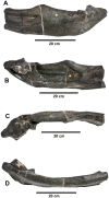
Posterior half of the left mandibular ramus in: (A) lateral view, (B) medial view, (C) dorsal view and (D) ventral view.
Figure 20
Figure 20. Plesiosuchus manselii, referred specimen NHMUK PV R1089.
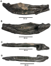
Posterior half of the right mandibular ramus in: (A) lateral view, (B) medial view, (C) dorsal view and (D) ventral view.
Figure 21
Figure 21. Plesiosuchus manselii, referred specimen NHMUK PV R1089.
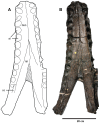
Mandibular symphysis in dorsal view, (A) line drawing and (B) photograph. Abbreviations: al, alveolus; co, coronoid, den, dentary; sp, splenial.
Figure 22
Figure 22. Plesiosuchus manselii, holotype NHMUK PV OR40103a.
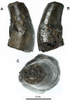
Isolated tooth crown in: (A) right lateral view, (B) left lateral view and (C) dorsal (apical) view.
Figure 23
Figure 23. Plesiosuchus manselii, holotype NHMUK PV OR40103a.
Close-up on the carinae of an isolated tooth crown, (A) anterior carina and (B) posterior carina.
Figure 24
Figure 24. Gracilineustes leedsi, holotype NHMUK PV R3540.
Snout in ventral (palatal) view, (A) photograph and (B) line drawing (thin grey lines represent breaks). Abbreviations: al, alveolus; max, maxilla; pal, palatine; ptg, pterygoid.
Figure 25
Figure 25. cf. Plesiosuchus manselii, MUJA-1004.
Isolated tooth in: (A) lingual view, (B) labial view, (C) distal view, and (D) basal view. Close-up on the denticulated carinae, showing: (E) middle of the distal carina, (F) base of the distal carina, and (G) middle of the mesial carina. Scale bar: 5 mm (A–D) and 0.5 mm (E–G). Figure modified from Figure 1 in Ruiz-Omeñaca et al. .
Figure 26
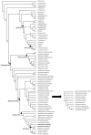
Figure 26. Strict consensus of 22 most parsimonious cladograms, showing the phylogenetic relationships of Plesiosuchus manselii and Dakosaurus maximus within Metriorhynchidae when all characters are unordered.
Note that while both are members of the subclade Geosaurini, P. manselii is in an unresolved position with Torvoneustes, Geosaurus and Dakosaurus (D. andiniensis + D. maximus). Length = 627; ensemble consistency index, CI = 0. 506; ensemble retention index, RI = 0. 860; rescaled consistency index, RC = 0. 435. The black arrow points to the only consistent difference in the 50% majority rule consensus topology: the slightly greater resolution in Rhacheosaurini. Bootstrap-values are given above or below the relevant node.
Figure 27
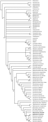
Figure 27. Strict consensus of 195 most parsimonious cladograms, showing the phylogenetic relationships of Plesiosuchus manselii and Dakosaurus maximus within Metriorhynchidae when 40 characters are ordered.
Length = 667; ensemble consistency index, CI = 0. 481; ensemble retention index, RI = 0. 863; rescaled consistency index, RC = 0. 415. Bootstrap-values are given above or below the relevant node.
Figure 28
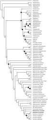
Figure 28. 50% majority rule consensus topology of 195 most parsimonious cladograms, showing the phylogenetic relationships of Plesiosuchus manselii and Dakosaurus maximus within Metriorhynchidae when 40 characters are ordered.
Length = 667; ensemble consistency index, CI = 0. 481; ensemble retention index, RI = 0. 863; rescaled consistency index, RC = 0. 415. The black circles at certain nodes denote clades that break in the strict consensus topology. Bootstrap-values are given above or below the relevant node.
Figure 29
Figure 29. Genus-level evolutionary relationships of Metriorhynchidae, based on the phylogenetic analysis presented herein.
The time-span of genera with question marks is uncertain, and the grey bars are range extensions.
Figure 30
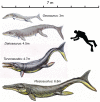
Figure 30. Life reconstructions showing the maximum body lengths for the four Geosaurini genera present in the late Kimmeridgian-early Tithonian of Western Europe.
The species from top to bottom are: Geosaurus giganteus, Dakosaurus maximus, Torvoneustes carpenteri and Plesiosuchus manselii. The maximum known body lengths of Torvoneustes and Geosaurus are from Young et al. , while those of Dakosaurus and Plesiosuchus are from this paper. The human diver is 1.8 m in height. All metriorhynchid life reconstructions are by Dmitry Bogdanov.
Figure 31
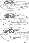
Figure 31. Lateral reconstructions of the skull of Plesiosuchus manselii, Dakosaurus maximus and D. andiniensis.
Line drawings: (A) Plesiosuchus manselii is an original skull reconstruction based on the mandible of NHMUK PV OR40103a, while the skull is a composite of NHMUK PV OR40103 and D. andiniensis (due to the holotype lacking part of the orbital and infratemporal regions of the skull, these regions are shown by broken lines); (B) Dakosaurus maximus is an original skull reconstruction based on the mandible of SMNS 8203 and SMNS 82043, while the skull is a composite of SMNS 8203, SMNS 10819b and D. andiniensis (due to the neotype and referred specimen lacking the lower orbital and infratemporal regions, these regions are shown by broken lines); (C) Dakosaurus andiniensis, redrawn from Pol & Gasparini .

References

    1. Young MT, Brusatte SL, Ruta M, Andrade MB (2010) The evolution of Metriorhynchoidea (Mesoeucrocodylia, Thalattosuchia): an integrated approach using geometrics morphometrics, analysis of disparity and biomechanics. Zool J Linn Soc 158: 801–859.
    1. Young MT, Andrade MB, Brusatte SL, Sakamoto M, Liston J (2012) The oldest known metriorhynchid super-predator: a new genus and species from the Middle Jurassic of England, with implications for serration and mandibular evolution in predacious clades. J Syst Palaeontol. In press.
    1. Lydekker R (1888) Catalogue of the Fossil Reptilia and Amphibia in the British Museum (Natural History), Cromwell Road, S.W., Part 1. Containing the Orders Ornithosauria, Crocodilia, Dinosauria, Squamata, Rhynchocephalia, and Proterosauria. London: British Museum of Natural History. 309 p.
    1. Fraas E (1902) Die Meer-Krocodilier (Thalattosuchia) des oberen Jura unter specieller berücksichtigung von Dacosaurus und Geosaurus . Palaeontographica 49: 1–72.
    1. Andrews CW (1913) A descriptive catalogue of the marine reptiles of the Oxford Clay, Part Two. London: British Museum (Natural History). 285 p.