. 2012;7(9):e45082.
doi: 10.1371/journal.pone.0045082. Epub 2012 Sep 18.

Phyllostachys edulis compounds inhibit palmitic acid-induced monocyte chemoattractant protein 1 (MCP-1) production

Affiliations

Phyllostachys edulis compounds inhibit palmitic acid-induced monocyte chemoattractant protein 1 (MCP-1) production

Jason K Higa et al. PLoS One. 2012.

Abstract

Background: Phyllostachys edulis Carriere (Poaceae) is a bamboo species that is part of the traditional Chinese medicine pharmacopoeia. Compounds and extracts from this species have shown potential applications towards several diseases. One of many complications found in obesity and diabetes is the link between elevated circulatory free fatty acids (FFAs) and chronic inflammation. This study aims to present a possible application of P. edulis extract in relieving inflammation caused by FFAs. Monocyte chemoattractant protein 1 (MCP-1/CCL2) is a pro-inflammatory cytokine implicated in chronic inflammation. Nuclear factor kappa-light-chain-enhancer of activated B cells (NF-κB) and activator protein 1 (AP-1) are transcription factors activated in response to inflammatory stimuli, and upregulate pro-inflammatory cytokines such as MCP-1. This study examines the effect of P. edulis extract on cellular production of MCP-1 and on the NF-κB and AP-1 pathways in response to treatment with palmitic acid (PA), a FFA.

Methodology/principal findings: MCP-1 protein was measured by cytometric bead assay. NF-κB and AP-1 nuclear localization was detected by colorimetric DNA-binding ELISA. Relative MCP-1 mRNA was measured by real-time quantitative PCR. Murine cells were treated with PA to induce inflammation. PA increased expression of MCP-1 mRNA and protein, and increased nuclear localization of NF-κB and AP-1. Adding bamboo extract (BEX) inhibited the effects of PA, reduced MCP-1 production, and inhibited nuclear translocation of NF-κB and AP-1 subunits. Compounds isolated from BEX inhibited MCP-1 secretion with different potencies.

Conclusions/significance: PA induced MCP-1 production in murine adipose, muscle, and liver cells. BEX ameliorated PA-induced production of MCP-1 by inhibiting nuclear translocation of NF-κB and AP-1. Two O-methylated flavones were isolated from BEX with functional effects on MCP-1 production. These results may represent a possible therapeutic application of BEX and its compounds toward alleviating chronic inflammation caused by elevated circulatory FFAs.


Conflict of interest statement

Competing Interests: The authors maintain that there is no conflict of interest with regards to the cited patent (Chinese invention patent, CN1287848A). The patent belongs to Golden Basin Bio-Tech, and we have no ownership of their patent. The bamboo extract used in this study was donated to us by Golden Basin. We have no ownership or financial investment in Golden Basin, nor are receiving any remuneration or compensation from Golden Basin for our studies. This does not alter the authors' adherence to all the PLOS ONE policies on sharing data and materials.

Figures

Figure 1
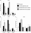
Figure 1. Effect of BEX on MCP-1 mRNA expression.
Levels of MCP-1 mRNA expression in 3T3-L1 (A), C2C12 (B), and Hepa6 (C) murine cells in response to palmitic acid (PA, 0.4 mM) in combination with BEX (125 µg/ml or 0.5%, v/v) or ethanol vehicle (0.5%, v/v) at different time points and differentiation states (3T3-L1 and C2C12). Fold change calculated via comparative cycle threshold (−ΔΔCt) normalized to beta-glucuronidase (GUSβ). Other housekeeping genes such as 18 S ribosomal RNA (18 S), hypoxanthine-guanine phosphoribosyltransferase (HPRT), and glyceraldehyde 3-phosphate dehydrogenase (GAPDH) were also used and yielded similar results as GUSβ. Mean ± standard deviation of at least 4 qRT-PCRs per cell type. Mean results were compared between treatments of the same cell type and at the same time point. Differences between means are statistically significant if columns do not share any common letters (p<0.05, one-way ANOVA with Tukey’s multiple comparison test).
Figure 2
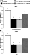
Figure 2. Protective effect of BEX on palmitic acid (PA)-induced lipotoxicity.
3T3-L1 (A), and Hepa6 cells (B) were grown to confluency in 96-well plates and treated with 0.4 mM PA for 24 hours in combination with BEX (125 µg/ml or 0.5%, v/v) or ethanol (0.5%, v/v) as a solvent control. Cell viability was measured using a MTS assay. Mean ± standard deviation over 5 trials, n ≥3 wells per trial. Differences between means are statistically significant if the columns do not share any common letters (p<0.001, one-way ANOVA, Tukey’s multiple comparison test).
Figure 3
Figure 3. Effect of BEX on lipotoxic MCP-1 production.
Production of MCP-1 was measured in the cell media supernatant (A–C) and cytosolic fraction (D–F) of 3T3-L1 (A, D), C2C12 (B, E), and Hepa6 (C, F) murine cells treated with palmitic acid (PA, 0.4 mM) in combination with BEX (125 µg/ml or 0.5%, v/v) or ethanol vehicle (0.5%, v/v). Concentrations were determined by cytometric bead array immunodetection against a reconstituted MCP-1 standard. Mean ± standard error over 3 trials, n ≥300 beads in any trial. Mean results were compared between treatments of the same cell type and at the same time point. Differences between means are statistically significant if columns do not share any common letters (p<0.05, one-way ANOVA with Bonferroni’s post hoc test).
Figure 4
Figure 4. Effect of BEX on nuclear translocation of NF-κB and AP-1 transcription factors over time.
Nuclear translocation of NF-κB (A) p50, (B) RelA/p65, AP-1 (C) c-Fos, and (D) phospho-c-Jun subunits in 3T3-L1 cells. Measurements are relative absorbance at 450 nm from a response-element DNA binding colormetric ELISA on nuclear extracts from cells treated with palmitic acid (PA, 0.4 mM) in combination with BEX (125 µg/ml or 0.5%, v/v) or ethanol vehicle (0.5%, v/v). Mean ± standard error over 3 trials, n ≥3 wells per trial. Mean results were compared between treatments at the same time point. Differences between means are statistically significant if columns do not share any common letters (p<0.05, one-way ANOVA with Tukey’s multiple comparison test).
Figure 5
Figure 5. Molecular structures of tricin and 7-O-methyl-tricin (7 MT), two compounds isolated from BEX.
Figure 6
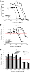
Figure 6. Dose-dependent effects of tricin, 7-O-methyl-tricin (7 MT), Fraction K, and BEX on lipotoxic MCP-1 production and cell viability.
(A) MCP-1 concentrations in cell culture media from 3T3-L1 cells cultured in 96 well plates and treated for 20 hours with PA (0.4 mM) and one of the following: tricin (black triangles), 7 MT (light grey hollow squares), Fraction K (grey diamonds), or BEX (dark grey crosses). Concentrations were determined by cytometric bead array immunodetection against a reconstituted MCP-1 standard. Mean ± standard error of 3 samples per dose, n ≥300 beads per sample. (B) Viability of 3T3-L1 cells treated with tricin (black triangles), 7 MT (light grey hollow squares), Fraction K (grey diamonds), or BEX (dark grey crosses) added to maintenance medium. All values are normalized to 3T3-L1 cells from the same plate treated with maintenance medium without flavonoids or PA (normalized average represented by a red dashed line). Mean ± standard error of three trials, n ≥3 per trial. (C) Viability of 3T3-L1 cells treated with ethanol vehicle (outlined white), Fraction K (grey), or BEX (black). Mean ± standard error of three trials, n ≥4 per trial. All values normalized to 3T3-L1 cells from the same plate treated with only maintenance medium (normalized average represented by a red dashed line). p values were obtained by performing a Student’s t-test between doses.

References

    1. Monteiro R, Azevedo I (2010) Chronic inflammation in obesity and the metabolic syndrome. Mediators Inflamm 2010. doi: 10.1155/2010/289645. - PMC - PubMed
    1. Boden G (1998) Free fatty acids (FFA), a link between obesity and insulin resistance. Front Biosci 3: d169–175. - PubMed
    1. Boden G, She P, Mozzoli M, Cheung P, Gumireddy K, et al. (2005) Free fatty acids produce insulin resistance and activate the proinflammatory nuclear factor-kappaB pathway in rat liver. Diabetes 54: 3458–3465. - PubMed
    1. Kanda H, Tateya S, Tamori Y, Kotani K, Hiasa K, et al. (2006) MCP-1 contributes to macrophage infiltration into adipose tissue, insulin resistance, and hepatic steatosis in obesity. J Clin Invest 116: 1494–1505. - PMC - PubMed
    1. Conti P, DiGioacchino M (2001) MCP-1 and RANTES are mediators of acute and chronic inflammation. Allergy Asthma Proc 22: 133–137. - PubMed

Publication types

MeSH terms