. 2012;7(9):e45183.
doi: 10.1371/journal.pone.0045183. Epub 2012 Sep 19.

The role of alpha-synuclein in melanin synthesis in melanoma and dopaminergic neuronal cells

Affiliations

The role of alpha-synuclein in melanin synthesis in melanoma and dopaminergic neuronal cells

Tianhong Pan et al. PLoS One. 2012.

Abstract

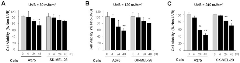
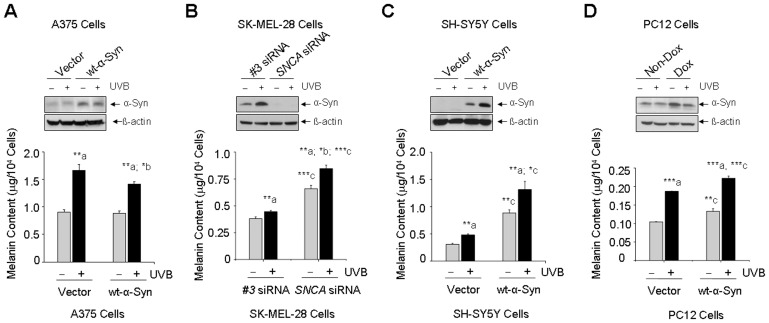
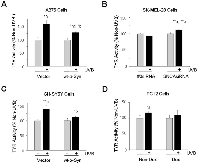
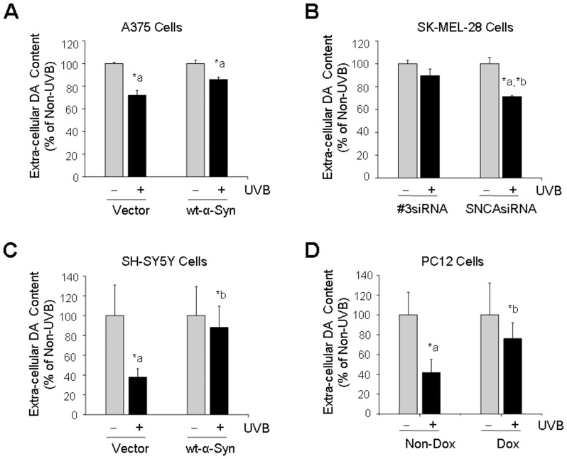
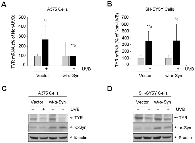
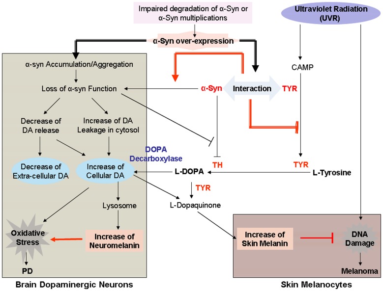
The relatively high co-occurrence of Parkinson's disease (PD) and melanoma has been established by a large number of epidemiological studies. However, a clear biological explanation for this finding is still lacking. Ultra-violet radiation (UVR)-induced skin melanin synthesis is a defense mechanism against UVR-induced damage relevant to the initiation of melanoma, whereas, increased neuromelanin (NM), the melanin synthesized in dopaminergic neurons, may enhance the susceptibility to oxidative stress-induced neuronal injury relevant to PD. SNCA is a PD-causing gene coding for alpha-Synuclein (α-Syn) that expresses not only in brain, but also in skin as well as in tumors, such as melanoma. The findings that α-Syn can interact with tyrosinase (TYR) and inhibit tyrosine hydroxylase (TH), both of which are enzymes involved in the biosynthesis of melanin and dopamine (DA), led us to propose that α-Syn may participate in the regulation of melanin synthesis. In this study, by applying ultraviolet B (UVB) light, a physiologically relevant stimulus of melanogenesis, we detected melanin synthesis in A375 and SK-MEL-28 melanoma cells and in SH-SY5Y and PC12 dopaminergic neuronal cells and determined effects of α-Syn on melanin synthesis. Our results showed that UVB light exposure increased melanin synthesis in all 4 cell lines. However, we found that α-Syn expression reduced UVB light-induced increase of melanin synthesis and that melanin content was lower when melanoma cells were expressed with α-Syn, indicating that α-Syn may have inhibitory effects on melanin synthesis in melanoma cells. Different from melanoma cells, the melanin content was higher in α-Syn-over-expressed dopaminergic neuronal SH-SY5Y and PC12 cells, cellular models of PD, than that in non-α-Syn-expressed control cells. We concluded that α-Syn could be one of the points responsible for the positive association between PD and melanoma via its differential roles in melanin synthesis in melanoma cells and in dopaminergic neuronal cells.


Conflict of interest statement

Competing Interests: The authors have declared that no competing interests exist.

Figures

Figure 1
Figure 1. UVB light-induced loss of cell viability.
A375 and SK-MEL-28 melanoma cells were exposed to UVB light at the doses of 30 mJ/cm2 (A), 120 mJ/cm2 (B) and 240 mJ/cm2(C) and post-cultured for 4 h, 24 h and 48 h (A, B, C). Cell viability was determined by MTT assay. The results were expressed as percentage of non-UVB control. *: p<0.05; **: p<0.01 as compared to its non-UVB control (0 h).
Figure 2
Figure 2. Roles of α-Syn in melanin synthesis in melanoma and dopaminergic neuronal cells.
A375 melanoma cells (A) and dopaminergic neuronal SH-SY5Y and PC12 cells (C, D) with or without wt-α-Syn over-expression, SK-MEL-28 melanoma cells (B) with or without suppression of endogenous α-Syn, were exposed to UVB light (120 mJ/cm2) or non-UVB light and post-cultured for 24 h. The protein levels of α-Syn were determined by western blot assay (A, B, C, D, upper panel). Melanin content was determined spectrophotometrically at 415 nm using iMark Microplate Reader (A, B, C, D, lower panel). *: p<0.05; **: p<0.01; *** p<0.001 as compared to its non-UVB control (a), non-α-Syn control (b), or vector/#3 siRNA control (c). Dox  =  doxycycline.
Figure 3
Figure 3. Effects of α-Syn on the activation of tyrosinase (TYR) induced by UVB light.
After cells were exposed to UVB light (120 mJ/cm2) and post-cultured for 24 h, the TYR activity was measured spectrophotometrically at 475 nm using iMark Microplate Reader. Cells without UVB light exposure (non-UVB) were used as a control. A: A375 melanoma cells; B: SK-MEL-28 melanoma cells; C: SH-SY5Y dopaminergic neuronal cells; D: PC12 dopaminergic neuronal cells. *: p<0.05; **: p<0.01 as compared to its non-UVB control (a) or non-α-Syn control (b). Dox  =  doxycycline.
Figure 4
Figure 4. Extra-cellular dopamine (DA) content as determined by HPLC assay.
After cells were exposed to UVB light (120 mJ/cm2) or non-UVB light and post-cultured for 24 h, cell culture medium from melanoma A375 cells (A) and SK-MEL-28 cells (B), dopaminergic neuronal SH-SY5Y cells (C) and PC12 cells (D) with or without α-Syn expression were subjected to HPLC assay for measuring DA level. *: p<0.05 as compared to its non-UVB control (a) or non-α-Syn control (b). Dox  =  doxycycline.
Figure 5
Figure 5. UVB light-induced changes in gene expression and protein level of TYR.
A375 (A) and SH-SY5Y cells (B) with or without α-Syn expression were exposed to UVB light (120 mJ/cm2) or non-UVB light and post-cultured for 24 h. Gene expression of TYR was determined by real-time quantitative PCR assay (A, B). *: p<0.05; **: p<0.01 as compared to its non-UVB control (a) or non-α-Syn control (b). The protein levels of TYR were determined by western blot assay (C, D).
Figure 6
Figure 6. Flow chat of the linkage between PD and melanoma mediated by α-Syn.
In skin melanocytes, UVR causes DNA damage, leading to the initiation of melanoma. UVR also induces melanin synthesis which is catalyzed by TYR. Increased melanin prevents UVR-induced DNA damage, reducing the vulnerability to development of melanoma. α-Syn expression in skin melanocytes may interact with TYR, inhibiting UVR-induced TYR activation, leading to the reduction of melanin synthesis, which may enhance the susceptibility of skin melanocytes to develop melanoma. In brain dopaminergic neurons, impaired degradation of α-Syn or α-Syn multiplications causes accumulation/aggregation of α-Syn, leading to the loss of α-Syn function, thereafter, the decrease of DA release and increase of cellular DA content, which may be converted into melanin in lysosome. The increased melanin content in dopaminergic neurons (neuromelanin) enhances the susceptibility to oxidative stress-induced neuronal injury relevant to PD.

References

    1. Liu R, Gao X, Lu Y, Chen H (2011) Meta-analysis of the relationship between Parkinson disease and melanoma. Neurology 76: 2002–2009. - PMC - PubMed
    1. Bertoni JM, Arlette JP, Fernandez HH, Fitzer-Attas C, Frei K, et al. (2010) Increased melanoma risk in Parkinson disease: a prospective clinicopathological study. Arch Neurol 67: 347–352. - PubMed
    1. Ferreira JJ, Neutel D, Mestre T, Coelho M, Rosa MM, et al. (2010) Skin cancer and Parkinson’s disease. Mov Disord 25: 139–148. - PubMed
    1. Gao X, Simon KC, Han J, Schwarzschild MA, Ascherio A (2009) Family history of melanoma and Parkinson disease risk. Neurology 73: 1286–1291. - PMC - PubMed
    1. Driver JA, Logroscino G, Buring JE, Gaziano JM, Kurth T (2007) A prospective cohort study of cancer incidence following the diagnosis of Parkinson’s disease. Cancer Epidemiol Biomarkers Prev 16: 1260–1265. - PubMed

Publication types

MeSH terms