. 2012;7(9):e45213.
doi: 10.1371/journal.pone.0045213. Epub 2012 Sep 19.

Attenuated Mycobacterium tuberculosis SO2 vaccine candidate is unable to induce cell death

Affiliations

Attenuated Mycobacterium tuberculosis SO2 vaccine candidate is unable to induce cell death

Adriana Aporta et al. PLoS One. 2012.

Abstract

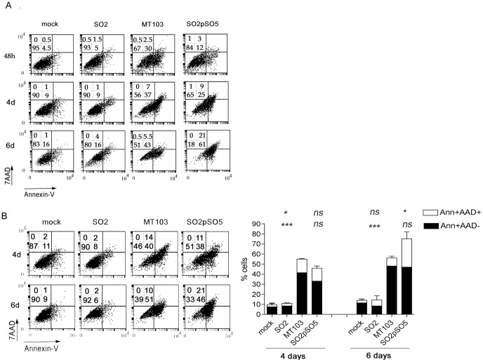
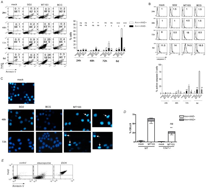
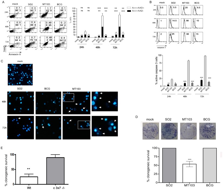
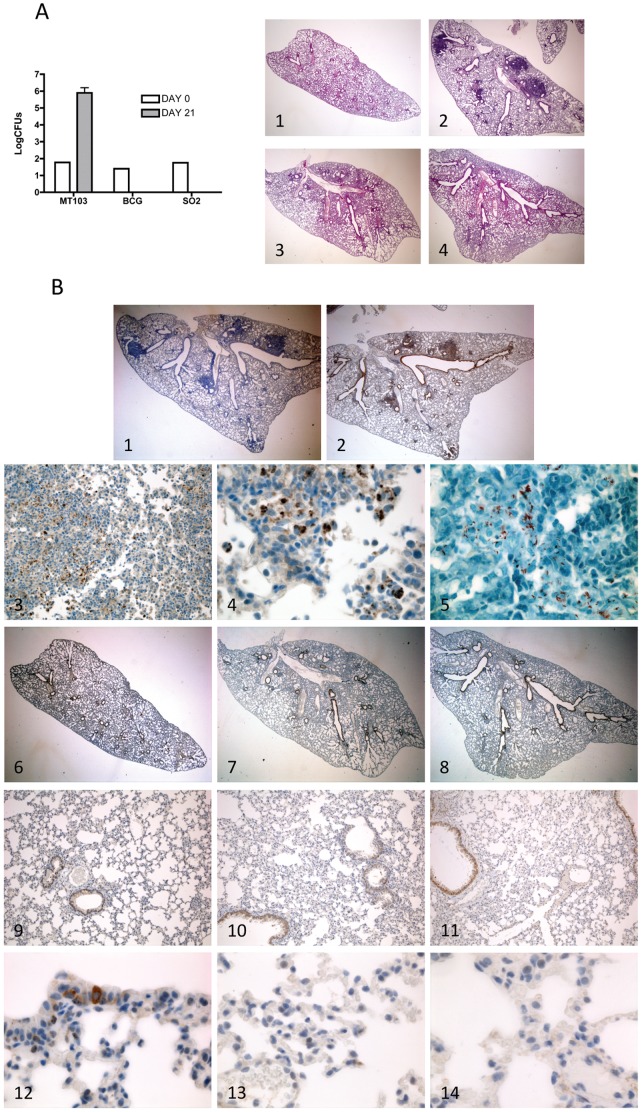
It has been proposed that Mycobacterium tuberculosis virulent strains inhibit apoptosis and trigger cell death by necrosis of host macrophages to evade innate immunity, while non-virulent strains induce typical apoptosis activating a protective host response. As part of the characterization of a novel tuberculosis vaccine candidate, the M. tuberculosis phoP mutant SO2, we sought to evaluate its potential to induce host cell death. The parental M. tuberculosis MT103 strain and the current vaccine against tuberculosis Bacillus Calmette-Guérin (BCG) were used as comparators in mouse models in vitro and in vivo. Our data reveal that attenuated SO2 was unable to induce apoptotic events neither in mouse macrophages in vitro nor during lung infection in vivo. In contrast, virulent MT103 triggers typical apoptotic events with phosphatidylserine exposure, caspase-3 activation and nuclear condensation and fragmentation. BCG strain behaved like SO2 and did not induce apoptosis. A clonogenic survival assay confirmed that viability of BCG- or SO2-infected macrophages was unaffected. Our results discard apoptosis as the protective mechanism induced by SO2 vaccine and provide evidence for positive correlation between classical apoptosis induction and virulent strains, suggesting apoptosis as a possible virulence determinant during M. tuberculosis infection.


Conflict of interest statement

Competing Interests: The authors have declared that no competing interests exist.

Figures

Figure 1
Figure 1. Attenuated SO2 strain does not induce PS translocation in primary mouse macrophages.
Mouse bone marrow-derived primary macrophages (BMDM) were mock-treated or infected at low MOI of 1∶1 (A) or 10∶1 (B) with the indicated strains. After 4 hours of infection, cells were washed and incubated with complete medium. At the indicated times post infection, both detached and adhered cells were pooled and PS exposure (annexin-V-FITC) and 7-AAD staining were analyzed by flow cytometry as described in Materials and Methods (A, B). Numbers in the dot-plot diagrams correspond to the percentage (%) of cells in each quadrant. A representative experiment is shown in the left panels. Data in the graph (figure B, right panel) is represented as mean±S.E.M. of at least three independent experiments. Statistical analyses were done with one-way ANOVA with Tukeýs post-test comparing every strain with MT103. Upper symbols  =  statistical analyses of Ann+AAD+ cells; lower symbols =  statistical analyses of Ann+AAD- cells. ns =  not statistically significant; *, **, ***  =  statistically significant; * p<0,05; ** p<0,01; *** p<0,001.
Figure 2
Figure 2. Attenuated SO2 and BCG strains do not induce PS translocation, caspase-3 activation and nuclear apoptosis in J774 mouse macrophages.
The mouse macrophage cell line J774 was mock-treated or infected at low MOI (up to 10∶1) with the indicated strains (A-C). Mouse embryonic fibroblasts (MEF) from wild type (WT) of caspase-3 and 7 deficient (c3x7) mice were infected with MT103 (D). After 4 hours, cells were washed and incubated with complete medium. At the indicated times post infection, both detached and adhered cells were pooled and PS exposure on plasma membrane (annexin-V-FITC) and 7-AAD uptake (A, D) or caspase-3 activation (B) were analyzed by flow cytometry as described in Materials and Methods. Numbers in the dot-plot diagrams correspond to the percentage (%) of cells in each quadrant. A representative experiment is shown in the left panels. Data in the graphs (right panels) are represented as mean±S.E.M. of at least three independent experiments. Statistical analyses were done with one-way ANOVA with Tukeýs post-test comparing BCG and SO2 strains with MT103 (A) or MEF.WT with MEF.C3x7−/− (D). Upper symbols  =  statistical analyses of Ann+AAD+ cells; lower symbols =  statistical analyses of Ann+AAD- cells. ns =  not statistically significant; *, **, ***  =  statistically significant; * p<0,05; ** p<0,01; *** p<0,001. Cells were stained with Hoetchs33258 and observed by fluorescence microscopy (C). Pictures from representative fields are shown. Original magnification: x400. Magnifications from the squared sections are shown on the right side. Arrows indicate cells with nuclear apoptotic morphology. (E) J774 cells were treated with 100 nM staurosporine for 24 h or 10% (v/v) ethanol for 90 min. Subsequently, both detached and adhered cells were pooled and PS exposure on plasma membrane (annexin-V-FITC) and 7-AAD uptake was analysed by flow cytometry.
Figure 3
Figure 3. Attenuated BCG or SO2 strains do not kill J774 macrophages at high MOI.
The mouse macrophage cell line J774 was mock-treated or infected at high MOI (100∶1) with the indicated strains (A-D). After 4 hours, cells were washed and incubated with complete medium. At the indicated times post infection, both detached and adhered cells were pooled and PS exposure on plasma membrane (annexin-V-FITC) and 7-AAD uptake (A) or caspase-3 activation (B) were analyzed by flow cytometry as described in Materials and Methods. Numbers in each dot-plot diagram correspond to the percentage (%) of cells in each quadrant. A representative experiment is shown in the left panels. Data in the graphs (right panels) are represented as mean±S.E.M. of at least three independent experiments. Statistical analyses were done with one-way ANOVA with Tukeýs post-test comparing BCG and SO2 strains with MT103. Upper symbols  =  statistical analyses of Ann+AAD+ cells; lower symbols =  statistical analyses of Ann+AAD- cells. ns =  not statistically significant; *, **, ***  =  statistically significant; * p<0,05; ** p<0,01; *** p<0,001. Cells were stained with Hoetchs33258 and observed by fluorescence microscopy (C). Pictures from representative fields are shown. Original magnification: x400. Magnifications from the squared sections are shown on the right side. Arrows indicate cells with nuclear apoptotic morphology. For clonogenic survival assay J774 cells were mock-treated or infected at high MOI (100∶1) with MT103, BCG or SO2 strains (D). 4 hours after infection, cells were washed, trypsinized and counted. 150 cells per well were seeded in triplicates (6-well plate) and incubated in fresh medium during 8 days. In the case of MEF.wt and MEF.c3x7−/− cells (E), they were infected with MT103 for seven days. After infection, cells were washed, trypsinized and counted. 150 cells per well were seeded in triplicates (6-well plate) and incubated in fresh medium for eight additional days. After incubation, cells were stained with Crystal Violet and colonies were counted as described in Materials and Methods. Images from single wells of a representative experiment are shown (D). Survival was calculated as percentage of colonies relative to the number of colonies in the non-infected controls (D, E). Values are represented as mean+/− SEM of 4 different experiments. Statistical analyses were done with one-way ANOVA with Tukeýs post-test by comparing MEF.WT with MEF.C3x7−/−. ** p<0,01; *** p<0,001.
Figure 4
Figure 4. Virulent MT103 strain, but not the attenuated SO2 and BCG strains, replicates in vivo, causes lung pathology and induces apoptosis in mouse lungs.
Groups of five C57BL/6 mice were intratracheally infected with a low dose (100 bacteria/mouse) of MT103, BCG or SO2 strains as described in Materials and Methods. Lungs were harvested and CFU counted at 21 days post-infection (A, left panel). Lung histopathology (hematoxilin/eosin) representative images (10x magnification) of mock-treated (A, right panel, image 1) or MT103-, BCG- or SO2-infected mice (A, right panel, images 2, 3, 4, respectively) at 3 weeks post inoculation. Representative images of active caspase 3 immunohistochemical and Ziehl-Neelsen staining of mock-treated or MT103-, BCG- or SO2-infected lungs at three weeks post inoculation (B): primary antibody control of MT103-infected lung section incubated only with secondary antibody (10x magnification) (image 1); active caspase-3 staining of MT103-infected lung section (10x magnification) (image 2); active caspase-3 staining of MT103-infected lung section (100x magnification) (image 3); active caspase-3 staining of MT103-infected lung section (600x magnification) (image 4); Ziehl-Neelsen staining of MT103-infected lung section (600x magnification) (image 5); active caspase-3 staining of mock-treated lung section (10x, 100x and 600x magnification, images 6, 9 and 12, respectively); active caspase-3 staining of BCG-infected lung section (10x, 100x and 600x magnification, images 7, 10 and 13, respectively); active caspase-3 staining of SO2-infected lung section (10x, 100x and 600x magnification, images 8, 11 and 14, respectively).

References

    1. Pieters J (2008) Mycobacterium tuberculosis and the macrophage: maintaining a balance. Cell Host Microbe 3: 399–407. - PubMed
    1. Finlay BB, McFadden G (2006) Anti-immunology: evasion of the host immune system by bacterial and viral pathogens. Cell 124: 767–782. - PubMed
    1. Kaufmann SH, Cole ST, Mizrahi V, Rubin E, Nathan C (2005) Mycobacterium tuberculosis and the host response. J Exp Med 201: 1693–1697. - PMC - PubMed
    1. Fan T, Lu H, Hu H, Shi L, McClarty GA, et al. (1998) Inhibition of apoptosis in chlamydia-infected cells: blockade of mitochondrial cytochrome c release and caspase activation. J Exp Med 187: 487–496. - PMC - PubMed
    1. Monack D, Falkow S (2000) Apoptosis as a common bacterial virulence strategy. Int J Med Microbiol 290: 7–13. - PubMed

Publication types

MeSH terms