. 2012;7(9):e45225.
doi: 10.1371/journal.pone.0045225. Epub 2012 Sep 18.

Deletion of IL-33R (ST2) abrogates resistance to EAE in BALB/C mice by enhancing polarization of APC to inflammatory phenotype

Affiliations

Deletion of IL-33R (ST2) abrogates resistance to EAE in BALB/C mice by enhancing polarization of APC to inflammatory phenotype

Marija Milovanovic et al. PLoS One. 2012.

Abstract

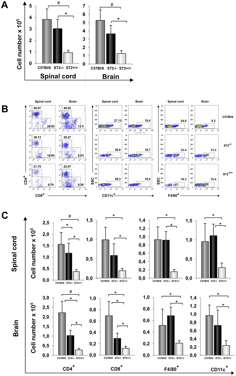
The administration of interleukin 33 and deletion of IL-33 receptor, ST2 molecule, affects the induction of autoimmunity in different experimental models of human autoimmune diseases. The aim of this study was to analyze the effect of ST2 deletion on the induction of experimental autoimmune encephalomyelitis (EAE) in resistant BALB/c mice. Mice were immunized with MOG(35-55) peptide or disease was induced by passive transfer of encephalitogenic singenic cells and EAE was clinically and histologically evaluated. Expression of intracellular inflammatory cytokines, markers of activation and chemokine receptors on lymphoid tissue and CNS infiltrating mononuclear cells was analyzed by flow cytometry. We report here that deletion of ST2(-/-) molecule abrogates resistance of BALB/c mice to EAE induction based on clinical and histopathological findings. Brain and spinal cord infiltrates of ST2(-/-) mice had significantly higher number of CD4(+) T lymphocytes containing inflammatory cytokines compared to BALB/c WT mice. Adoptive transfer of ST2(-/-) primed lymphocytes induced clinical signs of the disease in ST2(-/-) as well as in WT mice. MOG(35-55) restimulated ST2(-/-) CD4(+) cells as well as ex vivo analyzed lymph node cells had higher expression of T-bet and IL-17, IFN-γ, TNF-α and GM-CSF in comparison with WT CD4(+) cells. ST2(-/-) mice had higher percentages of CD4(+) cells expressing chemokine receptors important for migration to CNS in comparison with WT CD4(+) cells. Draining lymph nodes of ST2(-/-) mice contained higher percentage of CD11c(+)CD11b(+)CD8(-) cells containing inflammatory cytokines IL-6 and IL-12 with higher expression of activation markers. Transfer of ST2(-/-) but not WT dendritic cells induced EAE in MOG(35-55) immunized WT mice. Our results indicate that ST2 deficiency attenuates inherent resistance of BALB/c mice to EAE induction by enhancing differentiation of proinflammatory antigen presenting cells and consecutive differentiation of encephalitogenic T cells in the draining lymph node rather than affecting their action in the target tissue.


Conflict of interest statement

Competing Interests: The authors have declared that no competing interests exist.

Figures

Figure 1
Figure 1. ST2−/− BALB/c and C57BL/6 mice are susceptible while BALB/c mice are resistant to EAE.
C57BL/6, BALB/c ST2−/− and BALB/c WT mice were immunized with MOG35–55/CFA and were checked daily for (A) clinical signs of EAE. Data are representative of two independent experiments, twelve mice per group (means ± SEM). At the peak of the disease spinal cord and brain tissues were fixed, sectioned, and stained with hematoxylin and eosin. Histological scores were calculated from a total of 5 sections per group (B). Representative spinal cord and brain tissue sections are presented; magnification is 40 X (C). Statistical significance was tested by Student’s t-test.
Figure 2
Figure 2. ST2−/− BALB/c mice have similar infiltration in CNS as C57BL/6 mice.
Fourteen days after immunization mononuclear cells were isolated from spinal cords and brains and counted (A). Pooled mononuclear cells isolated from brains and spinal cords (three per group, four samples in group, 12 mice per group) were used for flow cytometric analysis of percentages (B) and total cell numbers (C) of CD4+, CD8+, CD11c+, and F4/80+ cells. Absolute numbers were calculated as the product of the average number of CNS mononuclear cells harvested per mouse pooled from brains and spinal cords. The data are from representative experiment (mean+SD *P<0.05 and #P<0.005). Statistical significance was tested by Student’s t-test.
Figure 3
Figure 3. Spinal cord and brain infiltrates from ST2−/− BALB/c mice contain inflammatory T helper lymphocytes.
Mononuclear cells were isolated from spinal cord (A) and brain tissue (B) of mice killed at the peak of the disease and analyzed by flow cytometry after intracellular staining for inflammatory cytokines. The total numbers of CD4+ cells containing IL-17, IFN-γ, TNF-α and GM-CSF are calculated as the product of the average number of CNS mononuclear cells harvested per mouse from pooled brains and spinal cords. Presented data are from representative experiment, 12 mice in each group (means+SD). Statistical significance was tested by Student’s t-test.
Figure 4
Figure 4. Primed ST2−/− T cells induce EAE in WT and ST2−/− recipient.
The donor cells were isolated from popliteal lymph nodes 9 days after foot pad immunization of ST2−/− BALB/c and BALB/c WT mice with MOG/CFA emulsion. Donor cells were restimulated with MOG35–55 peptide (50 µM) for 72 hours and 1×10⁷ were transfer to ST2−/− and WT BALB/c mice. Mice were observed every day after injection for disease onset and clinically scored. Data are presented as the mean of two independent experiments with 5 animals in each group in each experiment (n = 10 in both groups * P<0.05) (A). Lymph node's mononuclear cells harvested from donor mice and restimulated in vitro with MOG35–55 peptide (50 µM) for 72 hours are analyzed for expression of inflammatory cytokines (B) and markers of Th1 and Th2 differentiation (C). Data are presented as the mean+SD from representative experiment with 10 animals in each group. Histograms and mean values presented in the graphs are gated on CD4+ cells (C). Levels of proinflammatory cytokines were measured in cell culture supernatants by ELISA and CBA. Data are presented as the mean+SD of two independent experiments, 10 animals per group (D). Statistical significance was tested by Student’s t-test.
Figure 5
Figure 5. ST2 deletion leads to the induction of inflammatory T helper lymphocytes in draining lymph nodes.
Mononuclear cells from draining lymph nodes of immunized ST2−/− BALB/c and BALB/c WT mice were isolated on day 9. (A) Flow cytometry was used to evaluate IL-17, IFN-γ, TNF-α, GM-CSF and IL-10 containing CD4+ T cells. Data are presented as individual percentages of all mice, 10 mice per group. (B) Expression of activation marker CD69 on cells isolated from lymph nodes and peripheral blood 4 and 7 days after immunization was evaluated by flow cytometry. Plots are gated on lymphocyte population. Mean values of CD4+CD69+ cells are obtained from two experiments with 16 mice per group (C) Flow cytometric analysis of expression of chemokine receptors CCR6, CXCR3 and CXCR5 on CD4+ lymphocytes was performed on cells isolated from lymph nodes (4 days after immunization) and peripheral blood (7 days after immunization). Percentages of double positive cells CD4+CCR6+, CD4+CXCR3+ and CD4+CXCR5+ are presented (upper panel) and chemokine receptor expression in CD4+ population of lymph node cells (lower panel). Data are presented as the mean+SD, 16 mice per group (*P<0.05). Statistical significance was tested by Student’s t-test.
Figure 6
Figure 6. Absence of ST2 in dendritic cells enhances their APC capacity after EAE induction.
Mice were immunized with MOG35–55/CFA and cells were isolated from lymph nodes and spleens. (A) Ability of ST2−/− and WT dendritic cells isolated from spleen 8 days after immunization to affect proliferation of ST2−/− CD4+ and WT CD4+ cells without or with MOG35–55 specific re-stimulation was determined by MTT assay. Proliferation is presented as percentage calculated in relation to proliferation of CD4+ cells only. (B) ELISA was performed to determine the production of IL-10, IL-6, IL-12 and IL-23 cytokines by dendritic cells isolated from spleens 9 days after immunization and stimulation with TLR1/2 agonist. Data are presented from 2 independent experiments as the mean+SD of representative experiment (n = 16 in both groups *P<0.05). (C) Dendritic cells were isolated from ST2−/− and WT mice 4 and 8 days after MOG35–55 immunization. Active induction of EAE was performed on wild type BALB/c mice and after 4 and 8 days ST2−/− or WT dendritic cells were given intravenously. Mice were clinically scored daily. Data are shown as mean+SD; 5 mice per group (*P<0.05). Statistical significance was tested by Student’s t-test.
Figure 7
Figure 7. ST2 deletion leads to inflammatory phenotype of APC after EAE induction.
Mononuclear cells were isolated from lymph nodes of ST2−/− and WT BALB/c mice 4, 7 and 9 days after immunization and analyzed by flow cytometry. (A) Percentages of subpopulation of dendritic cells, CD11c+CD11b+CD8, in the lymph nodes 4, 7 and 9 days after immunization are presented. (B) Expression of markers of activation, CD86 and MHC II, on dendritic cells was performed at day 4. Histograms were gated on CD11c+CD11b+CD8 cells. (C) Percentages of CD11c+CD11b+CD8 cells positive for IL-1, IL-12 and IL-6 in lymph nodes 4 days after immunization is presented. Data are presented from 3 independent experiments as the mean+SD of representative experiment (n = 23 in both groups). Statistical significance was tested by Student’s t-test.

References

    1. Sospedra M, Martin R (2005) Immunology of multiple sclerosis. Annu Rev Immunol 23: 683–747. - PubMed
    1. Tominaga S, Jenkins NA, Gilbert DJ, Copeland NG, Tetsuka T (1991) Molecular cloning of the murine ST2 gene: characterization and chromosomal mapping. Biochim Biophys Acta 1090: 1–8. - PubMed
    1. Bergers G, Reikerstorfer A, Braselmann S, Graninger P, Busslinger M (1994) Alternative promoter usage of the Fos-responsive gene Fit-1 generates mRNA isoforms coding for either secreted or membrane-bound proteins related to the IL-1 receptor. EMBO J 13(5): 1176–1188. - PMC - PubMed
    1. Xu D, Chan WL, Leung BP, Huang F, Wheeler R, et al. (1998) Selective expression of a stable cell surface molecule on type 2 but not type 1 helper T cells. J Exp Med 187(5): 787–794. - PMC - PubMed
    1. Kropf P, Hearth S, Klemenz R, Muller I (2003) Signalling through the T1/ST2 molecule is not necessary for Th2 differentiation but is important for the regulation of type 1 responses in nonhealing Leishmania major infection. Infect Immun 71(4): 1961–1971. - PMC - PubMed

Publication types

MeSH terms