. 2012;7(9):e45234.
doi: 10.1371/journal.pone.0045234. Epub 2012 Sep 17.

Ectonucleotidase CD38 demarcates regulatory, memory-like CD8+ T cells with IFN-γ-mediated suppressor activities

Affiliations

Ectonucleotidase CD38 demarcates regulatory, memory-like CD8+ T cells with IFN-γ-mediated suppressor activities

Rajia Bahri et al. PLoS One. 2012.

Abstract

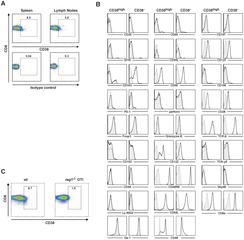
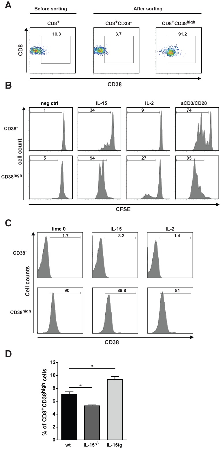
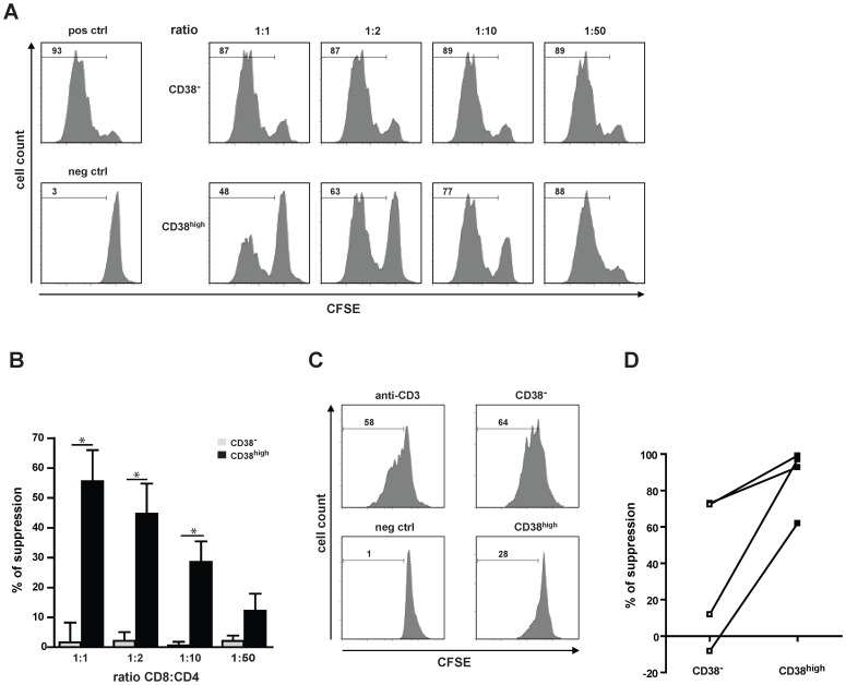
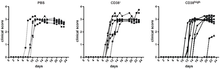
Regulatory CD8(+) T cells are critical for self-tolerance and restricting excessive immune responses. The variety of immune functions they fulfill, the heterogeneity of their phenotype, and the mechanism of action are still poorly understood. Here we describe that regulatory CD8(+) T cells exhibiting immunosuppressive actions in vitro and in vivo are recognized as CD38(high) T cells and present in naive mice. CD38 is a glycosylated membrane protein with ectonucleotidase properties. CD8(+)CD38(high) (CD44(+)CD122(+)CD62L(high)) lymphocytes suppress CD4(+) effector T-cell proliferation in an antigen-non specific manner via IFN-γ. While direct cell-to-cell contact is needed for this suppressor activity, it is independent of membrane-bound TGF-β and granzyme B release. IL-15 potentiates the suppressive activity of CD8(+)CD38(high) T cells and controls their survival and expansion. In humans CD8(+)CD38(high) T cells inhibit CD4(+) effector T cell proliferation. In vivo, CD8(+)CD38(high), but not CD8(+)CD38(-) T cells mitigate murine experimental autoimmune encephalomyelitis (EAE) by reducing the clinical score and delaying disease occurrence. EAE suppression is enhanced by pre-treatment of CD8(+)CD38(high) T cells with IL-15. These findings add evidence that the expression of ectoenzyme receptor family members positively correlates with suppressor functions and identifies CD8(+)CD38(high) T cells as potential inhibitors of excessive immune responses.


Conflict of interest statement

Competing Interests: The authors have declared that no competing interests exist.

Figures

Figure 1
Figure 1. Memory-like CD8+ T cells express CD38.
Negatively selected CD8+ T cells from spleens and lymph nodes were stained with CD8 and CD38 mAbs (1A, upper panel), CD8 and isotype matched control antibodies (1A, lower panel). Intracellular Foxp3, granzyme B, perforin and the indicated cell-surface markers on sorted CD8+ T cells from LN were determined by flow cytometry (B). Bold-line histograms show staining with specific antibodies and grey-filled histograms isotype-matched controls. Similar results were observed in at least three independent experiments for each marker (B). Negatively selected splenic CD8+ T cells from wt and rag1 −/−OTI mice were stained with CD8 and CD38 mAbs (C). Numbers represent percentage of cells in the indicated gate. One representative experiment from two is shown.
Figure 2
Figure 2. IL-15 promotes expansion and survival of CD8+CD38highT cells.
(A) After a CD8-negative selection by MACS the CD8+ T cells isolated from spleens and LN were stained with CD8 and CD38 mAbs and subsequently sorted into CD38high and CD38 T cells by FACS. Flow cytometric analysis of CD8 and CD38 expression on CD8+ T cells before and after FACS sorting are shown. (B) Sorted CD8+CD38high and CD8+CD38 T cells from spleens and LN were treated either with IL-15 (100 ng/mL), IL-2 (100 ng/mL), aCD3/CD28 (1 µg/mL) or left untreated (neg ctrl). After 72 h the proliferation of CD8+ T cells was detected by CFSE dilution. Numbers represent the percentage of proliferating cells (B) and CD38 positive cells (C). The results are representative of three independent experiments performed. (D) Spleen cells from wt (n = 4), IL-15−/− (n = 5), and IL-15 tg (n = 5) mice were stained with CD8 and CD38 mAbs and the percentage of CD38+CD8high T cells was determined by FACS. Values represent the mean±SEM of one experiment of two performed. *p<0.05 (Mann-Whitney test).
Figure 3
Figure 3. CD8+CD38high T cells suppress effector CD4+ T cell proliferation.
(A) CFSE-labelled CD4+ OTII T cells were used as responder and stimulated with OTII323–339 peptide in the presence of DCs. Either CD8+CD38high (CD38high) or CD8+CD38 (CD38) T cells were added to the culture at different CD8:CD4 ratios. Histograms show the CFSE dilution of the CD4+ OTII T cells after three days of culture (gated on CD4+ T cells). Numbers represent the percentage of proliferating cells. Proliferation of CD4+ T cells incubated with DC only, or in addition with OTII323–339 peptide are shown as a negative (neg ctrl) and positive control (pos ctrl), respectively. One of at least four independent experiments is shown. (B) Quantification of CD8+ T cell-mediated suppression of CD4+ T cell proliferation. Values represent the mean±SEM of four independent experiments. Percentage of suppression was calculated as: proliferation in the positive control-proliferation in the probe/proliferation in the positive control×100. *p<0.05 (Mann-Whitney test). (C) Suppression of human CD4+ T cell proliferation by CD8+CD38+ T cells. Anti-CD3 stimulated proliferation of CD4+ T cells (pos ctrl) in addition of CD8+38high or CD8+38 T cells, or left unstimulated (neg ctrl) was measured by CFSE dilution after four days of incubation. One out of four independent experiments is shown. (D) Quantification of CD8+ T cell-mediated suppression of CD4+ T cell proliferation. Values represent percentage of suppression of four independent experiments.
Figure 4
Figure 4. CD8+CD38high T cells are suppressive in vitro, their activity is dependent on IFN-γ secretion and requires cell-to-cell contact.
1×10⁵ CD4+ OTII T cells were stimulated with (pos ctrl) or without (neg ctrl) OTII323–339 peptide in the presence of DCs. CD8+CD38high (CD38high) or CD8+CD38 (CD38) T cells were added to the culture at a ratio 1∶1 (1×10⁵ cells) (A-E). (A) Apoptosis of CD4+ OTII T cells was measured by annexinV/PI staining. The percentage of CD4+ annexinV+ T cells is shown at indicated time points of co-culture with CD8+ T cells or in the presence of 100 ng/ml of IFN-γ (IFN-γ). The assay was performed in triplicates and shown is the mean±SEM. Results are representative of three independent experiments. (B) Concentrations of IL-2, IL-17 and IFN-γ in supernatants of 72 h co-cultures were measured by ELISA and the mean±SEM of one of three independent experiments is shown. (C) CD4+ OTII T cells were stimulated with OTII323–339 peptide in the presence of DCs. Either CD8+CD38high (CD38high) or CD8+CD38 (CD38) T cells were added to the culture at a CD8:CD4 ratio of 1 for 48 h. The last 6 h of incubation brefeldin (10 µg/mL), ionomycin (1 µg/mL) and PMA (100 ng/mL) were added to the co-cultures. Cells were stained with CD8 mAbs, fixed and intracellularly stained with IFN-γ mAbs or isotype matched control Abs. CD8+ T cells were gated and plots represent the expression of IFN-γ in CD38high or CD38 T cell populations. Numbers represent percentage of cells in the indicated gate from one representative experiment of two performed. (D) CD4+ OTII T cells were stimulated without (neg ctrl) or with (pos crtl) OTII323–339 peptide in the presence of DCs. Either CD8+CD38high (CD38high) or CD8+CD38 (CD38) T cells were added directly (direct contact) to the culture at a CD8:CD4 ratio of 1. CD4+ OTII T cells, DC and OTII323–339 peptide were placed in the lower chamber of the transwell, CD38high or CD38 T cells (Tw/CD8) or CD38high or CD38 T cells together with DCs (Tw/CD8+DC) were given into the upper chamber of the transwell (at a CD8:CD4 ratio of 1). (D) Antigen-specific proliferation of CD4+ OTII T cells was measured by CFSE dilution after four days of incubation. (E) IFN-γ concentration in the supernatants was measured by ELISA. Assay was performed in triplicates and the mean±SEM of one representative of three independent experiments is shown. (F) CD4+ OTII T cells were incubated with DCs with (pos ctrl) or without (neg ctrl) OTII323–339 peptide. CD8+CD38high (CD38high) and CD8+CD38 (CD38) T cells sorted from C57Bl/6j wt or ifn-γ −/− mice were added to the culture at the indicated CD8:CD4 ratios. The proliferation of CD4+ OTII T cells was measured in triplicates by CFSE dilution after four days of culture. The mean±SEM of one of two independent experiments is shown.
Figure 5
Figure 5. CD8+CD38high T cells are suppressive in vivo.
EAE was induced in C57/BL6j mice. On day 8 mice were injected i.v. either with CD8+CD38high cells, CD8+CD38 T cells (each 0.75×10⁵ cells/mouse) or with PBS in the negative control. Mice were monitored for disease associated symptoms every day during 23 days. The values represent the clinical scores for each individual mouse of two independent experiments. ctrl (n = 6), CD38 (n = 7), CD38high (n = 9). Similar results were observed in three independent experiments.

References

    1. Wei S, Kryczek I, Zou L, Daniel B, Cheng P, et al. (2005) Plasmacytoid dendritic cells induce CD8+ regulatory T cells in human ovarian carcinoma. Cancer Res 65: 5020–5026. - PubMed
    1. Najafian N, Chitnis T, Salama AD, Zhu B, Benou C, et al. (2003) Regulatory functions of CD8+CD28- T cells in an autoimmune disease model. J Clin Invest 112: 1037–1048. - PMC - PubMed
    1. Garba ML, Pilcher CD, Bingham AL, Eron J, Frelinger JA (2002) HIV antigens can induce TGF-beta(1)-producing immunoregulatory CD8+ T cells. J Immunol 168: 2247–2254. - PubMed
    1. Smith TR, Kumar V (2008) Revival of CD8+ Treg-mediated suppression. Trends Immunol 29: 337–342. - PubMed
    1. Chang CC, Ciubotariu R, Manavalan JS, Yuan J, Colovai AI, et al. (2002) Tolerization of dendritic cells by T(S) cells: the crucial role of inhibitory receptors ILT3 and ILT4. Nat Immunol 3: 237–243. - PubMed

Publication types

MeSH terms