. 2012;7(9):e45244.
doi: 10.1371/journal.pone.0045244. Epub 2012 Sep 18.

Distinct motifs in the intracellular domain of human CD30 differentially activate canonical and alternative transcription factor NF-κB signaling

Affiliations

Distinct motifs in the intracellular domain of human CD30 differentially activate canonical and alternative transcription factor NF-κB signaling

Sarah L Buchan et al. PLoS One. 2012.

Abstract

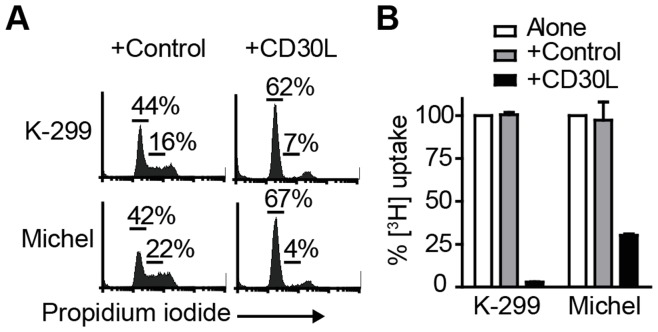
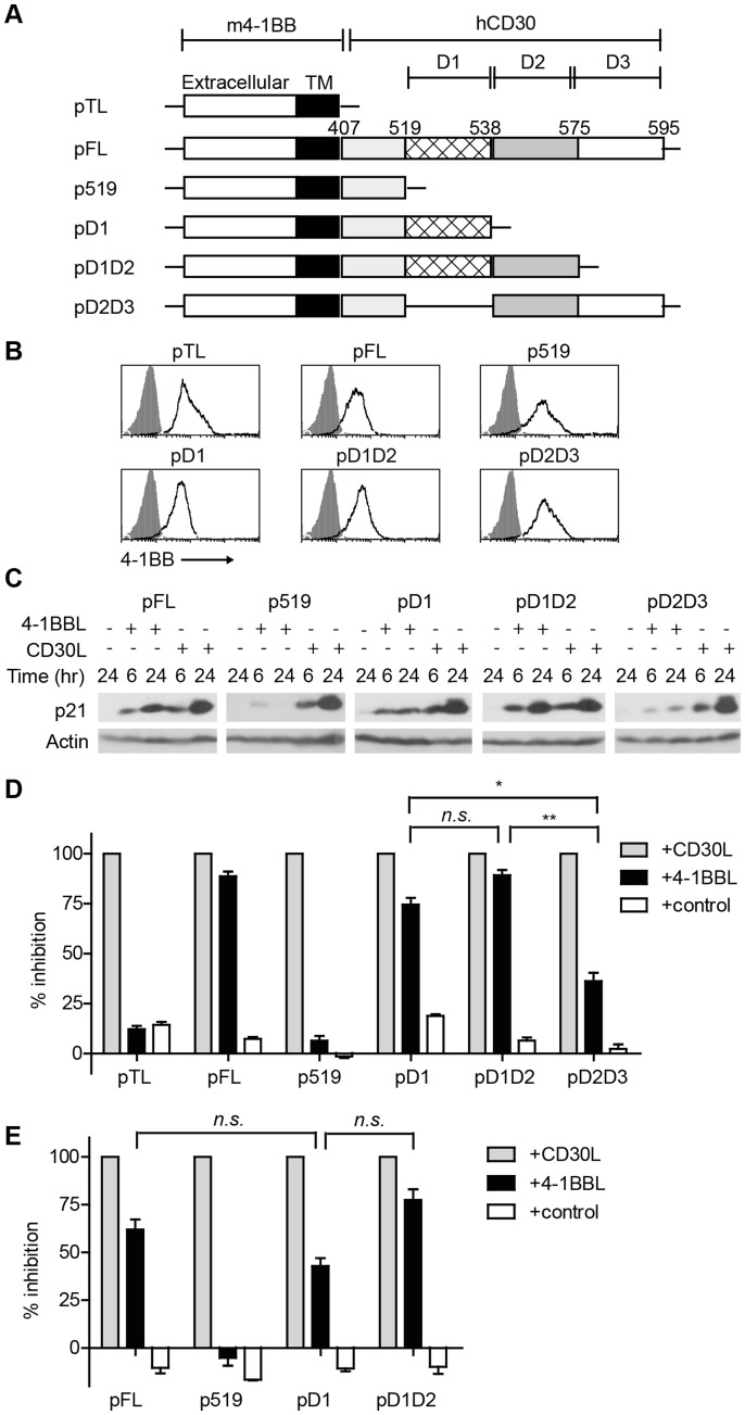
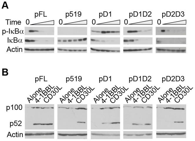
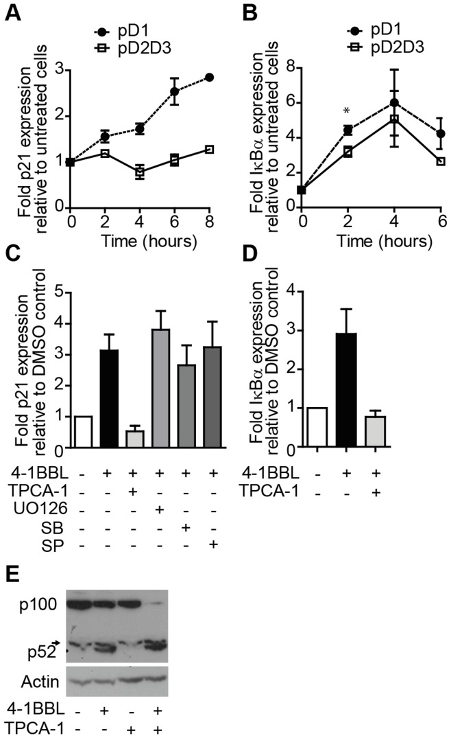
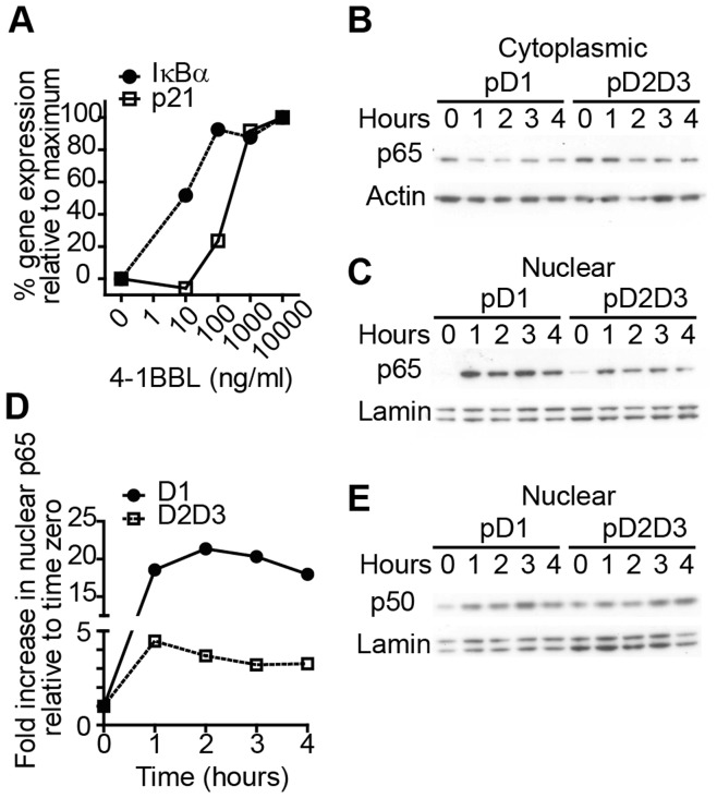
The TNF-receptor superfamily member CD30 is expressed on normal and malignant lymphocytes, including anaplastic large cell lymphoma (ALCL) cells. CD30 transmits multiple effects, including activation of NF-κB signaling, cell proliferation, growth arrest and apoptosis. How CD30 generates these pleiotropic effects is currently unknown. Herein we describe ALCL cells expressing truncated forms of the CD30 intracellular domain that allowed us to identify the key regions responsible for transmitting its biological effects in lymphocytes. The first region (CD30(519-537)) activated both the alternative and canonical NF-κB pathways as detected by p100 and IκBα degradation, IKKβ-dependent transcription of both IκBα and the cyclin-dependent kinase inhibitor p21(WAF1/CIP1) and induction of cell cycle arrest. In contrast, the second region of CD30 (CD30(538-595)) induced some aspects of canonical NF-κB activation, including transcription of IκBα, but failed to activate the alternative NF-κB pathway or drive p21(WAF1/CIP1)-mediated cell-cycle arrest. Direct comparison of canonical NF-κB activation by the two motifs revealed 4-fold greater p65 nuclear translocation following CD30(519-537) engagement. These data reveal that independent regions of the CD30 cytoplasmic tail regulate the magnitude and type of NF-κB activation and additionally identify a short motif necessary for CD30-driven growth arrest signals in ALCL cells.


Conflict of interest statement

Competing Interests: The authors have declared that no competing interests exist.

Figures

Figure 1
Figure 1. CD30L triggers growth arrest in two ALCL cell lines.
Karpas-299 (K-299) or Michel cells were incubated alone, with control (human IgG) or CD30L for (A) 24 hours and DNA content determined by propidium iodide staining; numbers indicate the percentage of cells in G0/G1 (left) or S-phase (right), or (B) 72 hours with [3H]-thymidine added for the final 16 hours. Bar graphs show the mean (+/−SEM) percentage of [3H]-thymidine uptake from triplicate wells in control or CD30L-stimulated cells; data are expressed as a percentage of [3H]-thymidine-uptake relative to untreated cells (normalised to 100%) and are representative of at least 3 independent experiments.
Figure 2
Figure 2. Signals from CD30 D1 drive increased expression of p21WAF1/CIP1 and growth arrest in ALCL cells.
(A) Schematic representation of all retroviral constructs showing the position of domains (D1, D2 and D3) of hCD30 downstream of m4-1BB extracellular and transmembrane (TM) domains. Numbers indicate CD30 residues relative to their position in full-length hCD30. (B) Karpas-299 cells were stably transduced with the constructs indicated and surface expression of m4-1BB detected by flow cytometry (open histograms); filled histograms represent staining with isotype control. (C) Karpas-299 transductants were stimulated for 6 or 24 hours with 4-1BBL or CD30L or left untreated for 24 hours and p21WAF1/CIP1 detected by immunoblot; actin is shown as a loading control. Blots are representative of two independent experiments. To determine the role of CD30 domains in triggering growth arrest, Karpas-299 (D) or Michel (E) cells transduced with the plasmids indicated, were stimulated for 72 hours and [3H]-thymidine uptake determined as a measure of cell cycling. Data are normalised to maximum growth inhibition (that induced by CD30L, set at 100%) and to minimum inhibition (medium alone; 0%) to show relative % inhibition induced by CD30L, 4-1BBL or control (CD70). Data in panels D and E are pooled from 3 of 3 (D) or 2 of 2 (E) experiments and show means and SEM. One-way ANOVA was performed on 4-1BBL-stimulated data sets across all experiments and was significant for Karpas-299 (p<0.0001) and Michel (p<0.018) cells. Statistical analyses using the Students two-tailed t-test were therefore performed comparing the means of individual groups; *p<0.05, **p<0.005, n.s. p>0.05.
Figure 3
Figure 3. CD30 D1 is necessary and sufficient for activation of the alternative NF-κB pathway.
(A) Karpas-299 transductants were incubated alone (0) or with 4-1BBL for 5, 10, 15, 20 or 30 mins and expression of phospho-IκBα (upper panels) or total IκBα (central panels) determined by immunoblot. (B) Karpas-299 transductants were incubated for 24 hours alone or in the presence of 4-1BBL or CD30L; p100 and its degradation product p52 were detected by immunoblot. Blots in (A) and (B) are representative of 2 independent experiments and actin expression (lower panels) is shown as a loading control.
Figure 4
Figure 4. Differential regulation of NF-κB-dependent genes by CD30 D1 and D2D3.
Karpas-299 cells stably transduced with pD1 (closed circles) or pD2D3 (open squares) were incubated with 4-1BBL, and p21WAF1/CIP1 (A) and IκBα (B) transcripts quantitated over time by qRT-PCR. Data show fold increase over untreated cells. (C and D) Karpas-299 pD1 transductants were incubated for 30 minutes with inhibitors of IKK-β (TPCA-1), ERK (UO126), p38 (SB203580; SB), JNK (SP600125; SP) or with an equal volume of DMSO (−), prior to addition of 4-1BBL where specified. After a further 7 hours p21WAF1/CIP1 (C) and IκBα (D) transcripts were quantitated by qRT-PCR. (E) In parallel whole cell lysates were assayed by immunoblot for p100/p52 and actin. Data are pooled from 2 (A and D) or 3 (B and C) experiments or are representative of 2 experiments (E). Graphs express the mean (+/−SEM)-fold increase in transcript expression over untreated (A and B) or DMSO-only treated (C and D) cells; *p<0.05 by two tailed Students t-test. The short arrow indicates a non-specific band above p52 (E).
Figure 5
Figure 5. CD30 D1 triggers greater nuclear p65 accumulation than D2D3.
(A) Karpas-299 cells stably transduced with pD1 were incubated with titrated doses of 4-1BBL for 5 hours and p21WAF1/CIP1 (open squares) and IκBα (closed circles) transcripts quantitated by qRT-PCR. Data represent the percentage fold-increase in expression relative to untreated (0%) and 10 µg/ml treated (100%) cells. (B, C, D and E) Karpas-299 cells stably transduced with pD1 or pD2D3 were incubated with 4-1BBL for the times indicated and cytoplasmic (B) or nuclear (C, D and E) fractions assayed by immunoblot for p65 (B, C and D), p50 (E) and actin and lamin as loading controls. D shows the fold-increase in nuclear p65 from pD1- (closed circles) and pD2D3- (open squares) expressing cells relative to time zero and normalised to lamin, calculated by densitometry and were obtained from a separate experiment to that shown in (C).

References

    1. Falini B, Pileri S, Pizzolo G, Durkop H, Flenghi L, et al. (1995) CD30 (Ki-1) molecule: a new cytokine receptor of the tumor necrosis factor receptor superfamily as a tool for diagnosis and immunotherapy. Blood 85: 1–14. - PubMed
    1. Al-Shamkhani A (2004) The role of CD30 in the pathogenesis of haematopoietic malignancies. Curr Opin Pharmacol 4: 355–359. - PubMed
    1. Lee SY, Park CG, Choi Y (1996) T cell receptor-dependent cell death of T cell hybridomas mediated by the CD30 cytoplasmic domain in association with tumor necrosis factor receptor-associated factors. J Exp Med 183: 669–674. - PMC - PubMed
    1. Telford WG, Nam SY, Podack ER, Miller RA (1997) CD30-regulated apoptosis in murine CD8 T cells after cessation of TCR signals. Cell Immunol 182: 125–136. - PubMed
    1. Dai Z, Nasr IW, Reel M, Deng S, Diggs L, et al. (2005) Impaired recall of CD8 memory T cells in immunologically privileged tissue. J Immunol 174: 1165–1170. - PubMed

Publication types

MeSH terms