. 2012;7(9):e45250.
doi: 10.1371/journal.pone.0045250. Epub 2012 Sep 18.

The neuron-astrocyte-microglia triad in normal brain ageing and in a model of neuroinflammation in the rat hippocampus

Affiliations

The neuron-astrocyte-microglia triad in normal brain ageing and in a model of neuroinflammation in the rat hippocampus

Francesca Cerbai et al. PLoS One. 2012.

Abstract

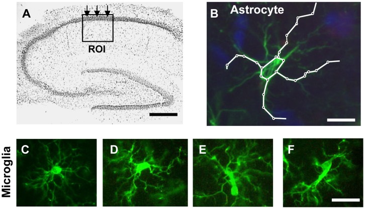
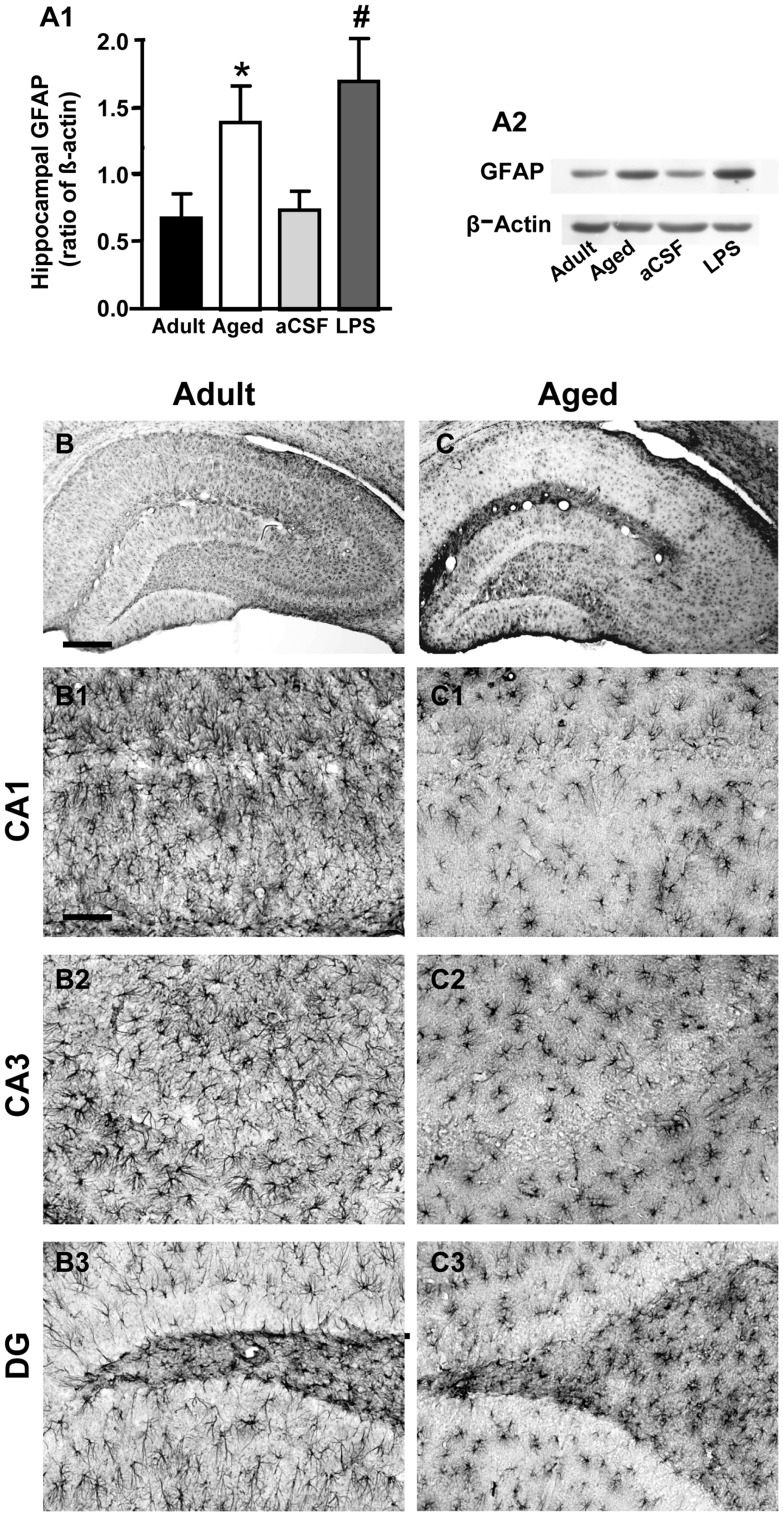
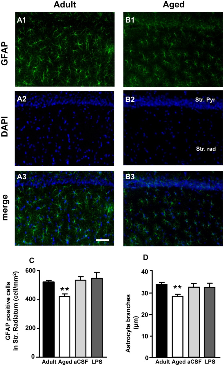
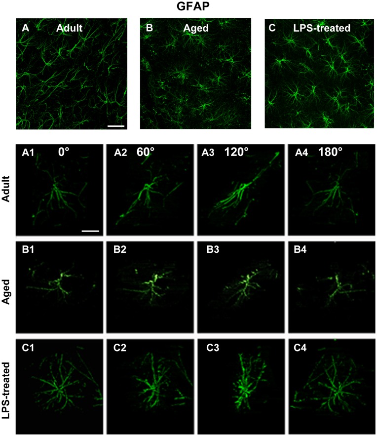
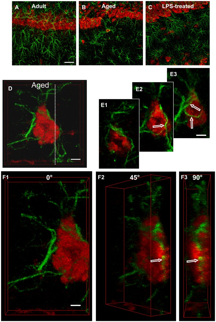
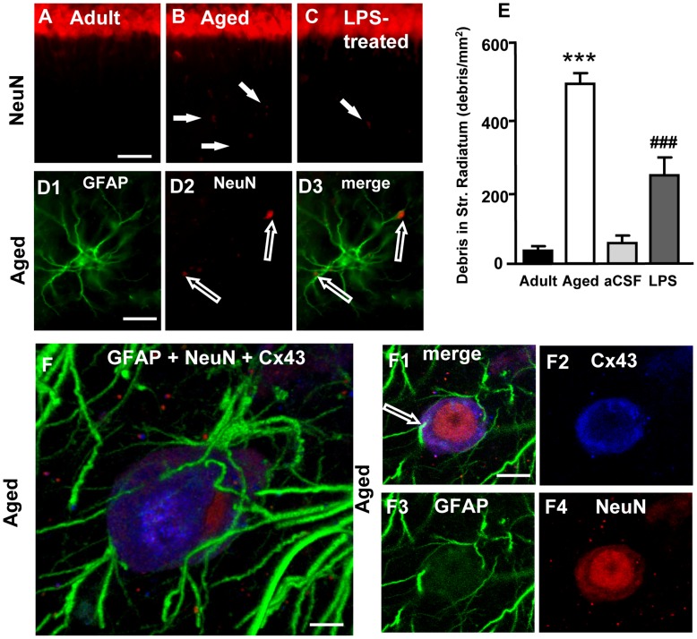
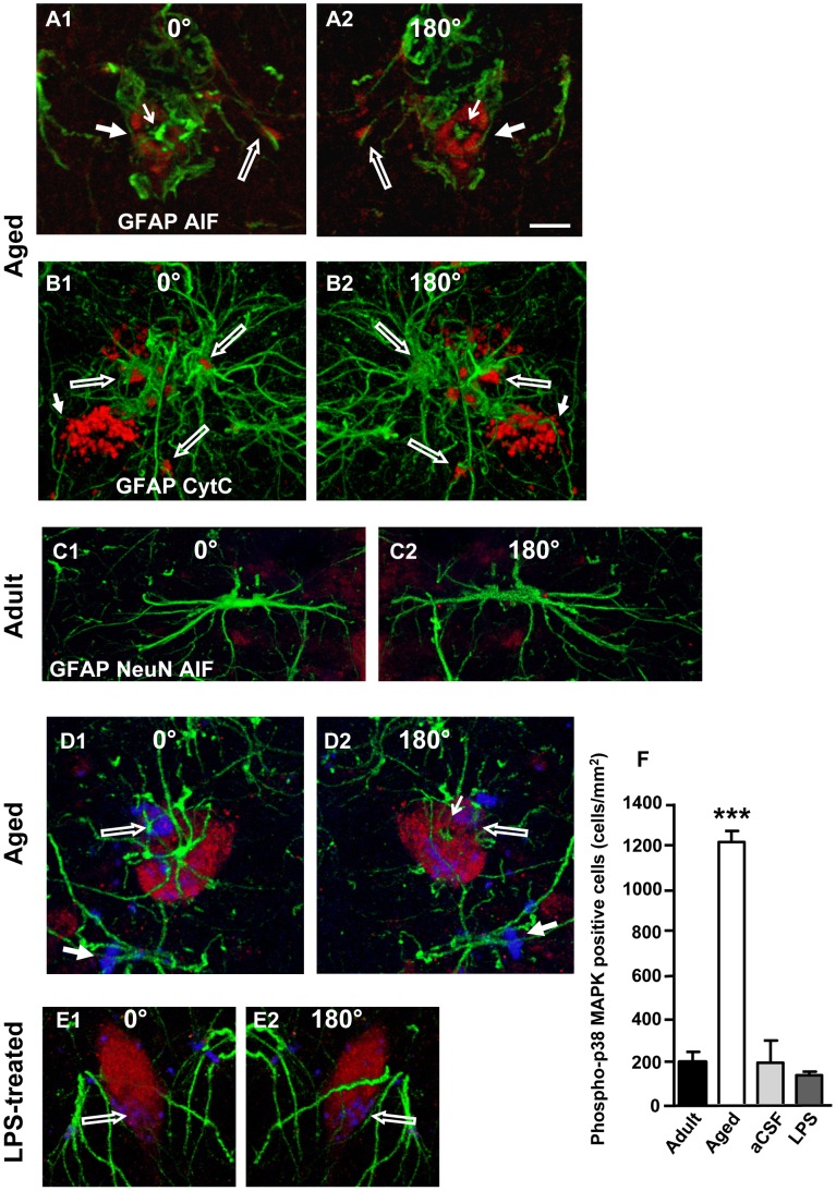
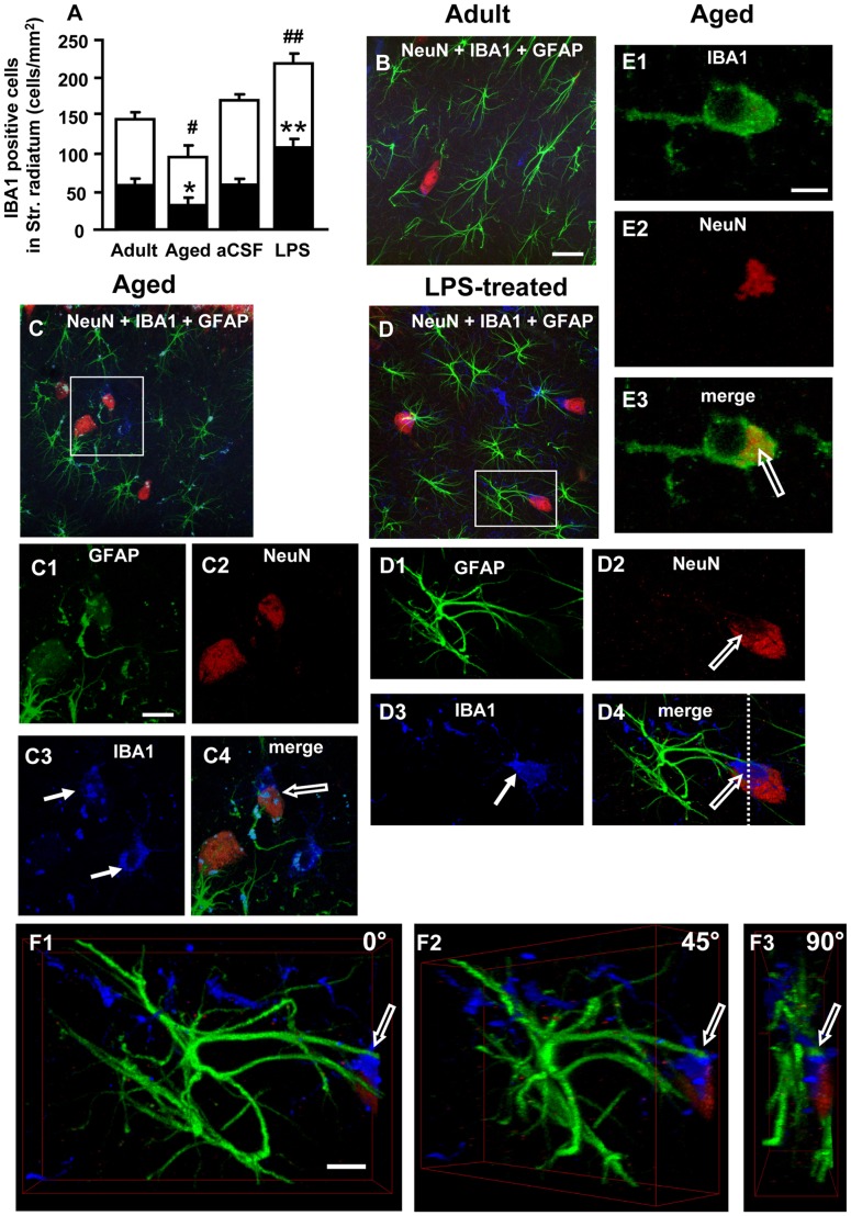
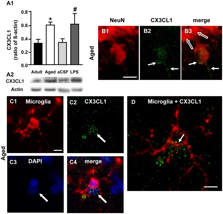
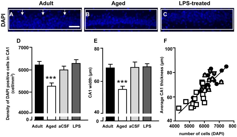
Ageing is accompanied by a decline in cognitive functions; along with a variety of neurobiological changes. The association between inflammation and ageing is based on complex molecular and cellular changes that we are only just beginning to understand. The hippocampus is one of the structures more closely related to electrophysiological, structural and morphological changes during ageing. In the present study we examined the effect of normal ageing and LPS-induced inflammation on astroglia-neuron interaction in the rat hippocampus of adult, normal aged and LPS-treated adult rats. Astrocytes were smaller, with thicker and shorter branches and less numerous in CA1 Str. radiatum of aged rats in comparison to adult and LPS-treated rats. Astrocyte branches infiltrated apoptotic neurons of aged and LPS-treated rats. Cellular debris, which were more numerous in CA1 of aged and LPS-treated rats, could be found apposed to astrocytes processes and were phagocytated by reactive microglia. Reactive microglia were present in the CA1 Str. Radiatum, often in association with apoptotic cells. Significant differences were found in the fraction of reactive microglia which was 40% of total in adult, 33% in aged and 50% in LPS-treated rats. Fractalkine (CX3CL1) increased significantly in hippocampus homogenates of aged and LPS-treated rats. The number of CA1 neurons decreased in aged rats. In the hippocampus of aged and LPS-treated rats astrocytes and microglia may help clearing apoptotic cellular debris possibly through CX3CL1 signalling. Our results indicate that astrocytes and microglia in the hippocampus of aged and LPS-infused rats possibly participate in the clearance of cellular debris associated with programmed cell death. The actions of astrocytes may represent either protective mechanisms to control inflammatory processes and the spread of further cellular damage to neighboring tissue, or they may contribute to neuronal damage in pathological conditions.


Conflict of interest statement

Competing Interests: The authors have declared that no competing interests exist.

Figures

Figure 1
Figure 1. Methods: ROI, scheme of measure of astrocytes branches lenght, examples of resting-reactive microglia.
A: localization and dimensions of the Region Of Interest (ROI) utilized to perform the quantitative analysis; scale bar: 500 µm. B: schematic diagram showing the method used to quantify principal astrocytes branches length; scale bar: 15 µm. C–F: different stages of microglia activation. C: a typical example of a resting microglia cell; D–F: typical examples of microglia in reactive states; scale bar: 20 µm.
Figure 2
Figure 2. Western Blot analysis of GFAP levels in hippocampus and immunohistochemistry of GFAP positive cells. A1:
quantification of GFAP by Western Blot from homogenates of whole hippocampus. Each column represents the levels of GFAP expressed as a ratio of β-actin expression run in the same gel (mean ± SEM; Adult, n = 8; aged, n = 5; aCSF, n = 6; LPS-treated, n = 6) A2: representative Western Blot runs of GFAP and β-actin. B,C: immunolabelling of astrocytes using anti GFAP antibody and DAB staining in whole hippocampal slices. B1–B3: higher magnification images of CA1 (B1), CA3 (B2) and DG (B3); C1–C3: higher magnification images of CA1 (C1), CA3 (C2) and DG (C3); B-B3: adult rat; C–C3: aged rat. Scale bar: B–C: 400 µm; B1–C3∶70 µm.
Figure 3
Figure 3. Quantitative analysis of astrocytes in CA1 Str. Radiatum.
Characterization of astrocytes in CA1 Str. Radiatum of adult and aged rats. Representative epifluorescent photomicrographs showing immunoreactivity of GFAP (green) and DAPI staining (blue) in CA1 Pyramidal cell layer and Str. Radiatum of adult (A1,A2,A3) and aged (B1,B2,B3) rats. A3 and B3 show the merged images. Scale bar: 50 µm. C: quantitative analysis of GFAP positive cells counted in CA1 Str. Radiatum of adult (n = 12), aged (n = 15), aCSF- (n = 5) and LPS-treated (n = 6) rats, expressed as GFAP positive cells/mm2 (mean±SEM); **P<0.01 vs all other groups. D: length of principal astrocyte branches in CA1 Str. Radiatum of adult (n = 12), aged (n = 15), aCSF- (n = 5) and LPS-treated (n = 6) rats; (mean±SEM), **P<0.01 vs all other groups.
Figure 4
Figure 4. Confocal microscopy 3D-analysis of astrocytes morphology.
Immunoreactivity of GFAP in the CA1 Str. Radiatum of adult (A, A1–A4), aged (B, B1–B4) and LPS-treated rats (C, C1–C4). Panels from A1 to C4 are obtained from the 3D stacks observed from different angles (0, 60, 120, 180 degrees) around the vertical axis. Scale bar: 40 µm (A,B,C) and 15 µm (A1–C4).
Figure 5
Figure 5. Characterization of astrocytes-neurons interplay.
Photos show confocal images of immunoreactivity of GFAP (green) and NeuN (red) in CA1 Pyramidal cell layer and CA1 Str. Radiatum of adult (A), aged (B and D, E1–F3) and LPS-treated rats (C). Scale bar: 60 µm (A,B,C). D: 3D stack of confocal scans of GFAP (green), and NeuN (red). Scale bar: 5 µm. E1–E3: each panel is obtained merging 2 consecutive confocal scans (total 0.738 µm). Scale bar: 5 µm. F1–F3∶3D stacks of the neuron shown in D, digitally cut along the white dotted line and rotated by 0, 45 and 90 degrees along the vertical axis. Scale bar: 3 µm.
Figure 6
Figure 6. Quantitative analysis of neuronal debris in Str. Radiatum, involvement of Cx43 in astrocytes-neuron interplay.
Images from CA1 Str. Pyramidalis and CA1 Str. Radiatum of an adult (A), aged (B) and LPS-treated rat (C) showing the presence of neuronal debris (arrows, B and C). Scale bar: 70 µm. D1–D3: higher magnification images of GFAP (green, D1) and NeuN (red, D2) staining and the merge of the two previous images (D3). Empty arrows show neuronal debris closely apposed to astrocyte branches. Scale bar: 15 µm. E: quantitative analysis of neuronal debris in CA1 Str. Radiatum of adult (n = 12), aged (n = 10), aCSF- (n = 5) and LPS-treated (n = 6) rats (mean±SEM; *** and ###P<0.001 vs all other groups). F–F4: Representative images of triple immunostaining of GFAP (green), NeuN (red) and Cx43 (blue) in the Str. Radiatum of an aged rat. F: 3D stack of 39 confocal scans (total 14.39 µm); F1: a “sub-slice” of the previous neuron (obtained stacking 6 consecutive scans, total 1.843 µm, starting at a depth of 5.899 µm into the cell) and separate staining of Cx43 (F2), GFAP (F3) and NeuN (F4). Scale bar: 5 µm (F); 10 µm (F1–F4).
Figure 7
Figure 7. Immunostaining of markers of apoptosis in cells surrounded by astrocyte branches in CA1 Str. Radiatum.
A1,A2: immunostaining for CytC (red) and GFAP (green). B1,B2: immunostaining for AIF (red) and GFAP (green). µm. Representative images of immunostaining for NeuN (red), AIF (blue) and GFAP (green) taken from the CA1 region of an adult (C1–C2), an aged (D1–D2) and an LPS-treated (E1–E2) rat. Note the presence of AIF staining within neurons of aged and LPS treated rats only (open arrows in D1,D2 and E1,E2). This effect was observed in all slices from aged and LPS-treated rats. Scale bar: 10. F: Quantification of phospho-p38MAPK positive cells in CA1 Str. Pyramidalis of adult (n = 11), aged (n = 16), aCSF- (n = 5) and LPS-treated (n = 5) rats (mean±SEM; ***P<0.001, vs all other groups).
Figure 8
Figure 8. Quantitative analysis of resting-reactive microglia cells, 3D-analysis of neuron-astrocyte-microglia interplay. A:
quantitative analysis of microglia cells (mean±SEM) in CA1 Str. Radiatum of adult (n = 12), aged (n = 12), aCSF- (n = 5) and LPS-treated (n = 6) rats. Black portion of columns represent reactive microglia cells, white portion of columns represent resting microglia cells, the entire columns represent total microglia cells; *at least P<0.05 vs all other groups; **P<0.01 vs all other groups; #at least P<0.05 vs all other groups; ##P<0.01 vs all other groups. B,C,D: Merged confocal images of triple immunostaining of neurons (NeuN, red), astrocytes (GFAP, green) and microglia (IBA1, blue) in the CA1 Str. Radiatum of adult (B), aged (C), and LPS-treated rats (D). Scale bar: 20 µm. C1–C4 and D1–D4: higher magnification confocal images of framed areas in C and D, respectively. Arrows in C3 and D3 indicate IBA1-positive reactive microglia cells. Open arrows in C4 and D4 show microglia cells phagocytising neurons. Scale bar: 7 µm. E1–E3: a confocal “sub-slice” (thickness 0.7 µm) acquired at depth 2.8 µm into a microglia cell (IBA1-positive, green) from the Str. Radiatum of an aged rat showing complete co-localization of a neuronal debris (E2, red, immunostained for NeuN) in the microglia cytoplasm (E3, yellow-orange, open arrow). Scale bar: 7 µm. F1–F3∶3D stacks of the neuron shown in D4, digitally cut along the white dotted line and rotated by 0, 45 and 90 degrees along the vertical axis. Scale bar: 10 µm.
Figure 9
Figure 9. Analysis of CX3CL1 expression in the hippocampus of adult, aged, aCSF- and LPS-treated rats.
A1: Western Blot analysis of CX3CL1 in whole hippocampus homogenates of adult (n = 7), aged (n = 4), aCSF- (n = 4) and LPS-treated (n = 4) rats. Each column in the graph represents the level of CX3CL1 expressed as mean±SEM, normalized to β-actin run in the same gel. *P<0.05 vs adult; #P<0.05 vs aCSF-tretated. A2: representative Western Blot runs of CX3CL1 and of β-actin. B1–B3: laser confocal microscopy immunohistochemistry of neurons (B1, NeuN, red), CX3CL1 (B2, green) and the merge of the two previous images (B3) from the CA1 region of an aged rat. Scale bar: 14 µm. C1–C4: epifluorescent microscopy images of a microglia cell (C1, red), CX3CL1 immunostaining (C2, green), DAPI staining of nuclei (C3, blue) and the merge of the three previous images (C4), indicating that CX3CL1 staining is localized on the surface of a cell, possibly a neuron (arrows). D: the image represents a confocal “sub-slice” (total thickness 2.233 µm) of the same microglia cell shown in C1–C4, acquired starting at a depth of 8.932 µm into the cell. The CX3CL1 positive cell (green) is partially colocalized with the microglia cell. Scale bar: 14 µm.
Figure 10
Figure 10. Quantitative analysis of CA1 pyramidal nuclei labelled with DAPI.
A–C: representative photomicrographs of DAPI staining in CA1 Str. Pyramidalis of adult (A), aged (B) and LPS-treated (C) rats. DAPI positive nuclei were counted within the framed areas, and thickness of CA1 Str. Pyramidalis was measured in correspondence to the vertical arrows. Scale bar: 60 µm. D: quantitative analysis of DAPI positive nuclei in CA1 Str. Pyramidalis of adult (n = 14), aged (n = 14), aCSF- (n = 5) and LPS-treated (n = 6) rats (cells/mm2, mean±SEM; ***P<0.01 vs all other groups). E: quantitative analysis of CA1 Str. Pyramidalis thickness of adult (n = 14), aged (n = 14), aCSF- (n = 5) and LPS-treated (n = 6) rats (mean±SEM). ***P<0.001 vs all other groups. F: linear regression analysis of CA1 pyramidal cells vs CA1 thickness; black circles: adult rats; light grey triangles: aCSF-treated; dark grey triangles: LPS-treated rats; white squares: aged rats.

References

    1. Barres BA (2008) The mystery and magic of glia: a perspective on their roles in health and disease. Neuron 60: 430–440. - PubMed
    1. Allen NJ, Barres BA (2009) Neuroscience: Glia - more than just brain glue. Nature 457: 675–677. - PubMed
    1. De Keyser J, Mostert JP, Koch MW (2008) Dysfunctional astrocytes as key players in the pathogenesis of central nervous system disorders. J Neurol Sci 267: 3–16. - PubMed
    1. Sofroniew MV (2009) Molecular dissection of reactive astrogliosis and glial scar formation. Trends Neurosci 32: 638–647. - PMC - PubMed
    1. Wenk GL, McGann K, Mencarelli A, Hauss-Wegrzyniak B, Del Soldato P, et al. (2000) Mechanisms to prevent the toxicity of chronic neuroinflammation on forebrain cholinergic neurons. Eur J Pharmacol 402: 77–85. - PubMed

Publication types

MeSH terms