. 2012;7(9):e45374.
doi: 10.1371/journal.pone.0045374. Epub 2012 Sep 18.

Peptide nanovesicles formed by the self-assembly of branched amphiphilic peptides

Affiliations

Peptide nanovesicles formed by the self-assembly of branched amphiphilic peptides

Sushanth Gudlur et al. PLoS One. 2012.

Abstract

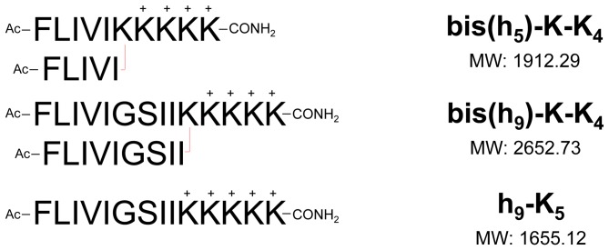
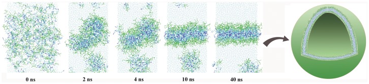
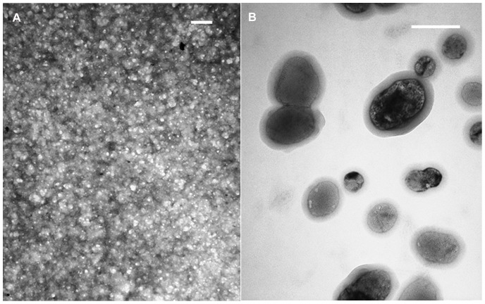
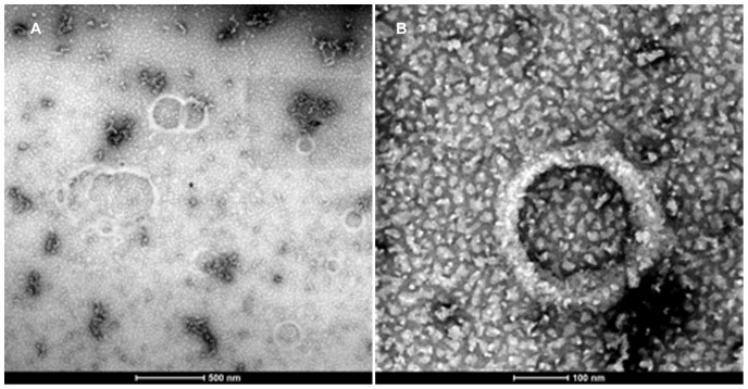
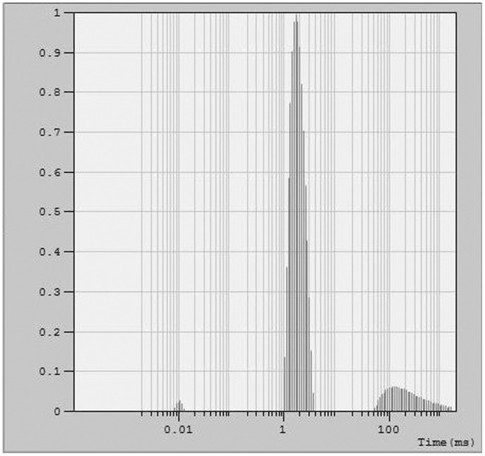
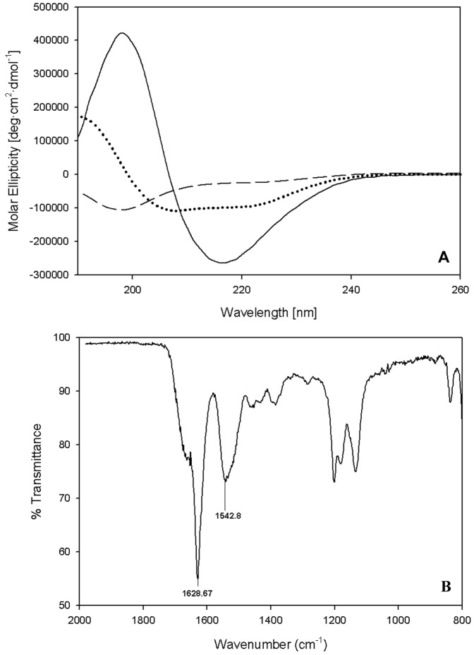
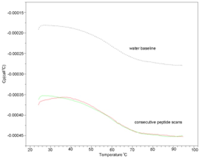
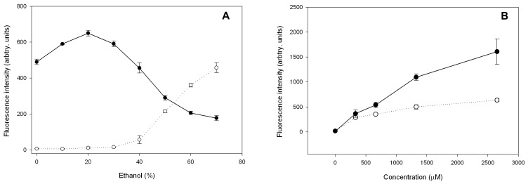
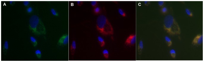
Peptide-based packaging systems show great potential as safer drug delivery systems. They overcome problems associated with lipid-based or viral delivery systems, vis-a-vis stability, specificity, inflammation, antigenicity, and tune-ability. Here, we describe a set of 15 & 23-residue branched, amphiphilic peptides that mimic phosphoglycerides in molecular architecture. These peptides undergo supramolecular self-assembly and form solvent-filled, bilayer delimited spheres with 50-200 nm diameters as confirmed by TEM, STEM and DLS. Whereas weak hydrophobic forces drive and sustain lipid bilayer assemblies, these all-peptide structures are stabilized potentially by both hydrophobic interactions and hydrogen bonds and remain intact at low micromolar concentrations and higher temperatures. A linear peptide lacking the branch point showed no self-assembly properties. We have observed that these peptide vesicles can trap fluorescent dye molecules within their interior and are taken up by N/N 1003A rabbit lens epithelial cells grown in culture. These assemblies are thus potential drug delivery systems that can overcome some of the key limitations of the current packaging systems.


Conflict of interest statement

Competing Interests: The authors have declared that no competing interests exist.

Figures

Figure 1
Figure 1. Assembling and control sequences.
The peptide assemblies are an equimolar mixture of the acetylated bis(h9)-K-K4 and bis(h5)-K-K4 sequences unless otherwise stated. The linear h9-K5 control sequence does not associate to give any type of structure.
Figure 2
Figure 2. A coarse-grained simulation of bis(h5)-K-K4 bilayer self-assembly.
All lysine residues are plotted in green ball-and-stick representations, the aromatic sidechains of phenylalanine residues in blue sticks, and all other residues in cyan. The snapshots were taken at 0 ns, 2 ns, 4 ns, 10 ns and 40 ns, respectively. A hypothetical peptide vesicle model is shown to the right.
Figure 3
Figure 3. Transmission Electron Micrograph (TEM) of h9h5-vesicles.
These peptides self-assemble into nano-sized vesicles. TEM images of (A) a concentrated and (B) a diluted sample of the peptide mixture, stained with 5% phosphotungstic acid and Osmium tetroxide (OsO4) vapors, respectively (200 nm scale bar).
Figure 4
Figure 4. Scanning Transmission Electron Micrograph (STEM).
Vesicles were prepared with 30% CH3-Hg label in both the h5 and h9 peptides at 0.1 mM concentration were negatively stained using a multi isotope 2% Uranyl acetate (Uranium bis(acetato)-O)dioxo-dihydrate) aqueous solution. The images were captured using annular dark field mode was then inverted to produce the final image.
Figure 5
Figure 5. Dynamic Light Scattering of h9h5-vesicles.
Distribution analysis of the data showing two major distributions centered around 1.68 ms and 119.4 ms and a mean correlation time of 1.85 ms and 275.7 ms respectively. These correlation times correspond to a hydrodynamic radius of 80 nm and 10 µm respectively.
Figure 6
Figure 6. Secondary Structural Characterizations.
(A) Circular dichroism (CD) spectroscopy shows that the h9h5-vesicles adopt a β-structure (solid line) at pH 7.0 in 5 mM NaHCO3, a helical structure (dotted line) in 40% TFE and remains unstructured (dashed line) when lacking the lysine branch point (h9-K5). (B) FTIR spectroscopy of dried material shows the characteristic 1629 cm−1 maximum corresponding to β- secondary structure.
Figure 7
Figure 7. Differential scanning calorimetry of 1.0 mg/ml peptide in water.
Heat capacity of solution is shown as a function of temperature. The instrument water baseline is shown together with the thermograms obtained during two consecutive upward temperature scans.
Figure 8
Figure 8. Encapsulation studies with 5(6)-Carboxyfluorescein.
(A) 5(6)-Carboxyfluorescein loaded peptide vesicles were prepared in different ethanol concentrations and separated over 30 kDa MWCO filters. Fluorescence intensities of the filtrate (open circles) and retentate (solid circles) were collected after separation and show that encapsulation efficiency decreases with increasing ethanol concentration. (B) h9h5-vesicles prepared with the dye present during vesicle formation (solid circles) or added after the vesicles are formed (open circles). Each data point represents an average of 3 separate experiments performed on different days. Data points are connected with straight lines through their midpoints.
Figure 9
Figure 9. Cellular uptake of preloaded h9h5-vesicles.
(A–C) h9h5-vesicles with one of the peptides labeled (bis(h5)-K-K4 labeled with Carboxytetramethylrhodamine) and loaded with 5(6)-Carboxyfluorescein were delivered to N/N 1003A rabbit lens epithelial cells. Green and red filter settings were used for the detection of 5(6)-Carboxyfluorescein (A) and Rhodamine labeled peptide (B) respectively. (C) is a merge of a & b showing co-localization of dye and h9h5-vesicles within the cell.

References

    1. Allen TM, Cullis PR (2004) Drug delivery systems: Entering the mainstream. Science 303(5665): 1818–1822. - PubMed
    1. Torchilin VP, Levchenko TS, Rammohan R, Volodina N, Papahadjopoulos-Sternberg B, et al. (2003) Cell transfection in vitro and in vivo with nontoxic TAT peptide-liposome-DNA complexes. Proc Natl Acad Sci U S A 100(4): 1972–1977. - PMC - PubMed
    1. Tanaka T, Legat A, Adam E, Steuve J, Gatot JS, et al. (2008) DiC14-amidine cationic liposomes stimulate myeloid dendritic cells through toll-like receptor 4. Eur J Immunol 38(5): 1351–1357. - PubMed
    1. Ouali M, Ruysschaert JM, Lonez C, Vandenbranden M (2007) Cationic lipids involved in gene transfer mobilize intracellular calcium. Mol Membr Biol 24(3): 225–232. - PubMed
    1. Koynova R, Tarahovsky YS, Wang L, MacDonald RC (2007) Lipoplex formulation of superior efficacy exhibits high surface activity and fusogenicity, and readily releases DNA. Biochim Biophys Acta 1768(2): 375–386. - PMC - PubMed

Publication types