

Globus Pallidus external segment neuron classification in freely moving rats: a comparison to primates
- PMID: 23028997
- PMCID: PMC3448641
- DOI: 10.1371/journal.pone.0045421
Globus Pallidus external segment neuron classification in freely moving rats: a comparison to primates
Abstract
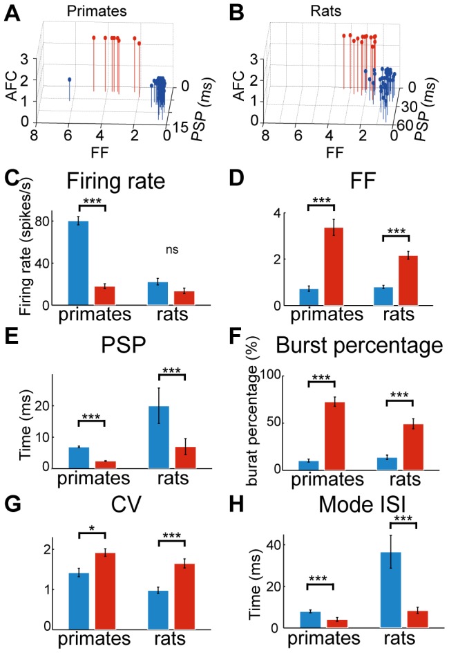
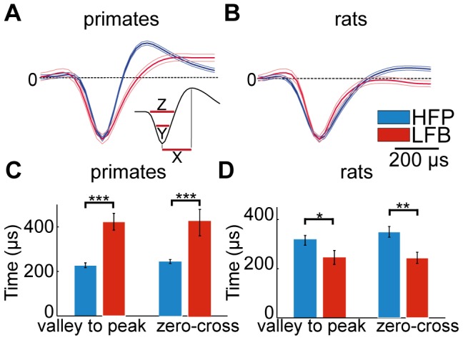
Globus Pallidus external segment (GPe) neurons are well-characterized in behaving primates. Based on their firing properties, these neurons are commonly divided into two distinct groups: high frequency pausers (HFP) and low frequency bursters (LFB). However, no such characterization has been made for behaving rats. The current study characterizes and categorizes extracellularly recorded GPe neurons in freely moving rats, and compares these results to those obtained by extracellular recordings in behaving primates using the same analysis methods. Analysis of our data recorded in rats revealed two distinct neuronal populations exhibiting firing-pattern characteristics that are similar to those obtained in primates. These characteristic firing patterns are conserved between species although the firing rate is significantly lower in rats than in primates. Significant differences in waveform duration and shape were insufficient to create a reliable waveform-based classification in either species. The firing pattern analogy may emphasize conserved processing properties over firing rate per-se. Given the similarity in GPe neuronal activity between human and non-human primates in different pathologies, our results encourage information transfer using complementary studies across species in the GPe to acquire a better understanding of the function of this nucleus in health and disease.
Conflict of interest statement
Figures

References
-
- Haber SN (2003) The primate basal ganglia: parallel and integrative networks. J Chem Neuroanat 26: 317–330. - PubMed
-
- Alexander GE, DeLong MR, Strick PL (1986) Parallel organization of functionally segregated circuits linking basal ganglia and cortex. Annu Rev Neurosci 9: 357–381. - PubMed
-
- Alexander GE, Crutcher MD (1990) Functional architecture of basal ganglia circuits: neural substrates of parallel processing. Trends Neurosci 13: 266–271. - PubMed
-
- Albin RL, Young AB, Penney JB (1989) The functional anatomy of basal ganglia disorders. Trends Neurosci 12: 366–375. - PubMed
-
- Kita H, Kitai ST (1994) The morphology of globus pallidus projection neurons in the rat: an intracellular staining study. Brain Res 636: 308–319. - PubMed
Publication types
MeSH terms
LinkOut - more resources
Full Text Sources
