. 2012;7(9):e45510.
doi: 10.1371/journal.pone.0045510. Epub 2012 Sep 17.

Annexin A10 in human oral cancer: biomarker for tumoral growth via G1/S transition by targeting MAPK signaling pathways

Affiliations

Annexin A10 in human oral cancer: biomarker for tumoral growth via G1/S transition by targeting MAPK signaling pathways

Toshihiro Shimizu et al. PLoS One. 2012.

Abstract

Background: Annexins are calcium and phospholipid binding proteins that form an evolutionary conserved multigene family. Considerable evidence indicates that annexin A10 (ANXA10) is involved in tumoral progression, although little is known about its role in human oral carcinogenesis. In this study, we investigated the involvement of ANXA10 in oral squamous cell carcinoma (OSCC).

Methodology/principal findings: ANXA10 mRNA and protein expressions were assessed by quantitative reverse transcriptase polymerase chain reaction and immunoblotting, and we conducted a proliferation assay and cell-cycle analysis in ANXA10 knockdown cells in vitro. We evaluated the correlation between the ANXA10 expression status in 100 primary OSCCs and the clinicopathological features by immunohistochemistry. ANXA10 mRNA and protein expression levels were up-regulated in all cellular lines examined (n = 7, p<0.05). ANXA10 knockdown cells showed that cellular proliferation decreased by inactivation of extracellular regulated kinase (ERK) (p<0.05), and cell-cycle arrest at the G1 phase resulted from up-regulation of cyclin-dependent kinase inhibitors. ANXA10 protein expression in primary OSCCs was also significantly greater than in normal counterparts (p<0.05), and higher expression was correlated with tumoral size (p = 0.027).

Conclusions/significance: Our results proposed for the first time that ANXA10 is an indicator of cellular proliferation in OSCCs. Our results suggested that ANXA10 expression might indicate cellular proliferation and ANXA10 might be a potential therapeutic target for the development of new treatments for OSCCs.


Conflict of interest statement

Competing Interests: The authors have declared that no competing interests exist.

Figures

Figure 1
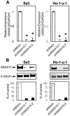
Figure 1. Evaluation of ANXA10 expression in OSCC-derived cellular lines.
(A) Quantification of ANXA10 mRNA levels in OSCC-derived cellular lines by qRT-PCR analysis. Significant up-regulation of ANXA10 mRNA is seen in the seven OSCC-derived cellular lines compared with that in the HNOKs. Data are expressed as the means ± SEM of values from three assays (*p<0.05; Mann-Whitney U test). (B) Immunoblotting analysis of ANXA10 protein in the OSCC-derived cellular lines and HNOKs. ANXA10 protein expression (molecular weight, 37 kDa) is up-regulated in the OSCC-derived cellular lines compared with that in the HNOKs. Densitometric ANXA10 protein data are normalized to α-tubulin protein levels. The values are expressed as a percentage of the HNOKs.
Figure 2
Figure 2. Expression of ANXA10 in shANXA10-transfected cells.
(A) qRT-PCR shows that ANXA10 mRNA expression in the shANXA10-transfected cells (Sa3- and Ho-1-u-1-derived transfectants; 2 clones each) are significantly lower than in the shMock-transfected cells (*p<0.05; Mann-Whitney U test). (B) Immunoblotting analysis shows that the ANXA10 protein levels in shANXA10-transfected cells (Sa3- and Ho-1-u-1-derived transfectants; 2 clones each) also have decreased markedly compared with that in the shMock-transfected cells.
Figure 3
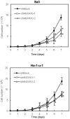
Figure 3. Effect of ANXA10 knockdown in shANXA10-transfected cells.
To determine the effect of shANXA10 on cellular proliferation, shANXA10- and shMock-transfected cells are seeded in 6-well plates at a density of 1×10⁴ viable cells/well. Both transfectants were counted on 7 consecutive days. The cellular growth of shANXA10-transfected cells (Sa3- and Ho-1-u-1-derived transfectants; 2 clones each) is significantly inhibited compared with shMock-transfected cells after 7 days (168 hours). The results are expressed as the means ± SEM of values from three assays. The asterisks indicate significant differences between the shANXA10- and shMock-transfected cells (*p<0.05; Mann-Whitney U test).
Figure 4
Figure 4. ANXA10 knockdown inhibits ERK activation.
ANXA10 knockdown causes decreased levels of phosphorylated ERK (pERK) compared with the shMock-transfected cells (Sa3- and Ho-1-u-1-derived transfectants; 2 clones each); the ERK level is unchanged. Densitometric pERK/ERK protein data are normalized to α-tubulin protein levels.
Figure 5
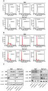
Figure 5. shANXA10 promotes G1 arrest.
To investigate cell-cycle progression, we analyzed flow cytometric determination of DNA content by a FACScalibur in the G0–G1, S, and, G2–M phases. We determined the expression level of CDKIs (p21Cip1and p27Kip1), cyclin D1, cyclin E, CDK2, CDK4, and CDK6 to identify the mechanism by which ANXA10 inhibits cell-cycle progression in the G1 phase. (A) After synchronizing at G0/G1 phase with deprived of serum, flow cytometric analysis was performed to investigate the cell cycle in the shANXA10- and shMock-transfected cells. The percentage of the G1 phase in the shANXA10-transfected cells (Sa3- and Ho-1-u-1-derived transfectants; 2 clones each) has increased markedly compared with the mock-transfected cells (p<0.05, Mann-Whitney's U test). (B) After synchronizing at G2/M phase to treated with nocodazol, flow cytometric analysis was performed to investigate the cell cycle in the shANXA10- and shMock-transfected cells. The percentage of the G1 phase in the shANXA10-transfected cells (Sa3- and Ho-1-u-1-derived transfectants; 2 clones each) has also increased markedly compared with the mock-transfected cells (p<0.05, Mann-Whitney's U test). (C) Immunoblotting analysis shows up-regulation of p21Cip1and p27Kip1 and down-regulation of cyclin D1, cyclin E, CDK2, CDK4, and CDK6 in the shANXA10-transfected cells (Sa3- and Ho-1-u-1-derived transfectants; 2 clones each) compared with the shMock-transfected cells.
Figure 6
Figure 6. Evaluation of ANXA10 protein expression in primary OSCCs.
Representative IHC results for ANXA10 protein in normal oral tissue (A, B) and primary OSCC (C, D) (A, C) Original magnification, ×100. Scale bars, 50 μm. (B, D) Original magnification, ×400. Scale bars, 10 μm). Strong ANXA10 immunoreactivity is detected in primary OSCCs; normal oral tissues show almost negative immunostaining. (E) The state of ANXA10 protein expression in primary OSCCs (n = 100) and the normal counterparts. The ANXA10 IHC scores are calculated as follows: IHC score  = 1× (number of weakly stained cells in the field) +2× (number of moderately stained cells in the field) +3× (number of intensely stained cells in the field). The ANXA10 IHC scores for normal oral tissues and OSCCs ranged from 27.5 to 132.4 (median, 93.5) and 60.5 to 230.4 (median, 150.6), respectively. ANXA10 protein expression levels in OSCCs are significantly (*p<0.001, Mann-Whitney's U test) higher than in normal oral tissues.

References

    1. Chang KS, Wang G, Freireich EJ, Daly M, Naylor SL, et al. (1992) Specific expression of the annexin VIII gene in acute promyelocytic leukemia. Blood 79: 1802–10. - PubMed
    1. Pencil SD, Toth M (1998) Elevated levels of annexin I protein in vitro and in vivo in rat and human mammary adenocarcinoma. Clin Exp Metastasis 16: 113–21. - PubMed
    1. Sarkar A, Yang P, Fan YH, Mu ZM, Hauptmann R, et al. (1994) Regulation of the expression of annexin VIII in acute promyelocytic leukemia. Blood 84: 279–86. - PubMed
    1. Theobald J, Hanby A, Patel K, Moss SE (1995) Annexin VI has tumour-suppressor activity in human A431 squamous epithelial carcinoma cells. Br J Cancer 71: 786–8. - PMC - PubMed
    1. Masaki T, Tokuda M, Ohnishi M, Watanabe S, Fujimura T, et al. (1996) Enhanced expression of the protein kinase substrate annexin in human hepatocellular carcinoma. Hepatology 24: 72–81. - PubMed

MeSH terms