. 2012;7(9):e45562.
doi: 10.1371/journal.pone.0045562. Epub 2012 Sep 20.

Glioblastoma cell-secreted interleukin-8 induces brain endothelial cell permeability via CXCR2

Affiliations

Glioblastoma cell-secreted interleukin-8 induces brain endothelial cell permeability via CXCR2

Julie Dwyer et al. PLoS One. 2012.

Abstract

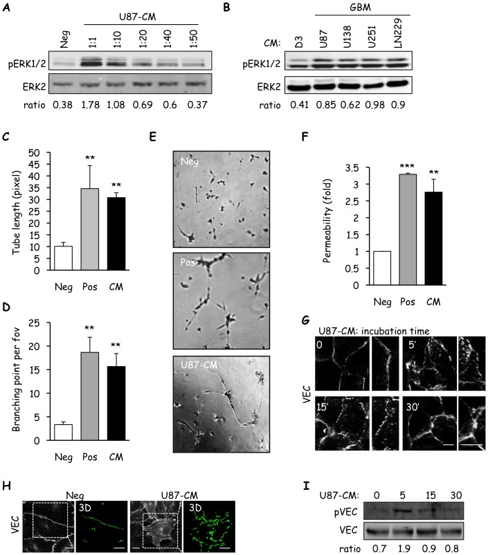
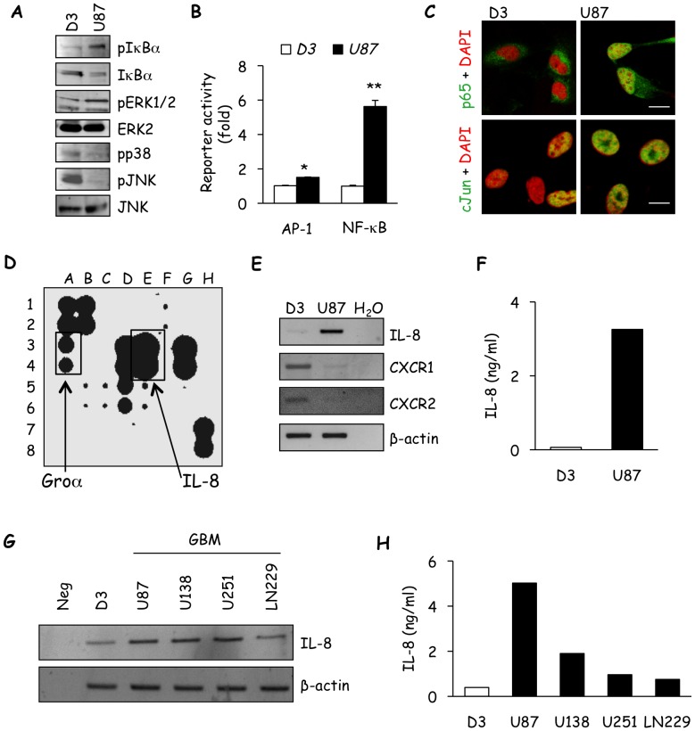
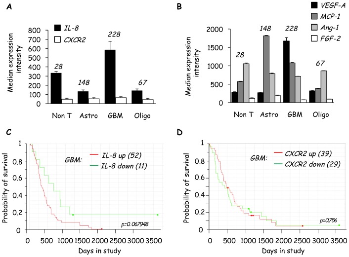
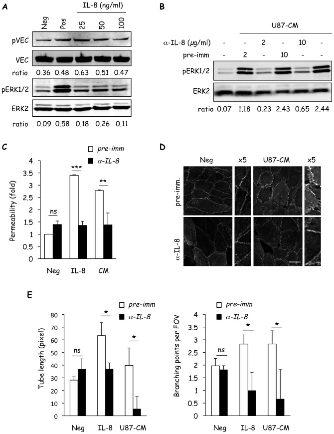
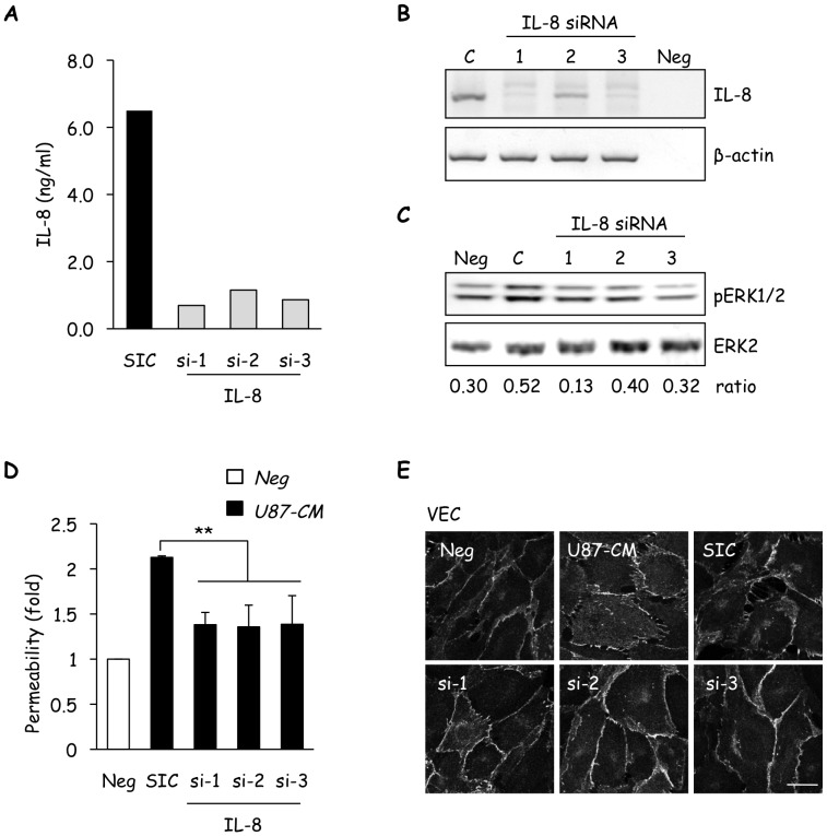
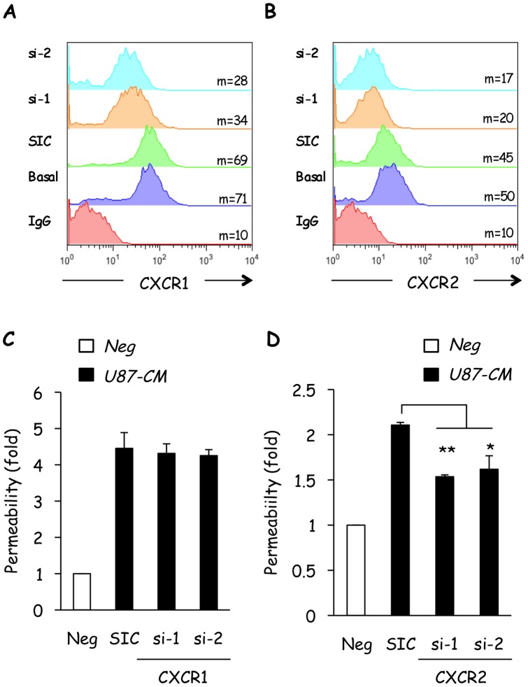
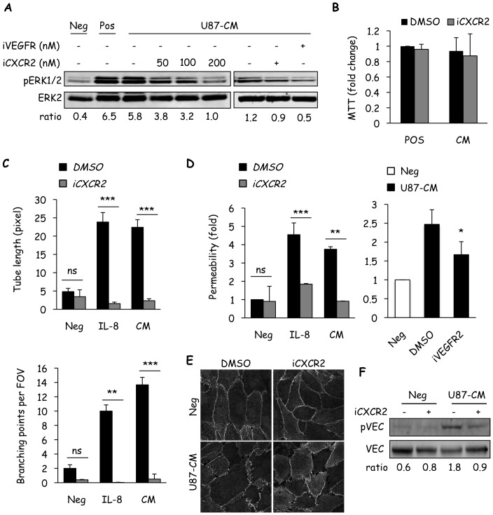
Glioblastoma constitutes the most aggressive and deadly of brain tumors. As yet, both conventional and molecular-based therapies have met with limited success in treatment of this cancer. Among other explanations, the heterogeneity of glioblastoma and the associated microenvironment contribute to its development, as well as resistance and recurrence in response to treatments. Increased vascularity suggests that tumor angiogenesis plays an important role in glioblastoma progression. However, the molecular crosstalk between endothelial and glioblastoma cells requires further investigation. To examine the effects of glioblastoma-derived signals on endothelial homeostasis, glioblastoma cell secretions were collected and used to treat brain endothelial cells. Here, we present evidence that the glioblastoma secretome provides pro-angiogenic signals sufficient to disrupt VE-cadherin-mediated cell-cell junctions and promote endothelial permeability in brain microvascular endothelial cells. An unbiased angiogenesis-specific antibody array screen identified the chemokine, interleukin-8, which was further demonstrated to function as a key factor involved in glioblastoma-induced permeability, mediated through its receptor CXCR2 on brain endothelia. This underappreciated interface between glioblastoma cells and associated endothelium may inspire the development of novel therapeutic strategies to induce tumor regression by preventing vascular permeability and inhibiting angiogenesis.


Conflict of interest statement

Competing Interests: The authors have declared that no competing interests exist.

Figures

Figure 1
Figure 1. Glioblastoma cell-secreted factors induce loss of brain endothelial monolayer integrity.
A) Starved human cerebral microvascular endothelial cells (hCMEC/D3) were incubated for 5 min with serum-free media (Neg) or U87-conditioned media (CM), diluted as indicated. Protein lysates were examined by western-blot for phosphorylated (p) ERK1/2 and ERK2. Ratios between pERK1/2 and ERK2 intensities are indicated below scans. B) Similarly, ERK1/2 phosphorylation was assessed in hCMEC/D3 stimulated with CM derived from glioblastoma cell lines (GBM, U87, U138, U251 and LN229), where hCMEC/D3-CM (D3) served as control. C–E) In vitro tubulogenesis of hCMEC/D3 was scored after incubation in matrigel with serum free-media (Neg), 50 ng/ml each VEGF/bFGF (Pos) or U87-CM (U87). After 8 h of incubation, cells were photographed and both tube length (C) and number of branch points per field of view (FOV) (D) were quantified. Representative images are shown. F) Using similar conditions, permeability to FITC-dextran was measured after 1 h incubation on hCMEC/D3 monolayers. Graph shows the mean fold-increase normalized to Neg conditions. G–H) Localization of VE-cadherin in response to stimulation with U87-CM at indicated time points was analyzed by confocal microscopy (G), and three-dimensional (3D) images were reconstructed using z-stacks (H). Scale bars: 5 µm (G) and 20 µm (H). I) VE-cadherin phosphorylation on S665 (pVEC) was analyzed by western-blot in hCMEC/D3 stimulated with U87-CM for the indicated times. Total VEC served as a loading control. One out of three independent experiments is shown. Ratios between pVEC and VEC intensities are indicated below scans. T test on 3 independent experiments: *p<0.05; **p<0.01; ***p<0.001.
Figure 2
Figure 2. The glioblastoma cell secretome contains high levels of IL-8.
A) The status of the NF-κB (pIκBα and IκBα) and MAPK (pERK1/2, ERK2, pp38, pJNK, and JNK) signaling pathways were assessed in lysates from serum-starved hCMEC/D3 (D3) and U87 by western-blot. B) Promoter activity of both AP-1 and NF-κB in D3 and U87 were determined by luciferase reporter assays. C) Immunofluorescent staining for NF-κB p65 (green) and c-Jun (green) was performed as readout of NF-κB and AP-1 signaling activity, respectively, in serum-starved D3 and U87. DAPI (red) was used to stain nuclei, scale bars: 10 µm. D) The angiogenic secretome profile was determined in U87 conditioned medium (CM) using antibody array, IL-8 and its closely related family member, Gro-α, are indicated. E) The mRNA levels of IL-8, CXCR1 and CXCR2 cells were determined in U87 and D3 by RT-PCR. β-actin was used as a control for loading and PCR efficacy. F) IL-8 secretion in conditioned media from U87 and D3 was examined through ELISA. G) Expression of the IL-8 transcript was examined in D3, U87 and additional glioblastoma cell lines (GBM, U138, U251 and LN229) by RT-PCR. H) Similarly, IL-8 secretion was measured in these cell lines by ELISA. One out of three independent experiments is shown. T test on 3 independent experiments: *p<0.05; **p<0.01.
Figure 3
Figure 3. Expression of IL-8, but not CXCR2, is elevated in human glioblastoma.
A–D) Retrospective gene array analysis was performed using the REMBRANDT (Repository of Molecular Brain Neoplasia Data) collection from National Cancer Institute (NCI) and the National Institute of Neurological Disorders and Stroke (NINDS). Median expression intensities are reported as a graph for IL-8 and CXCR2 genes in (A) and for VEGF-A, MCP-1 (Monocyte Chemotactic Protein), Ang-1 (angiopoeitin) and FGF-2 (fibroblast growth factor) genes in (B), on Non T: non tumor tissue; Astro: astrocytoma; GBM: glioblastoma multiforme; Oligo: oligodendroglioma. C–D) Probability of survival is shown for GBM divided into groups with either high (3-fold increase) or low (3-fold decrease) gene expression of IL-8 (C) and CXCR2 (D). p values (p) are provided. N indicates sample number in each category. All data were obtained using the REMBRANDT database accessed on June 11th 2012.
Figure 4
Figure 4. IL-8 mimics the effects of the glioblastoma secretome on brain endothelial cells.
A) The status of VE-cadherin (pVEC and VEC) and ERK (pERK1/2 and ERK2) were examined by western-blot in hCMEC/D3 stimulated with recombinant IL-8 (25–100 ng/ml, 5 min). Starvation media and serum-containing media were used for negative (Neg) and positive (Pos) controls, respectively. Ratios between pVEC and VEC and pERK1/2 and ERK2 intensities are indicated below scans. B) Two doses of anti-IL-8 blocking antibody (a-IL-8) and of pre-immune serum (pre-imm.) were tested for their effect on U87-CM-induced activation of ERK (pERK1/2) by western-blot. Total ERK2 was used as a control for loading. Ratios between pERK1/2 and ERK2 intensities are indicated below scans. C–E) Similarly, the effects of IL-8 blocking antibody (2 µg/ml) were examined on recombinant IL-8- or U87-CM-induced permeability (C), VE-cadherin localization (D) or tubulogenesis (E).
Figure 5
Figure 5. Glioblastoma cell-secreted IL-8 modulates brain endothelial cell properties.
A–B) IL-8 secretion was decreased in U87 using three independent IL-8-targeting siRNA (si-1, si-2, and si-3) with the non-silencing sequence (SIC) serving as control. IL-8 siRNA efficiency was confirmed by ELISA (A) and RT-PCR (B). C–E) Three day-old starved human cerebral microvascular endothelial cells (hCMEC/D3) were stimulated with U87 conditioned medium (CM, 1/20 diluted) collected 72 h after transfection with IL-8 siRNAs, and analyzed for ERK activation (pERK1/2) by western-blot (C), permeability to FITC-dextran (D), and VE-cadherin (VEC) immunolocalization (E). Ratios between pERK1/2 and ERK2 intensities are indicated below scans. Scale bar: 10 µm. One out of three independent experiments is shown. T test on 3 independent experiments: **p<0.01.
Figure 6
Figure 6. CXCR2, but not CXCR1, is required for U87-CM-induced permeability.
A–B) Human cerebral microvascular endothelial cells (hCMEC/D3) were transfected with CXCR1- and CXCR2-targeting duplexes (si), and non-targeting control siRNA (SIC). Flow cytometric analysis of CXCR1 and CXCR2 expression was performed 72 h later. Non-transfected cells (basal) and IgG alone served as controls. Mean fluorescence intensity is indicated (m). Offset histograms were prepared for comparison of all conditions. C–D) Similarly transfected cells were seeded on collagen-coated permeability inserts and permeability to FITC-dextran was measured 72 h later. Graph shows the mean fold-increase normalized to serum-starved cells (Neg). T test on 3 independent experiments: **p<0.01; *p<0.05.
Figure 7
Figure 7. Endothelial CXCR2 conveys glioblastoma cell-driven effects.
A) Human cerebral microvascular endothelial cells (hCMEC/D3) were pre-incubated for 45 min with SB225002 (iCXCR2, 200 nM unless indicated) or Ki8751 (iVEGFR2, 10 nM) prior to incubation with U87-CM for 5 min and subsequent determination of ERK activation (pERK). B) The effect of U87-CM, with or without iCXCR2, on the proliferation of hCMEC/D3 was tested by MTT. C–D) The effects of iCXCR2 on U87-CM- or recombinant IL-8-induced tubulogenesis (C) and permeability (D) were investigated. Alternatively, iVEGFR2 was also used in permeability assay. E–F) VE-cadherin (VEC) localization and phosphorylation was examined by confocal (E) and western-blot (F), respectively, after treatment with U87-CM in the presence of iCXCR2 or vehicle (DMSO). For western-blots, ratios between pERK1/2 and ERK2 or pVEC and VEC are indicated below scans. Scale bar: 10 µm. One out of three independent experiments is shown. T test on 3 independent experiments: **p<0.01; ***p<0.001.

References

    1. Jain RK, di Tomaso E, Duda DG, Loeffler JS, Sorensen AG, et al. (2007) Angiogenesis in brain tumours. Nat Rev Neurosci 8: 610–622. - PubMed
    1. Plate KH, Breier G, Weich HA, Mennel HD, Risau W (1994) Vascular endothelial growth factor and glioma angiogenesis: coordinate induction of VEGF receptors, distribution of VEGF protein and possible in vivo regulatory mechanisms. Int J Cancer 59: 520–529. - PubMed
    1. Carmeliet P, Jain RK (2011) Molecular mechanisms and clinical applications of angiogenesis. Nature 473: 298–307. - PMC - PubMed
    1. Calabrese C, Poppleton H, Kocak M, Hogg TL, Fuller C, et al. (2007) A perivascular niche for brain tumor stem cells. Cancer Cell 11: 69–82. - PubMed
    1. Batchelor TT, Sorensen AG, di Tomaso E, Zhang WT, Duda DG, et al. (2007) AZD2171, a pan-VEGF receptor tyrosine kinase inhibitor, normalizes tumor vasculature and alleviates edema in glioblastoma patients. Cancer Cell 11: 83–95. - PMC - PubMed

Publication types

MeSH terms