

Involvement of the tubular ClC-type exchanger ClC-5 in glomeruli of human proteinuric nephropathies
- PMID: 23029130
- PMCID: PMC3454393
- DOI: 10.1371/journal.pone.0045605
Involvement of the tubular ClC-type exchanger ClC-5 in glomeruli of human proteinuric nephropathies
Abstract
Glomerular protein handling mechanisms have received much attention in studies of nephrotic syndrome. Histopathological findings in renal biopsies from severely proteinuric patients support the likelihood of protein endocytosis by podocytes. ClC-5 is involved in the endocytosis of albumin in the proximal tubule.
Aim: To investigate whether ClC-5 is expressed in the glomerular compartment and whether it has a role in proteinuric nephropathies. ClC-5 expression was studied using Real-time PCR in manually- and laser-microdissected biopsies from patients with type 2 diabetes (n 37) and IgA nephropathy (n 10); in biopsies of membranous glomerulopathy (MG) (n 14) immunohistochemistry for ClC-5 (with morphometric analysis) and for WT1 was done.
Controls: cortical tissue (n 23) obtained from unaffected parts of tumor-related nephrectomy specimens.
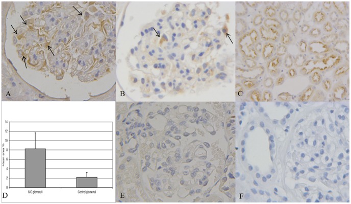
Results: ClC-5 was expressed at glomerular level in all biopsies. Glomerular ClC-5 levels were significantly higher in diabetic nephropaty and MG at both mRNA and protein level (p<0.002; p<0.01). ClC-5 and WT1 double-staining analysis in MG showed that ClC-5 was localized in the podocytes. ClC-5 ultrastructural immunolocalization was demonstrated in podocytes foot processes. Our study is the first to demonstrate that ClC-5 is expressed in human podocytes. The ClC-5 overexpression found in biopsies of proteinuric patients suggests that proteinuria may play a part in its expression and that podocytes are likely to have a key role in albumin handling in proteinuric states.
Conflict of interest statement
Figures


References
-
- Pavenstadt H, Kriz W, Kretzler M (2003) Cell biology of the glomerular podocyte. Physiol Rev 83: 253–307. - PubMed
-
- Birn H, Christensen EI (2006) Renal albumin absorption in physiology and pathology. Kidney Int 69: 440–449. - PubMed
-
- Hryciw DH, Ekberd J, Pollock CA, Poronnik P (2006) ClC-5: a chloride channel with multiple roles in renal tubular albumin uptake. Int J Biochem Cell Biol 38: 1036–1042. - PubMed
-
- Wellhauser L, D’Antonio Christina, Bear CE (2010) ClC transporter: discoveries and challenges in defining the mechanisms underlying function and regulation of ClC-5. Eur J Physiol 460: 543–557. - PubMed
-
- Yoshika N, Ito H, Akamatsu R, Hazikano H, Okada S, et al. (1986) Glomerular podocyte vacuolization in focal segmental glomerulosclerosis. Arch Pathol Lab Med 110: 394–398. - PubMed
Publication types
MeSH terms
Substances
LinkOut - more resources
Full Text Sources
Medical
Miscellaneous
