. 2012;7(9):e45695.
doi: 10.1371/journal.pone.0045695. Epub 2012 Sep 20.

Role of vegetation-associated protease activity in valve destruction in human infective endocarditis

Affiliations

Role of vegetation-associated protease activity in valve destruction in human infective endocarditis

Ghada Al-Salih et al. PLoS One. 2012.

Abstract

Aims: Infective endocarditis (IE) is characterized by septic thrombi (vegetations) attached on heart valves, consisting of microbial colonization of the valvular endocardium, that may eventually lead to congestive heart failure or stroke subsequent to systemic embolism. We hypothesized that host defense activation may be directly involved in tissue proteolytic aggression, in addition to pathogenic effects of bacterial colonization.

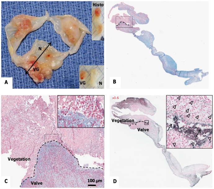
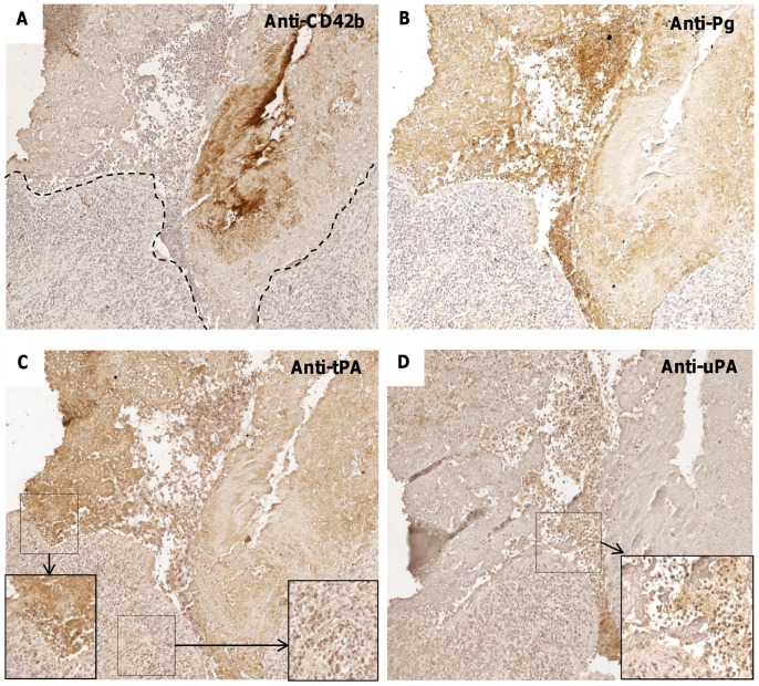
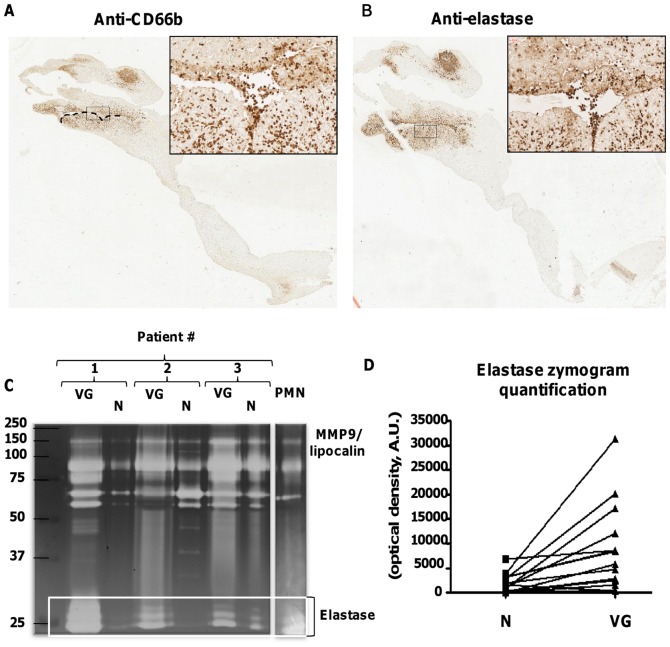
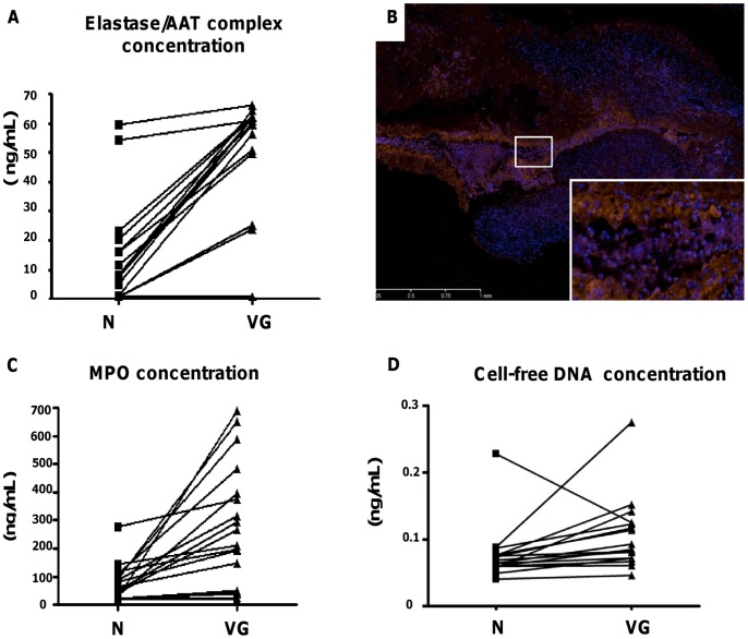
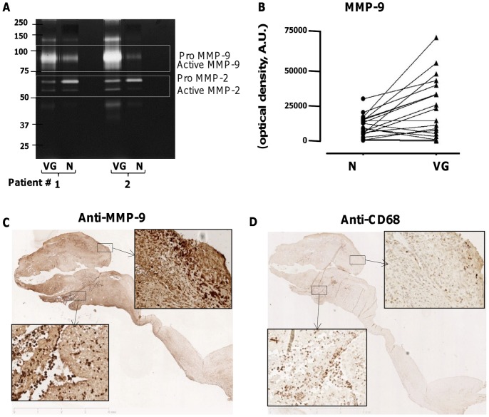
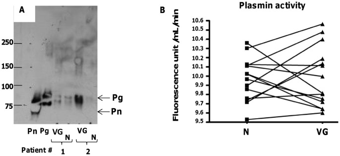
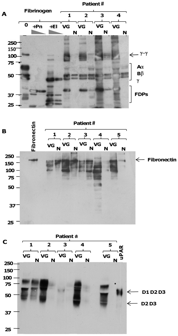
Methods and results: IE valve samples collected during surgery (n = 39) were dissected macroscopically by separating vegetations (VG) and the surrounding damaged part of the valve from the adjacent, apparently normal (N) valvular tissue. Corresponding conditioned media were prepared separately by incubation in culture medium. Histological analysis showed an accumulation of platelets and polymorphonuclear neutrophils (PMNs) at the interface between the VG and the underlying tissue. Apoptotic cells (PMNs and valvular cells) were abundantly detected in this area. Plasminogen activators (PA), including urokinase (uPA) and tissue (tPA) types were also associated with the VG. Secreted matrix metalloproteinase (MMP) 9 was also increased in VG, as was leukocyte elastase and myeloperoxidase (MPO). The presence of neutrophil extracellular traps (NETs) associating MPO and externalized nucleosomes, was shown by immunostaining in the VG. Both MPO and cell-free DNA were released in larger amounts by VG than N samples, suggesting bacterial activation of PMNs within the vegetation. Finally, evidence of proteolytic tissue damage was obtained by the release of fragments of extracellular matrix components such as fibrinogen and fibronectin, as well as protease-sensitive receptors such as the uPA receptor.

Conclusion: Our data obtained using human IE valves suggest that septic vegetations represent an important source of proteases originating from massive leukocyte recruitment and activation of the host plasminergic system. The latter forms a potential therapeutic target to minimize valvular tissue degradation independently from that induced by bacterial proteases.


Conflict of interest statement

Competing Interests: The authors have declared that no competing interests exist.

Figures

Figure 1
Figure 1. Characterization of the excised valve(s) containing infected vegetations.
(A) Macroscopic view of human endocarditic valves, sectioned for histological analysis (“Histo”) as indicated by the double-headed arrow. Vegetations (VG) and adjacent macroscopically normal (N) parts of the valve were then incubated in culture medium for preparation of “conditioned medium”. (B) Alcian blue staining with nuclear fast red counterstaining of a vegetation and underlying valvular tissue. Blue areas correspond to mucoid degeneration interfacing with infected thrombus (rectangle, magnified in C). Red staining shows inflammatory cell nuclei as well as the septic thrombus. (C) Higher magnifications of the interface between the vegetation and the valvular tissue (Alcian blue staining). Inset shows an accumulation of cells with polylobed nuclei. (D) Detection of fragmented DNA by TUNEL showing apoptotic cells (arrowheads) and extracellular fragmented DNA (arrows). Nuclei were counterstained with nuclear fast red.
Figure 2
Figure 2. Immunostaining for platelet markers and PAs in human endocarditic valves.
(A,B) Immunostaining for CD42b/GPIb and for plasminogen, respectively, with hemalun nuclear counterstaining (x10), showing positive immunostaining in the VG. (C) Immunostaining for tissue-type PA (tPA). Insets: higher magnification (x20) of the interface VG/valve (left-hand side) and within the valvular tissue (right-hand side). (D) Immunostaining for urokinase (uPA). Inset: higher magnification (x20) of the interface VG/valve showing an intense staining of polymorphonuclear cells.
Figure 3
Figure 3. Immunocolocalization of PMNs and leukocyte elastase in human endocarditic valves.
(A,B) Immunostaining for CD66b and for elastase, respectively (x1.2), highlighting the presence of numerous PMNs at the interface VG/valve. Inset: higher magnification of these areas (x20). (C) Representative zymogram of conditioned media of VG and N from three patients. Supernatant of activated PMNs (20 µL) was used as a control for secreted elastase. Positions in the gel of molecular mass standard proteins used for calibration are indicated on the left-hand side. (D) Quantification of zymograms: areas of gelatinolysis corresponding to elastase were quantified by densitometry for 17 pairs of conditioned media. The mean optical intensity is 1572±460 versus 7549±2075 for N and VG samples, respectively, (P = 0.0001).
Figure 4
Figure 4. Characterization of protease activity in the vegetation.
(A) Diagram illustrating the variation of concentrations of elastase/α1-antitrypsin complexes between N and VG for 16 pairs of conditioned media. The mean of concentrations is 0.0769±0.0106 versus 0.107±0.014 ng/mL for N and VG respectively, (P = 0.004). (B) Immunostaining of MPO (red fluorescence) coupled to DNA detection by DAPI (blue fluorescence) (x20) showing both cellular and extracellular staining at the interface VG/valve. Co-localization of MPO and DAPI staining was observed in extracellular filamentous DNA NETs. (C) Diagram illustrating the variation in extracellular MPO concentrations in the conditioned media between N and VG. The mean of concentrations is 12±4 versus 39±6 ng/mL for N and VG respectively, (P<0.001). (D) Concentrations of cf-DNA in the conditioned media of N and VG samples. The mean of concentrations is 73±14 versus 252±49 ng/mL for N and VG respectively (P<0.001).
Figure 5
Figure 5. Identification and activity of MMPs and plasmin in human endocarditic valves.
(A) Detection of the different forms of MMP-9 and MMP-2 in conditioned media of VG and N, in representative zymogram from 2 patients. Gelatinolytic bands corresponding to proMMP-9, MMP-9, proMMP-2 and MMP-2 were quantified and expressed in arbitrary optical density units. (B) Diagram illustrating the variation of MMP-9 activity between N and VG for 19 pairs of conditioned media (P<0.01). (C,D) Immunostaining of MMP-9 and macrophages respectively (x1.2) in human endocarditic valves. (C) Strong immunostaining of MMP-9 in the VG and adjacent valve tissue. Insets: higher magnification (x20) of the interface VG/valve (bottom left) and within the valvular tissue (top right). (D) Presence of numerous macrophages at the interface VG/valve where only few of them could be observed in the upper area. Insets: higher magnification (x20) of the interface VG/valve (bottom left) and within the valvular tissue (top right).
Figure 6
Figure 6. Plasmin activity in the conditioned media.
(A) Western blot analysis for detection of plasmin(ogen) with a monoclonal anti-human plasminogen antibody, in conditioned medium obtained from the VG and N samples of 2 representative patients (Pn and Pg: purified human plasmin, 0.25 µg per well, and purified human plasminogen 0.2 µg per well, respectively, used as reference). Positions in the gel of molecular mass standard proteins used for calibration are indicated on the left. (B) Plasmin activity quantified in the conditioned media of N and VG, using a selective chromogenic substrate (P = 0.8).
Figure 7
Figure 7. Extracellular matrix and pericellular proteolysis.
(A) Western blot analysis of fibrinogen and proteolytic fragments in conditioned media of VG and N. Human fibrinogen (100 µg/mL) was incubated with saline (0), plasmin (Pn, 500 nM and 100 nM) and elastase (El, 100 nM and 20 nM) for 2 h at 37°C, to generate reference fibrinogen degradation products (FDPs). Pairs of conditioned media from 4 patients were analyzed under conditions of reduced disulfide bonds using a rabbit anti-human fibrinogen antibody, which recognizes both the intact Aα, Bβ, and γ chains, γ-γ dimers, and FDPs (positions indicated by brackets on the right-hand side). (B) Western blot analysis of fibronectin and its proteolytic fragments in pairs of conditioned media from 5 patients using a polyclonal anti-fibronectin antibody. Purified fibronectin (0.5 µg per well) was used as a control. (C) Western blot analysis of shedding of soluble species of uPAR in pairs of conditioned media from 5 patients, using a mouse monoclonal anti-uPAR D2 domain antibody. Purified recombinant human uPAR (15 ng per well) was used as a reference, and the position of the intact three-domain (D1D2D3) and truncated two-domain (D2D3) species under reducing conditions is indicated on the right. For all Western blots, positions in the gel of molecular mass standard proteins used for calibration are indicated on the left.

References

    1. Delahaye F, Goulet V, Lacassin F, Ecochard R, Selton-Suty C, et al. (1995) Characteristics of infective endocarditis in France in 1991. A 1-year survey. Eur Heart J 16: 394–401. - PubMed
    1. Hoen B, Alla F, Selton-Suty C, Beguinot I, Bouvet A, et al. (2002) Changing profile of infective endocarditis: results of a 1-year survey in France. Jama 288: 75–81. - PubMed
    1. Moreillon P, Que YA (2004) Infective endocarditis. Lancet 363: 139–149. - PubMed
    1. Fontaine V, Jacob MP, Houard X, Rossignol P, Plissonnier D, et al. (2002) Involvement of the mural thrombus as a site of protease release and activation in human aortic aneurysms. Am J Pathol 161: 1701–1710. - PMC - PubMed
    1. Fontaine V, Touat Z, Mtairag el M, Vranckx R, Louedec L, et al. (2004) Role of leukocyte elastase in preventing cellular re-colonization of the mural thrombus. Am J Pathol 164: 2077–2087. - PMC - PubMed

Substances