. 2012;7(9):e46036.
doi: 10.1371/journal.pone.0046036. Epub 2012 Sep 28.

Implication of DNA demethylation and bivalent histone modification for selective gene regulation in mouse primordial germ cells

Affiliations

Implication of DNA demethylation and bivalent histone modification for selective gene regulation in mouse primordial germ cells

Kentaro Mochizuki et al. PLoS One. 2012.

Abstract

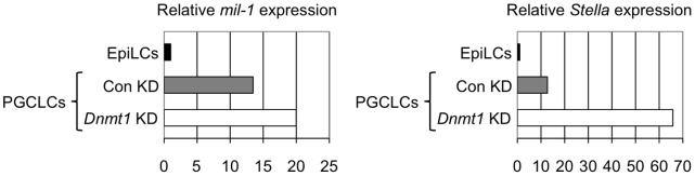
Primordial germ cells (PGCs) sequentially induce specific genes required for their development. We focused on epigenetic changes that regulate PGC-specific gene expression. mil-1, Blimp1, and Stella are preferentially expressed in PGCs, and their expression is upregulated during PGC differentiation. Here, we first determined DNA methylation status of mil-1, Blimp1, and Stella regulatory regions in epiblast and in PGCs, and found that they were hypomethylated in differentiating PGCs after E9.0, in which those genes were highly expressed. We used siRNA to inhibit a maintenance DNA methyltransferase, Dnmt1, in embryonic stem (ES) cells and found that the flanking regions of all three genes became hypomethylated and that expression of each gene increased 1.5- to 3-fold. In addition, we also found 1.5- to 5-fold increase of the PGC genes in the PGCLCs (PGC-like cells) induced form ES cells by knockdown of Dnmt1. We also obtained evidence showing that methylation of the regulatory region of mil-1 resulted in 2.5-fold decrease in expression in a reporter assay. Together, these results suggested that DNA demethylation does not play a major role on initial activation of the PGC genes in the nascent PGCs but contributed to enhancement of their expression in PGCs after E9.0. However, we also found that repression of representative somatic genes, Hoxa1 and Hoxb1, and a tissue-specific gene, Gfap, in PGCs was not dependent on DNA methylation; their flanking regions were hypomethylated, but their expression was not observed in PGCs at E13.5. Their promoter regions showed the bivalent histone modification in PGCs, that may be involved in repression of their expression. Our results indicated that epigenetic status of PGC genes and of somatic genes in PGCs were distinct, and suggested contribution of epigenetic mechanisms in regulation of the expression of a specific gene set in PGCs.


Conflict of interest statement

Competing Interests: The authors have declared that no competing interests exist.

Figures

Figure 1
Figure 1. The regulatory region of mil-1 becomes hypomethylated during PGC development.
Bisulfite sequencing analysis of the regulatory region of mil-1 was performed using epiblasts, PGCs/somatic cells purified as GFP positive/negative cells from embryos at each embryonic day (E). The rectangle containing mil-1 in the top line represents an exon, and the numbers with ‘kb (kilobase)’ under the line indicate distance from the transcription start site (TSS). The box outlined in green represents the regulatory element required for PGC-specific expression and the Ifitm genes consensus element (ICE) is shown in more detail in Figure 4A. Each circle corresponds to a CpG site in the regulatory region, and the degree of gray in each circle corresponds to the level of DNA methylation.
Figure 2
Figure 2. The expression of mil-1, Blimp1, and Stella become upregulated during PGC development.
(A, B, C) Quantitative RT-PCR analysis of the expression of (A) mil-1, (B) Blimp1, and (C) Stella was performed using epiblasts (E6.0) and PGCs (E7.5 and E9.0). Histograms represent relative expression levels of these three genes at each developmental stage. The averages of expression levels in the epiblasts (E6.0) were set as 1.0. Gapdh PCR signal was used as an internal control to measure relative. The data were obtained from three individual embryos. *p<0.05. Error bars represent SEM.
Figure 3
Figure 3. DNA demethylation of the regulatory region of mil-1 resulted in upregulation of its expression in ES cells.
(A) Bisulfite sequencing analysis of the regulatory region of mil-1 and (B) quantitative RT-PCR analysis of mil-1 expression were performed on embryonic stem (ES) cells with or without siRNA-mediates Dnmt1 knockdown (Dnmt1 KD/Con KD). (A) The regulatory region became more hypomethylated following Dnmt1 knockdown. (B) Histogram represents the relative expression level of mil-1 in the Dnmt1-knockdown ES cells. The expression level in the control ES cells (Con KD) was set as 1.0. Gapdh PCR signal was used an internal control to measure relative expression. The data were obtained from four independent experiments. *p<0.05. Error bars represent SEM. (C) Luciferase activity of the reporter vectors with methylated or unmethylated regulatory region of mil-1 in ES cells. Luciferase activity was normalized against the activity of a cotransfected Renilla construct. The liciferase activity of the methylated construct (Methylated 3.0k-pCpGL) was set as 1.0. The data were obtained from six independent experiments. *p<0.05. Error bars represent SEM.
Figure 4
Figure 4. Flanking regions of the PGC-specific genes also become hypomethylated during PGC development.
(A) Comparison of the Ifitm genes consensus element (ICE) of mil-1/Ifitm3 with the homologous sequences found in the putative regulatory regions flanking Blimp1/Prdm1, Prdm14, Stella/Dppa3, and Nanos3. (B) Bisulfite sequencing analysis of the flanking regions of Blimp1, Stella, and Dazl was performed using epiblasts (E6.0 and E6.75) and PGCs (E7.5 and E10.5). The flanking regions of Blimp1 and Stella, like those in mil-1, were also progressively demethylated during PGC development, whereas that of Dazl was maintained hypermethylated in PGCs at E10.5.
Figure 5
Figure 5. Knockdown of Dnmt1 causes hypomethylation of Blimp1 and Stella flanking regions and upregulation of Blimp1 and Stella expression in ES cells.
(A) Bisulfite sequencing analysis of the flanking regions of Blimp1 and Stella and (B) quantitative RT-PCR analysis of Blimp1 and Stella expression were performed using ES cells with or without Dnmt1 knockdown treatment (Dnmt1 KD/Con KD).
Figure 6
Figure 6. Knockdown of Dnmt1 causes upregulation of mil-1 and Stella expression in PGCLCs.
Quantitative RT-PCR analysis of mil-1 and Stella expression in PGCLCs (PGC-like cells) with or without Dnmt1 knockdown (Dnmt1 KD/Con KD). The expression level in EpiLCs was set as 1.0. Shown is a representative data from two independent experiments.
Figure 7
Figure 7. Repression of somatic gene expression does not depend on DNA methylation in PGCs.
(A) Bisulfite sequencing analysis of the flanking regions of Hoxa1, Hoxb1, and Gfap was performed using epiblasts (E6.0), epiblast stem cells (EpiSCs), and PGCs (E13.5), showing hypomethylation in PGCs. (B) Bisulfite sequencing analysis of the regulatory region of Gfap and (C) quantitative RT-PCR analysis of the Gfap expression were performed using ES cells with or without Dnmt1 knockdown treatment (Dnmt1 KD/Con KD).
Figure 8
Figure 8. Bivalent histone modification on the somatic genes in PGCs.
(A, B) ChIP analysis with the H3K4me3 or H3K27me3 antibodies for the promoter regions of somatic genes (Hoxa1, Hoxb1, and Gfap) and of the PGC-specific genes (mil-1, Blimp1, and Stella) was performed on EpiSCs (A), and male and female PGCs at E13.5 (B), showing the bivalent histone modification. Histogram represents ratios of the immuno-precipitated chromatin to the input chromatin, which was quantified by quantitative PCR analysis. Also shown are results using beads only as a no antibody control (NAC). Shown is a representative data from two independent experiments.

References

    1. Ohinata Y, Payer B, O’Carroll D, Ancelin K, Ono Y, et al. (2005) Blimp1 is a critical determinant of the germ cell lineage in mice. Nature 436: 207–213. - PubMed
    1. Kurimoto K, Yabuta Y, Ohinata Y, Shigeta M, Yamanaka K, et al. (2008) Complex genome-wide transcription dynamics orchestrated by Blimp1 for the specification of the germ cell lineage in mice. Genes Dev 22: 1617–1635. - PMC - PubMed
    1. Yamaji M, Seki Y, Kurimoto K, Yabuta Y, Yuasa M, et al. (2008) Critical function of Prdm14 for the establishment of the germ cell lineage in mice. Nat Genet 40: 1016–1022. - PubMed
    1. Yabuta Y, Kurimoto K, Ohinata Y, Seki Y, Saitou M (2006) Gene expression dynamics during germline specification in mice identified by quantitative single-cell gene expression profiling. Biol Reprod 75: 705–716. - PubMed
    1. Okamura D, Tokitake Y, Niwa H, Matsui Y (2008) Requirement of Oct3/4 function for germ cell specification. Dev Biol 317: 576–584. - PubMed

Publication types

MeSH terms