Comparative Study
. 2012;7(9):e46106.
doi: 10.1371/journal.pone.0046106. Epub 2012 Sep 28.

Comparison of optical and power Doppler ultrasound imaging for non-invasive evaluation of arsenic trioxide as a vascular disrupting agent in tumors

Affiliations
Comparative Study

Comparison of optical and power Doppler ultrasound imaging for non-invasive evaluation of arsenic trioxide as a vascular disrupting agent in tumors

Mustafa K Alhasan et al. PLoS One. 2012.

Abstract

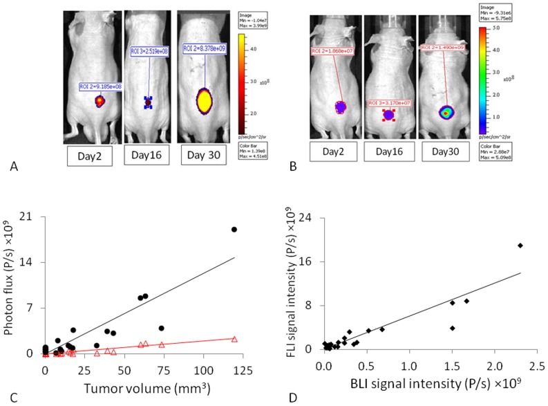
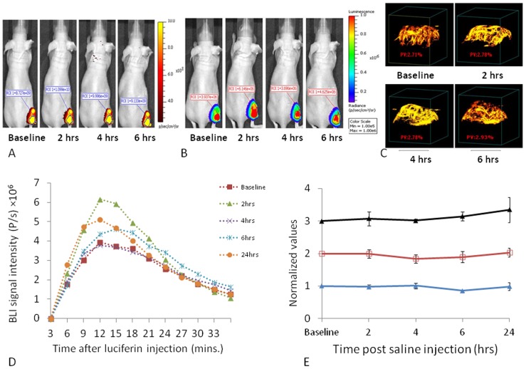
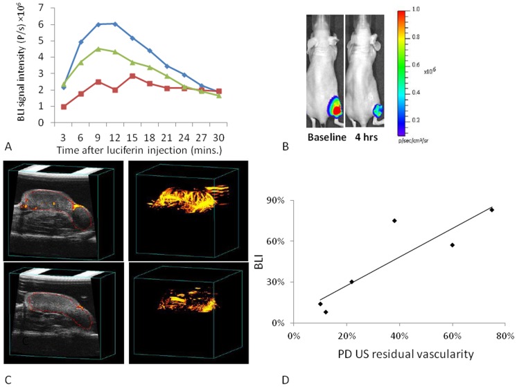
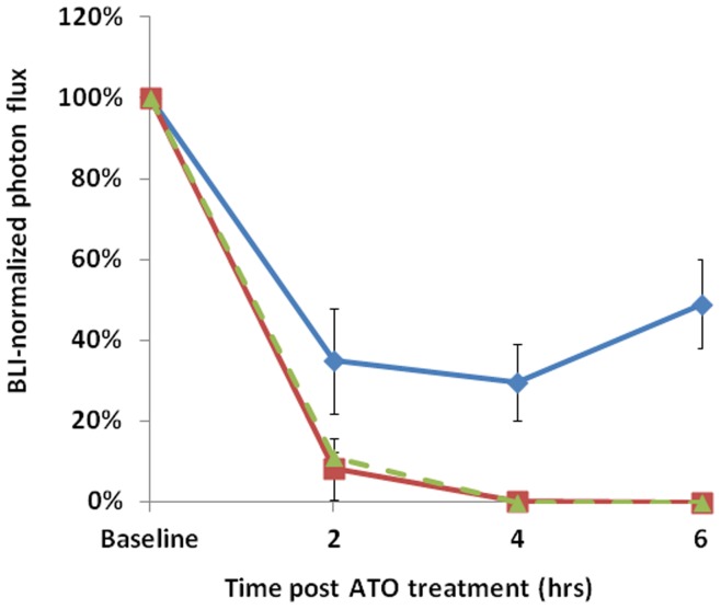
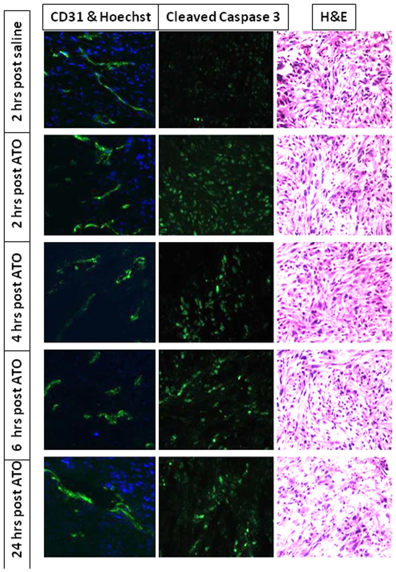
Small animal imaging provides diverse methods for evaluating tumor growth and acute response to therapy. This study compared the utility of non-invasive optical and ultrasound imaging to monitor growth of three diverse human tumor xenografts (brain U87-luc-mCherry, mammary MCF7-luc-mCherry, and prostate PC3-luc) growing in nude mice. Bioluminescence imaging (BLI), fluorescence imaging (FLI), and Power Doppler ultrasound (PD US) were then applied to examine acute vascular disruption following administration of arsenic trioxide (ATO).During initial tumor growth, strong correlations were found between manual caliper measured tumor volume and FLI intensity, BLI intensity following luciferin injection, and traditional B-mode US. Administration of ATO to established U87 tumors caused significant vascular shutdown within 2 hrs at all doses in the range 5 to 10 mg/kg in a dose dependant manner, as revealed by depressed bioluminescent light emission. At lower doses substantial recovery was seen within 4 hrs. At 8 mg/kg there was >85% reduction in tumor vascular perfusion, which remained depressed after 6 hrs, but showed some recovery after 24 hrs. Similar response was observed in MCF7 and PC3 tumors. Dynamic BLI and PD US each showed similar duration and percent reductions in tumor blood flow, but FLI showed no significant changes during the first 24 hrs.The results provide further evidence for comparable utility of optical and ultrasound imaging for monitoring tumor growth, More specifically, they confirm the utility of BLI and ultrasound imaging as facile assays of the vascular disruption in solid tumors based on ATO as a model agent.


Conflict of interest statement

Competing Interests: The authors have declared that no competing interests exist.

Figures

Figure 1
Figure 1. Optical detection of U87-mCherry-luc tumor growth.
A) FLI (λEx = 570 nm and λEm = 620 nm) showed tumor growth on the back of a nude mouse. B) Corresponding BLI acquired 10 mins after administration of sodium D-luciferin (80 µL, 40 mg/ml). C) Correlation between signal intensities and caliper measured tumor volume for a group of six U87-mCherry-Luc tumors; FLI (•; R2>0.82) and BLI (Δ; R2>0.86); D) Correlation between FLI and BLI photon signal intensities at each measurement time (R2>0.86).
Figure 2
Figure 2. Optical assessment of vascular disruption in U87-mCherry-luc tumors.
Optical imaging was performed at various times before and after administration of ATO. On each occasion FLI was performed first and then D-luciferin was administered SC in the neck and BLI was performed over a period of 16 mins. A) Sequential FLI with respect to a dose of 8 mg/kg ATO administered IP, B) Corresponding time course of bioluminescent signal evolution in this mouse following luciferin injection at respective times, C) BLI signal intensity at the 10 minute time point following luciferin administration, D) Normalized BLI signal intensity at 10 minute time point after administration of luciferin indicating vascular shutdown following various doses of ATO.
Figure 3
Figure 3. Reproducibility of sequential images in MCF7-mCherry-luc tumors.
A group of nine MCF7-mCherry-luc tumors was repeatedly observed by FLI, BLI, and PD US following injection of 100 µl saline IP. A) Sequential FLI for a representative mouse, B) BLI from the same mouse showing images acquired 10 mins after administration of fresh luciferin on each occasion, c) PD US showing MIP (maximum intensity projection) observed at 40 MHz, D) Variation in dynamic bioluminescent signal intensity from the same tumor as in A,B and C, on five sequential occasions over 24 hrs, E) Comparison of signal stability based on FLI (red ), BLI (blue ▴) and PD-US (black ▴) normalized values are presented mean ± SE for the whole group.
Figure 4
Figure 4. Vascular disruption assessed by BLI and PD-US in MCF7-mCherry-luc tumors with respect to ATO (8 mg/kg).
A) Variation in bioluminescent signal intensity from a representative tumor on sequential occasions over 24 hrs: blue ♦ baseline; red ▪ 4 hrs; green ▴ 24 hrs. B) BLI acquired 10 mins after administration of luciferin. C) PD US images are presented as single slice (left) and PD maximum intensity projection (right) before and 4 hrs after treatment with ATO. D) Comparison of PD US and BLI in MCF7-mCherry-luc tumors as fractional signal versus baseline.
Figure 5
Figure 5. Comparative vascular shutdown following ATO (8 mg/kg) to mice bearing various tumors.
Relative BLI signal intensity observed for tumors 10 mins after administration of luciferin with respect to drug treatment in three different types: blue ♦ MCF7-mCherry-luc; green ▴U87-mCherry-luc; red ▪ PC3-luc; mean values ± SE.
Figure 6
Figure 6. Histology showing vascular impairment and apoptosis in MCF7-mCherry-luc tumors.
Sections were obtained from a series of mice sacrificed at various times after ATO (8 mg/kg). Hoechst stain shows reduced perfusion 2–6 hrs following ATO and H&E stain shows increased necrosis after 24 hrs. Left column: vascular extent (CD31; green) and perfusion (Hoechst 33342; blue). Middle column: Caspase-3 activity indicating apoptosis. Right column: H&E.

References

    1. Thorpe PE (2004) Vascular Targeting Agents as Cancer Therapeutics. Clin Cancer Res 10: 415–427. - PubMed
    1. Mason RP, Zhao D, Liu L, Trawick ML, Pinney KG (2011) A Perspective on Vascular Disrupting Agents that Interact with Tubulin: Preclinical Tumor Imaging and Biological Assessment. Integrat Biol 3: 375–387. - PMC - PubMed
    1. Weis SM, Cheresh DA (2011) Tumor angiogenesis: molecular pathways and therapeutic targets. Nature Med 17: 1359–1370. - PubMed
    1. Lew YS, Kolozsvary A, Brown SL, Kim JH (2002) Synergistic interaction with arsenic trioxide and fractionated radiation in locally advanced murine tumor. Cancer Res 62: 4202–4205. - PubMed
    1. Kim JH, Lew YS, Kolozsvary A, Ryu S, Brown SL (2003) Arsenic trioxide enhances radiation response of 9L glioma in the rat brain. Radiat Res 160: 662–666. - PubMed

Publication types

MeSH terms