. 2012;7(9):e46134.
doi: 10.1371/journal.pone.0046134. Epub 2012 Sep 24.

The Relationship between fenestrations, sieve plates and rafts in liver sinusoidal endothelial cells

Affiliations

The Relationship between fenestrations, sieve plates and rafts in liver sinusoidal endothelial cells

Dmitri Svistounov et al. PLoS One. 2012.

Abstract

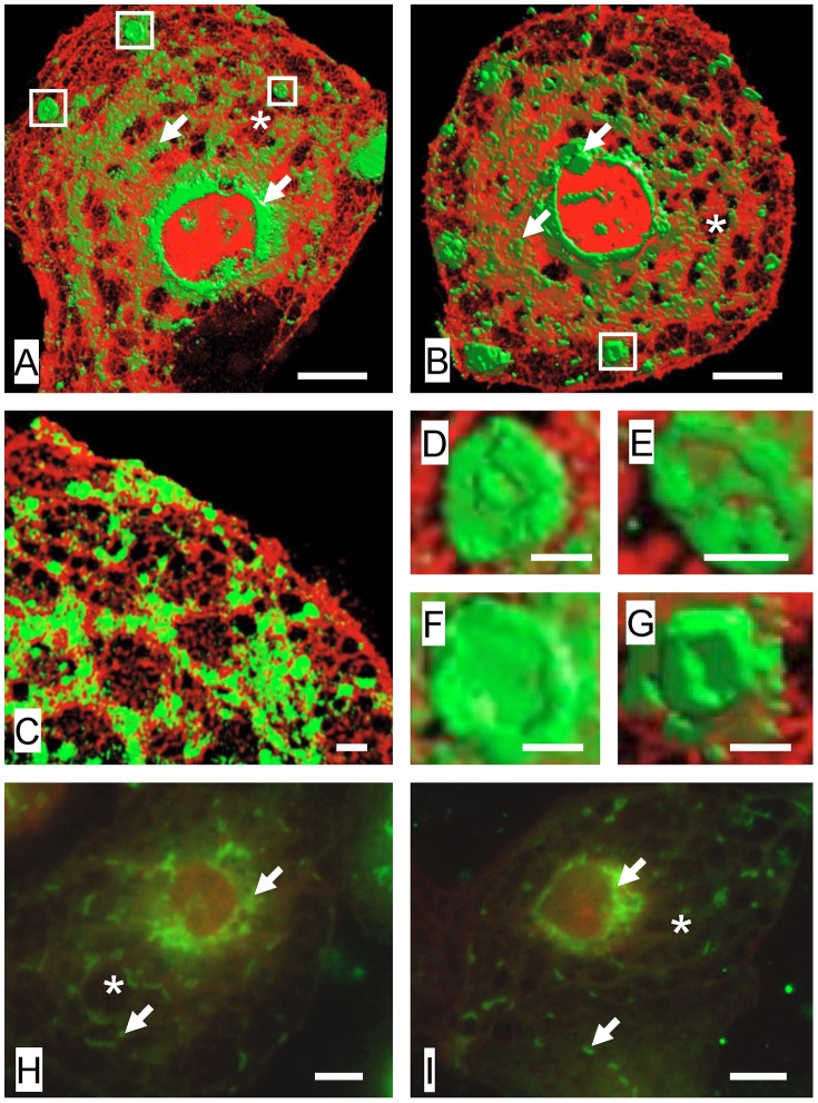
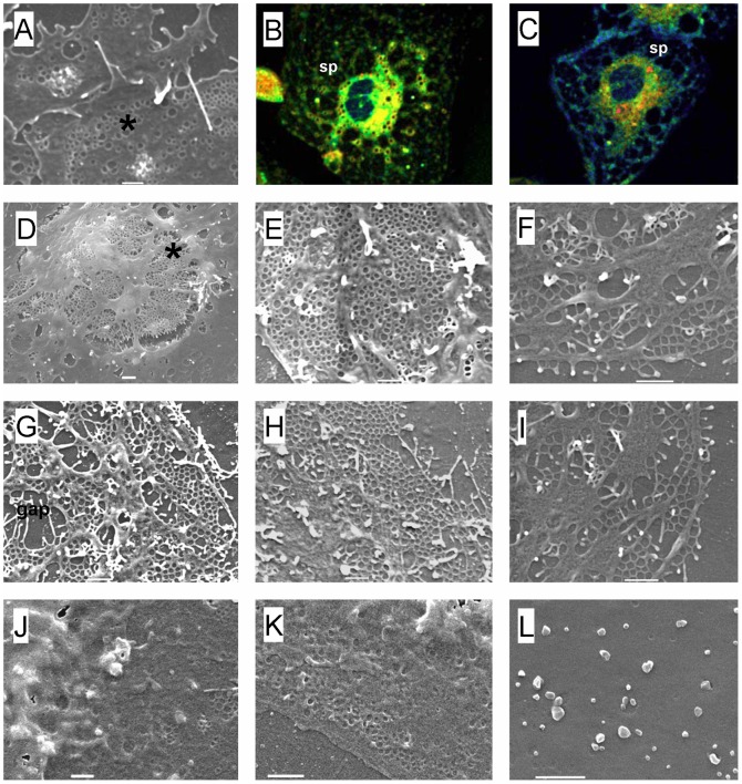
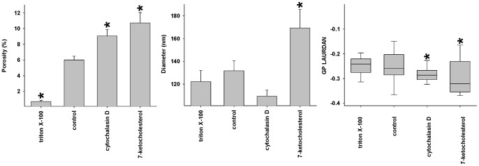
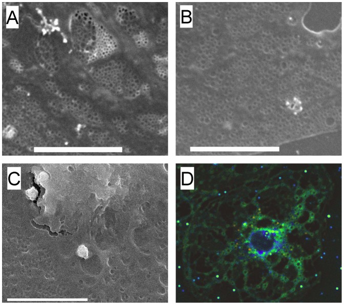
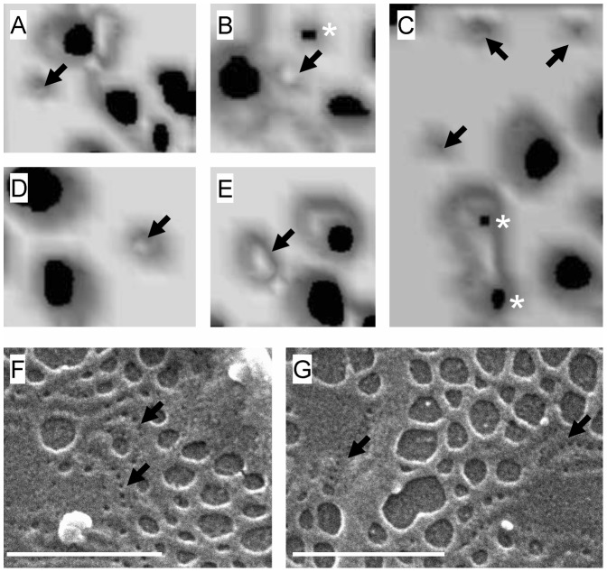
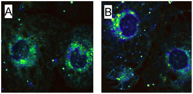
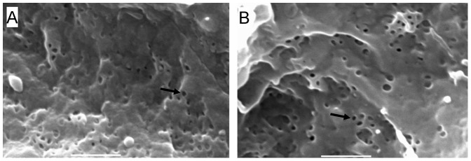
Fenestrations are transcellular pores in endothelial cells that facilitate transfer of substrates between blood and the extravascular compartment. In order to understand the regulation and formation of fenestrations, the relationship between membrane rafts and fenestrations was investigated in liver sinusoidal endothelial cells where fenestrations are grouped into sieve plates. Three dimensional structured illumination microscopy, scanning electron microscopy, internal reflectance fluorescence microscopy and two-photon fluorescence microscopy were used to study liver sinusoidal endothelial cells isolated from mice. There was an inverse distribution between sieve plates and membrane rafts visualized by structured illumination microscopy and the fluorescent raft stain, Bodipy FL C5 ganglioside GM1. 7-ketocholesterol and/or cytochalasin D increased both fenestrations and lipid-disordered membrane, while Triton X-100 decreased both fenestrations and lipid-disordered membrane. The effects of cytochalasin D on fenestrations were abrogated by co-administration of Triton X-100, suggesting that actin disruption increases fenestrations by its effects on membrane rafts. Vascular endothelial growth factor (VEGF) depleted lipid-ordered membrane and increased fenestrations. The results are consistent with a sieve-raft interaction, where fenestrations form in non-raft lipid-disordered regions of endothelial cells once the membrane-stabilizing effects of actin cytoskeleton and membrane rafts are diminished.


Conflict of interest statement

Competing Interests: The authors have declared that no competing interests exist.

Figures

Figure 1
Figure 1. Visualization of membrane rafts and fenestrations.
(A–C) 3D-SIM of LSECs stained with Bodipy FL C5 ganglioside GM1, a marker for rafts (green) and Cell-Mask Orange, a cell membrane marker (orange). There is an inverse distribution between liver sieve plates and membrane rafts. Membrane rafts are mostly perinuclear while sieve plates are mostly peripheral. Some sieve plates are identified by an asterix (*) and fenestrations can be resolved within the sieve plates. Rafts are shown with arrows (→). The areas marked in a box (clustered rafts) are further magnified in (D–G). (D–G) Magnification of areas in Figure 1(A–B) showing clustered membrane rafts with raised perimeters. (H) TIRFM of LSEC stained with NBD-cholesterol (green), a marker for rafts, and CellMask Orange (orange) showing perinuclear distribution of rafts (arrows). Fenestrations are not resolved within the sieve plates (*) with TIRFM. (I) TIRFM of LSEC stained with Bodipy FL C5 ganglioside GM1, a marker for rafts, and CellMask Orange (orange) confirming mostly perinuclear distribution of rafts. Scale bar 5 µm (A, B, H, I), 1 µm (C–G).
Figure 2
Figure 2. Effects of manipulating membrane rafts on fenestrations.
(A) SEM of normal LSEC. Fenestrations (*) are clustered in sieve plates. (B) Two-photon fluorescence microscopy of normal LSEC stained with LAURDAN, a stain which changes from red/yellow in raft regions to blue/green in non-raft regions. There is widespread red and yellow staining consistent with membrane rafts in the cell membrane between the sieve plates (sp). Fenestrations cannot be resolved with two-photon fluorescence microscopy. (C) Two-photon fluorescence microscopy of LSEC stained with LAURDAN following treatment with 7KC. There is increased blue staining consistent with increased lipid-disordered, non-raft regions. (D–I ) SEMs of LSEC following treatment with 7KC which depletes rafts by inducing lipid disorder. There is an increased number of fenestrations and gaps are visible presumably where rafts have been disrupted, including the perinuclear region (*). (J,K,L) SEM of LSEC after treatment with Triton X-100. There is a marked decrease in fenestrations. (scale bar = 1 µM).
Figure 3
Figure 3. Effects of manipulating rafts on porosity.
The effects of 7KC, Triton X-100 and cytochalasin D on the porosity and diameter of fenestrations (determined using SEM) and generalized polarization (GP, following staining with LAURDAN imaged using two-photon microscopy) (* significantly different from control values P<0.05, each data point represents average ± SEM of 7–28 images and 83–2840 fenestrations).
Figure 4
Figure 4. Effects of manipulating actin on fenestrations.
(A) SEM of control LSEC showing fenestrations clustered in sieve plates. (B) SEM of LSEC following treatment with cytochalasin D showing an increase in fenestrations. (C) SEM of LSEC following treatment with Triton X-100 and cytochalasin D. Triton X-100 ameliorated the effects of cytochalasin D on fenestrations. (D) Two-photon fluorescence microscopy of LSEC following treatment with cytochalasin D and stained with LAURDAN. Numerous sieve plates are apparent. (scale bar 5 µM).
Figure 5
Figure 5. Pores and fenestrations.
(A–E) Isorendered 3D-SIM reconstructions of fenestrations from LSECs. Around the sieve plates are a few pores (→) and some early fenestrations can be identified forming at the base of the pores (*). (F) Scanning electron microscopy isolated LSECs. Pores (→) similar to those seen on 3D-SIM are present towards the periphery of the sieve plates. (scale bar 1 µm).
Figure 6
Figure 6. Effects of VEGF on membrane rafts.
(A) Two-photon fluorescence microscopy of LSECs stained with LAURDAN. (B) Two-photon fluorescence microscopy of LSECs stained with LAURDAN following treatment with VEGF, showing increased blue staining consistent with increased non-raft lipid disordered membrane.
Figure 7
Figure 7. Effects of 7KC on the perfused mouse liver.
(A) Scanning electron micrograph of a mouse perfused liver sinusoid. (B) Scanning electron micrograph of a mouse liver sinusoid following perfusion with 7KC (scale bar 1 µM, → fenestration).

Comment in

References

    1. Cogger VC, Le Couteur DG (2009) Fenestrations in the liver sinusoidal endothelial cell. In: Arias I, Wolkoff A, Boyer J, Shafritz D, Fausto N, et al., editors. The Liver: Biology and Pathobiology. 5th ed. Hokoben NJ: John Wiley & Sons, Ltd. 387–404.
    1. Hilmer SN, Cogger VC, Fraser R, McLean AJ, Sullivan D, et al. (2005) Age-related changes in the hepatic sinusoidal endothelium impede lipoprotein transfer in the rat. Hepatology 42: 1349–1354. - PubMed
    1. Le Couteur DG, Fraser R, Hilmer S, Rivory LP, McLean AJ (2005) The hepatic sinusoid in aging and cirrhosis - Effects on hepatic substrate disposition and drug clearance. Clin Pharmacokinet 44: 187–200. - PubMed
    1. Furrer K, Rickenbacher A, Tian Y, Jochum W, Bittermann AG, et al. (2011) Serotonin reverts age-related capillarization and failure of regeneration in the liver through a VEGF-dependent pathway. Proc Natl Acad Sci U S A 108: 2945–2950. - PMC - PubMed
    1. Warren A, Le Couteur DG, Fraser R, Bowen DG, McCaughan GW, et al. (2006) T lymphocytes interact with hepatocytes through fenestrations in murine liver sinusoidal endothelial cells. Hepatology 44: 1182–1190. - PubMed

Publication types

MeSH terms