. 2012;7(9):e46213.
doi: 10.1371/journal.pone.0046213. Epub 2012 Sep 28.

Molecular characterisation of endogenous Vangl2/Vangl1 heteromeric protein complexes

Affiliations

Molecular characterisation of endogenous Vangl2/Vangl1 heteromeric protein complexes

Edwige Belotti et al. PLoS One. 2012.

Abstract

Background: Mutations in the Planar Cell Polarity (PCP) core gene Vangl2 cause the most severe neural tube defects (NTD) in mice and humans. Genetic studies show that the Vangl2 gene genetically interacts with a close homologue Vangl1. How precisely Vangl2 and Vangl1 proteins interact and crosstalk has remained a difficult issue to address, with the main obstacle being the accurate discrimination of the two proteins, which share close sequence homology. Experimental evidence previously presented has been sparse and addressed with ectopically expressed proteins or with antibodies unable to biochemically discriminate Vangl1 from Vangl2, therefore giving rise to unclear results.

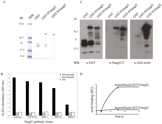
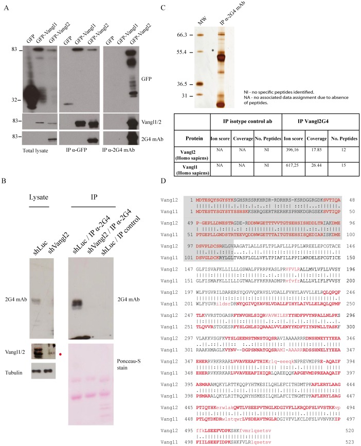
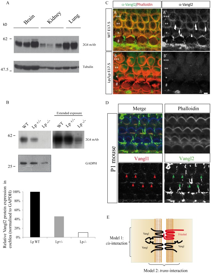
Methodology and main findings: A highly specific monoclonal anti-Vangl2 antibody was generated and rigorously tested on both recombinant and extracted Vangl2 using surface plasmon resonance (SPR) analysis, western blot, and immunoprecipitation experiments. This antibody efficiently affinity-purified Vangl2 from cell lysates and allowed the unambiguous identification of endogenous Vangl2 by proteomic analysis. Vangl1 was also present in Vangl2 immunoprecipitates, establishing the first biochemical evidence for the existence of Vangl2/Vangl1 heterodimers at an endogenous level. Epitope-tagged Vangl2 and Vangl1 confirmed that both proteins interact and colocalize at the plasma membrane. The Vangl2 antibody is able to acutely assess differential expression levels of Vangl2 protein in culture cell lines, as corroborated with gene expression analysis. We characterised Vangl2 expression in the cochlea of homozygous and heterozygous Lp mutant mice bearing a point mutation within the C-terminal Vangl2 region that leads to profound PCP defects. Our antibody could detect much lower levels of Vangl2(Lp) protein in mutant mice compared to the wild type mice.

Conclusion: Our results provide an in-depth biochemical characterisation of the interaction observed between Vangl paralogues.


Conflict of interest statement

Competing Interests: The authors have declared that no competing interests exist.

Figures

Figure 1
Figure 1. Generation of a highly specific anti-Vangl2 monoclonal antibody.
(A) Expression of GST, GST-NVangl1 and GST-NVangl2 fusion proteins verified by SDS-PAGE and Coomassie staining. (B) Selected hybridomas were screened using an ELISA against GST, GST-NVangl1 and GST-NVangl2. mAb: monoclonal antibody. (C) Specificity of 2G4 mAb for GST-NVangl2 shown by western blot experiments, using antibodies specific for GST, Vangl2 and Vangl1/2. (D) SPR analysis using 2G4 mAb (10 µg/ml) injected over immobilized GST, GST-NVangl1 or GST-NVangl2. Sensorgrams show total signal from GST fusion proteins normalised with the non-specific signal (GST).
Figure 2
Figure 2. Identification of an endogenous protein complex containing the Vangl2 and Vangl1 proteins.
(A) Specificity of 2G4 mAb was verified in western blot using T47D cell extracts expressing GFP, GFP-Vangl1, Vangl2 or GFP. Proteins were separated on SDS-PAGE and analyzed with western blot with specific antibodies. Specificity of 2G4 mAb in immunoprecipitation experiments GFP-Vangl1, GFP-Vangl2 or GFP expressed in T47D cells. Western blotting was carried out using 2G4 mAb, anti-GFP and Vangl1/2 antibodies. (B) SKBR7 cells were treated with shLuc or shVangl2 and assessed for Vangl expression. Vangl2 was immunoprecipitated from cell lysates and detected with the same 2G4 antibody. (C) Proteins were subsequently separated using Bis-Tris gradient gels and silver stained. Bands specific to 2G4 mAb were excised followed by in-gel trypsin digestion, chromatographic separation and orbitrap analysis. An asterisk indicates the bands corresponding to Vangl2. (D) Endogenous Vangl2 and Vangl1 were identified with the presence of multiple peptides and associated with a high probability Mascot scores (at least 40 arbitrary units). Bold characters indicate the peptides identified by mass spectrometry as shown with alignment of the two sequences. Regions shaded in grey correspond to the amino-terminal regions of Vangl1 and Vangl2.
Figure 3
Figure 3. Interaction of epitope-tagged Vangl2 and Vangl1 proteins.
(A) Schematic diagram of Vangl1 and full-length and truncated mutants of Vangl2 used for this study. (B) Transient co-expression of myc-Vangl1 with GFP-Vangl2, GFP-Vangl2ΔN, GFP-Vangl2ΔC, GFP-Vangl2ΔNΔC a control protein (GFP in T47D cells and immunoprecipitation with myc antibody shows co-immunoprecitation of Vangl2 with Vangl1). (C) Colocalization of ectopically expressed Myc-Vangl1 and GFP-Vangl2 in T47D cells and analysis by immunofluorescence and confocal analysis. All images were taken under a 40×objective. Scale bar corresponds to 10 µm and is labelled in white. (D) Vangl2 homodimerization is displayed by immunoprecipitation with Myc antibody shows co-immunoprecitation of Myc-Vangl2 with GFP-Vangl2. Protein complexes were separated using SDS-PAGE and western blot analysis.
Figure 4
Figure 4. Differential expression of Vangl2 in different murine tissues and mouse cochlea.
(A) Expression profile of Vangl2 from murine tissues (brain, kidney and lung) detected with 2G4 antibody in western blot. (B) Vangl2 protein expression in mouse cochlear tissue deriving from homozygous Lp and heterozygous Vangl2Lp/+ heterozygous mice compared to wild-type littermates, using 2G4 mAb antibody. Corresponding densitometry measurements representing relative Vangl2 protein expression normalized to GADPH protein levels. (C) Absence of staining with 2G4 mAb in Vangl2 mutant. Surface views of cochleae from WT (A-A’) and Looptail homozygote (B-B’) from E17.5 mice processed for immunocytochemistry with 2G4 mAb. At this stage, the cochlea comprises a single row of inner hair cells (#) and three rows of outer Hair Cells (OHC1 *, OHC2 **, OHC3 ***) surrounded by supporting cells. Phalloidin staining (actin, red) reveals hair cells borders. The image is taken at the level of the zonula adherens for the second and third row of OHC2 and OHC3, where Vangl2 accumulates. In the WT sample, we observe accumulation of Vangl2 at the junction between hair cells and supporting cells (A-A’, arrows). In contrast, in Lp/Lp cochlea, there is a complete absence of Vangl2 staining at the membrane of the cells (B,B’). Scale bar = 3 µm. (D) Co-localization of staining with 2G4 mAb and Vangl1 Ab. Surface view of a cochlea from a newborn mouse processed for immunocytochemistry with 2G4 mAb and Vangl1 Ab. The two antibodies reveal a co-localization of Vangl1 (A”) and Vangl2 (A’”) proteins at the junction between hair cells and supporting cells (arrows). Phalloidin is in blue, Vangl1 in red, and Vangl2 in green. Scale bar = 3 µm. Note: in the green channel, remains of the tectorial membrane covering normally the cochlear epithelium lead spots of non-specific green labeling. (E) Schematic diagram representing the two possible models of Vangl1/Vangl2 interaction.

References

    1. Doudney K, Ybot-Gonzalez P, Paternotte C, Stevenson RE, Greene ND, et al. (2005) Analysis of the planar cell polarity gene Vangl2 and its co-expressed paralogue Vangl1 in neural tube defect patients. Am J Med Genet A 136: 90–92. - PubMed
    1. Murdoch JN, Doudney K, Paternotte C, Copp AJ, Stanier P (2001) Severe neural tube defects in the loop-tail mouse result from mutation of Lpp1, a novel gene involved in floor plate specification. Hum Mol Genet 10: 2593–2601. - PubMed
    1. Park M, Moon RT (2002) The planar cell-polarity gene stbm regulates cell behaviour and cell fate in vertebrate embryos. Nat Cell Biol 4: 20–25. - PubMed
    1. Copp AJ, Greene ND, Murdoch JN (2003) The genetic basis of mammalian neurulation. Nat Rev Genet 4: 784–793. - PubMed
    1. Lake BB, Sokol SY (2009) Strabismus regulates asymmetric cell divisions and cell fate determination in the mouse brain. J Cell Biol 185: 59–66. - PMC - PubMed

Publication types

MeSH terms