. 2012;7(9):e46241.
doi: 10.1371/journal.pone.0046241. Epub 2012 Sep 27.

Differential effects of IL-12 on Tregs and non-Treg T cells: roles of IFN-γ, IL-2 and IL-2R

Affiliations

Differential effects of IL-12 on Tregs and non-Treg T cells: roles of IFN-γ, IL-2 and IL-2R

Jingxian Zhao et al. PLoS One. 2012.

Abstract

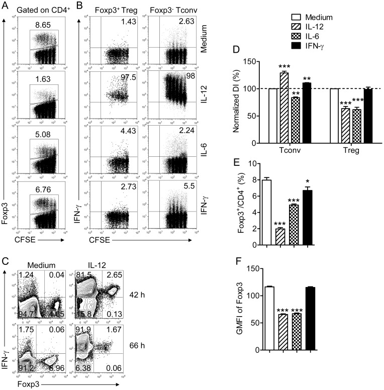
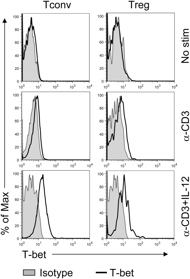
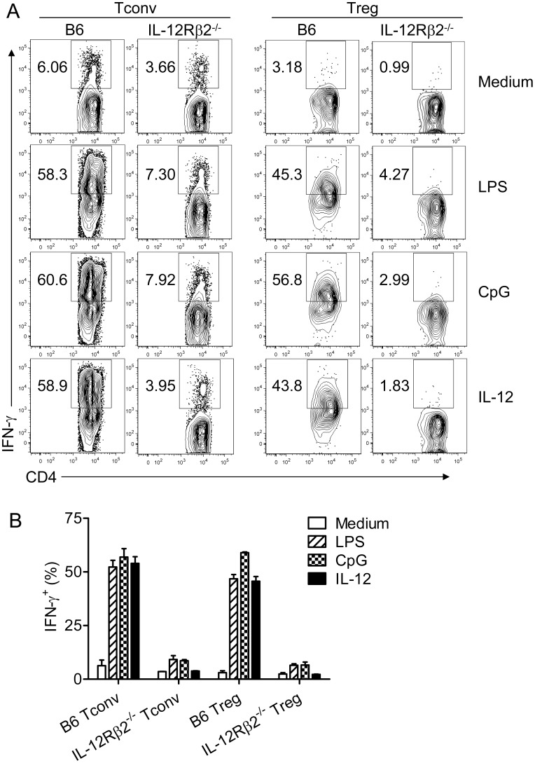
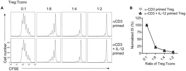
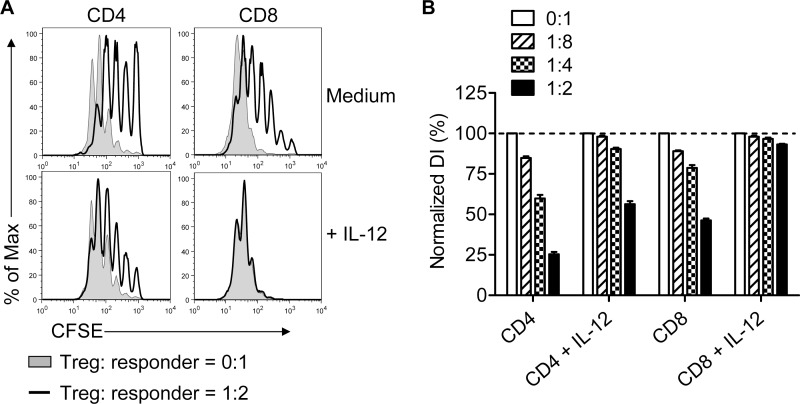
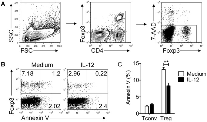
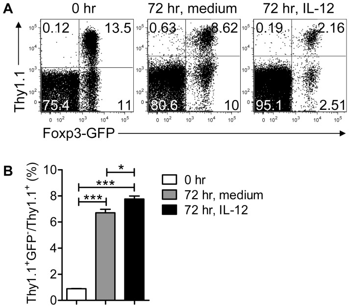
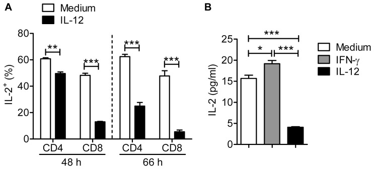
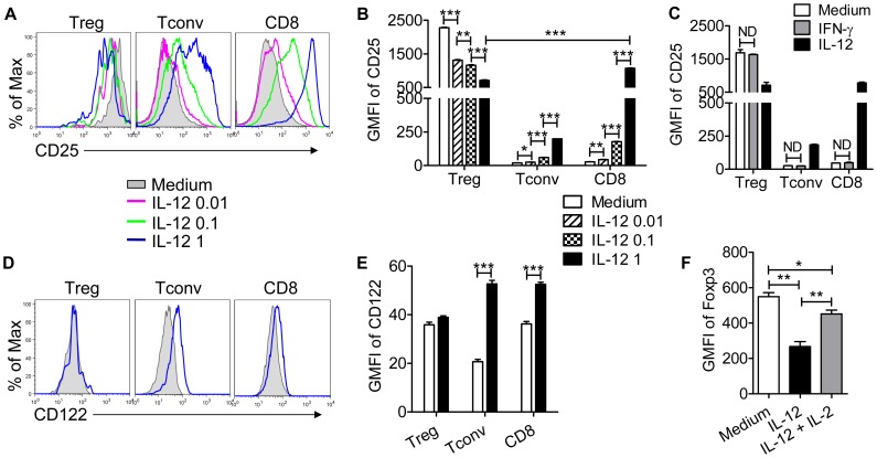
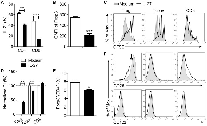
Complex interactions between effector T cells and Foxp3(+) regulatory T cells (Treg) contribute to clinical outcomes in cancer, and autoimmune and infectious diseases. Previous work showed that IL-12 reversed Treg-mediated suppression of CD4(+)Foxp3(-) T cell (Tconv) proliferation. We and others have also shown that Tregs express T-bet and IFN-γ at sites of Th1 inflammation and that IL-12 induces IFN-γ production by Tregs in vitro. To investigate whether loss of immunosuppression occurs when IFN-γ is expressed by Tregs we treated mouse lymphocyte cultures with IL-12. IFN-γ expression did not decrease the ability of Tregs to suppress Tconv proliferation. Rather, IL-12 treatment decreased Treg frequency and Foxp3 levels in Tregs. We further showed that IL-12 increased IL-2R expression on Tconv and CD8 T cells, diminished its expression on Tregs and decreased IL-2 production by Tconv and CD8 T cells. Together, these IL-12 mediated changes favored the outgrowth of non-Tregs. Additionally, we showed that treatment with a second cytokine, IL-27, decreased IL-2 expression without augmenting Tconv and CD8 T cell proliferation. Notably, IL-27 only slightly modified levels of IL-2R on non-Treg T cells. Together, these results show that IL-12 has multiple effects that modify the balance between Tregs and non-Tregs and support an important role for relative levels of IL-2R but not for IFN-γ expression in IL-12-mediated reversal of Treg immunosuppression.


Conflict of interest statement

Competing Interests: The authors have declared that no competing interests exist.

Figures

Figure 1
Figure 1. IL-12, but not IL-6 or IFN-γ induces IFN-γ expression by Foxp3+ Tregs.
Lymphocytes were prepared from lymph nodes of naïve B6 mice, labeled with CFSE and stimulated with anti-CD3 mAb and the indicated cytokine for 66 hr. Cells were then stimulated with PMA and ionomycin for 4 hr. (A) Percentage and CFSE dilution of Foxp3+ Tregs in the CD4 T cell population. (B) IFN-γ expression by Tconvs (Foxp3) or Tregs (Foxp+) after exposure to IL-12 (1 ng/ml), IL-6 (25 ng/ml) or IFN-γ (100 ng/ml). (C) IFN-γ expression by Tconvs (Foxp3) and Tregs (Foxp+) after exposure to IL-12 for 42 or 66 hr. (D) Division indices (DI) were calculated for Tconvs and Tregs and normalized to the groups treated with medium. Data are from three independent experiments. (E) Percentage of CD4 T cells expressing Foxp3 after treatment. Data were acquired from 4–9 independent experiments. (F) Geometric mean fluorescent intensity (GMFI) of Foxp3 in Tregs after indicated treatment, measured in triplicate. Data are from one experiment representative of 3 independent experiments. *P<0.05, **P<0.01, ***P<0.001 compared to cells exposed to medium, Student’s two-tailed paired t tests (D) or un-paired t tests (E, F).
Figure 2
Figure 2. IL-12 induces T-bet expression by Tconvs and Tregs.
Lymphocytes were prepared as in Figure 1 and stimulated with anti-CD3 mAb for 66 hr in the presence or absence of IL-12 or not stimulated. T-bet expression in Tconvs and Tregs was measured by flow cytometry. Data are from one experiment representative of 3 independent experiments.
Figure 3
Figure 3. TLR agonists induction of IFN-γ expression by Tconvs and Tregs is IL-12-dependent.
Purified T cells were prepared from naïve B6 and IL-12Rβ2−/− mice and mixed with splenic DCs as described in Materials and Methods. Cells were stimulated for 72 hr with anti-CD3 mAb and LPS (1 µg/ml), CpG (1 µM), IL-12 or medium. LPS and CpG induced IFN-γ expression by B6 but not IL-12Rβ2−/− Tconvs and Tregs. (A) Representative FACS plots are shown. (B) Summary data combined from three independent experiments are shown.
Figure 4
Figure 4. IFN-γ- and IFN-γ+ Tregs are equivalently suppressive.
Lymphocytes from naïve Foxp3gfp mice were treated with anti-CD3 with or without IL-12 for 66 hr. Tregs were sorted and co-cultured with CFSE-labeled Tconvs at the indicated ratios for 66 hr in the presence of irradiated splenocytes and anti-CD3 mAb. Tconvs were then analyzed for CFSE dilution. Samples were analyzed in triplicate. (A) Representative histograms are shown. (B) Normalized DI were calculated for each group. Data are from one experiment representative of 3 independent experiments.
Figure 5
Figure 5. Treg-mediated suppression is reduced in the presence of IL-12.
Tregs were isolated from naïve B6 mice and mixed with CFSE-labeled T cells at the indicated ratios. Cells were cultured for 66 hr in the presence of irradiated splenocytes and anti-CD3 mAb with or without IL-12. Samples were analyzed in triplicate. (A) Representative histograms are shown after gating on CD4 or CD8 T cells. (B) DI were calculated and normalized to samples incubated in the absence of Tregs in each group. Data are from one experiment representative of 3 independent experiments.
Figure 6
Figure 6. IL-12 does not induce apoptosis in Tconvs or Tregs.
Lymphocytes were prepared from Foxp3gfp mice and cultured in the presence of anti-CD3 mAb with or without IL-12 for 72 hr. (A) Gating strategy to identify live CD4 T cells is shown. (B) Representative FACS plots are shown. (C) Summary of data from 3 independent experiments, each performed in triplicate, is shown. ***P<0.01, Student’s two-tailed un-paired t tests.
Figure 7
Figure 7. IL-12 does not induce substantial Treg conversion.
Tregs were sorted from Foxp3gfp/Thy1.1 mice and mixed with lymphocytes harvested from naïve Foxp3gfp mice (Thy1.2). Samples were incubated for 72 hr with anti-CD3 mAb in the presence or absence of IL-12. (A) Representative FACS plots are shown after gating on CD4 T cells. (B) Summary of data showing GFP- fraction of Thy1.1+ cells. Data are from 3 independent experiments, each performed in triplicate. *P<0.05, ***P<0.001, Student’s two-tailed un-paired t tests.
Figure 8
Figure 8. IL-12 inhibits IL-2 expression by T cells.
Lymphocytes were prepared from naïve Foxp3gfp mice and treated with anti-CD3 mAb and IL-12 or IFN-γ. (A). The fraction of CD4 and CD8 T cells expressing IL-2 was diminished after 48 and 66 hr. Data are from 3–5 independent experiments. (B) Levels of IL-2, measured by ELISA, were reduced in IL-12 but not IFN-γ-treated cultures at 48 hr. Data are representative of 2 independent experiments performed in triplicate for stimulation, and each sample plated in duplicate for ELISA. *P<0.05, ***P<0.001, Student’s two-tailed un-paired t tests.
Figure 9
Figure 9. IL-12 has opposite effects on IL-2R expression by Tregs vs non-Treg T cells.
Lymphocytes were prepared from naïve Foxp3gfp mice and treated with anti-CD3 mAb and IL-12 or IFN-γ for 66 hr. (A, B) CD25 expression was decreased on Tregs and increased on Tconvs and CD8 T cells by IL-12 in a dose-dependent fashion. Representative histograms are shown in (A) and summary data are shown in (B). (C) IFN-γ did not affect CD25 expression by Tregs and non-Treg T cells (ND-no difference). (D, E) IL-12 increased CD122 expression on Tconvs and CD8 T cells but not on Tregs. Representative histograms are shown in (D) and data are summarized in (E). (F) IL-12 diminished Foxp3 GMFI in Tregs and this was largely reversed by IL-2 (100 U/ml) treatment. Data are representative of three independent experiments performed in triplicate. *P<0.05, **P<0.01, ***P<0.001, Student’s two-tailed un-paired t tests.
Figure 10
Figure 10. IL-27 inhibits IL-2 production, but only modestly affects IL-2R expression by Tconv and CD8 T cells.
Lymphocytes were prepared from naïve B6 mice and treated with anti-CD3 mAb and IL-27 (100 ng/ml) for 66 hr. (A) The fraction of CD4 and CD8 T cells expressing IL-2 was diminished. Data are from 3 independent experiments. (B) IL-27 diminished Foxp3 GMFI in Tregs. Samples were analyzed in triplicate. Data are from one experiment representative of 3 independent experiments. (C, D, E) Lymphocytes were labeled with CFSE prior to incubation. IL-27 reduced proliferation of Tregs and Tconvs but not CD8 T cells (C, D), resulting in a modest decrease in the percentage of Foxp3+ Tregs (E). Data are from 3 independent experiments. (F) IL-27 treatment resulted in small changes in CD25 expression on Tregs and CD8 T cells but not Tconvs and in a small decrease in CD122 expression on all three cell types. Data are representative of 3 independent experiments. *P<0.05, **P<0.01, ***P<0.001, Student’s two-tailed un-paired t tests (A, B, E) or paired t tests (D).

References

    1. Vignali DA, Collison LW, Workman CJ (2008) How regulatory T cells work. Nat Rev Immunol 8: 523–532. - PMC - PubMed
    1. Belkaid Y (2007) Regulatory T cells and infection: a dangerous necessity. Nat Rev Immunol 7: 875–888. - PubMed
    1. Wing K, Sakaguchi S (2010) Regulatory T cells exert checks and balances on self tolerance and autoimmunity. Nat Immunol 11: 7–13. - PubMed
    1. Curiel TJ, Coukos G, Zou L, Alvarez X, Cheng P, et al. (2004) Specific recruitment of regulatory T cells in ovarian carcinoma fosters immune privilege and predicts reduced survival. Nat Med 10: 942–949. - PubMed
    1. Oldenhove G, Bouladoux N, Wohlfert EA, Hall JA, Chou D, et al. (2009) Decrease of Foxp3+ Treg cell number and acquisition of effector cell phenotype during lethal infection. Immunity 31: 772–786. - PMC - PubMed

Publication types

MeSH terms