. 2012;7(9):e46399.
doi: 10.1371/journal.pone.0046399. Epub 2012 Sep 27.

Lactobacillus reuteri maintains a functional mucosal barrier during DSS treatment despite mucus layer dysfunction

Affiliations

Lactobacillus reuteri maintains a functional mucosal barrier during DSS treatment despite mucus layer dysfunction

Johan Dicksved et al. PLoS One. 2012.

Abstract

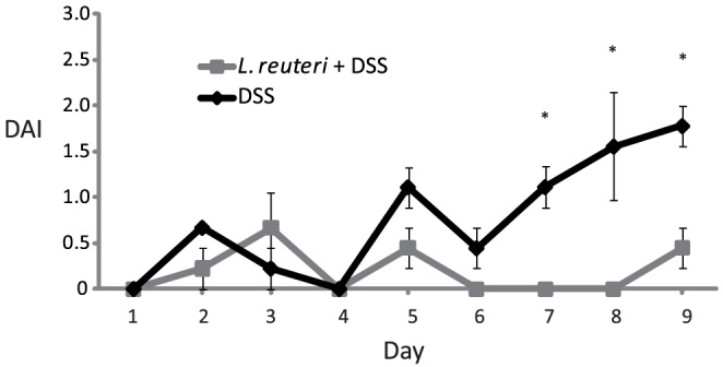
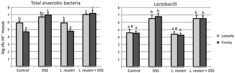
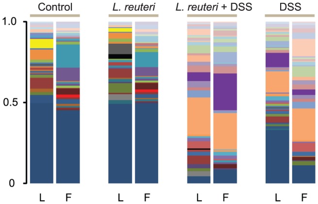
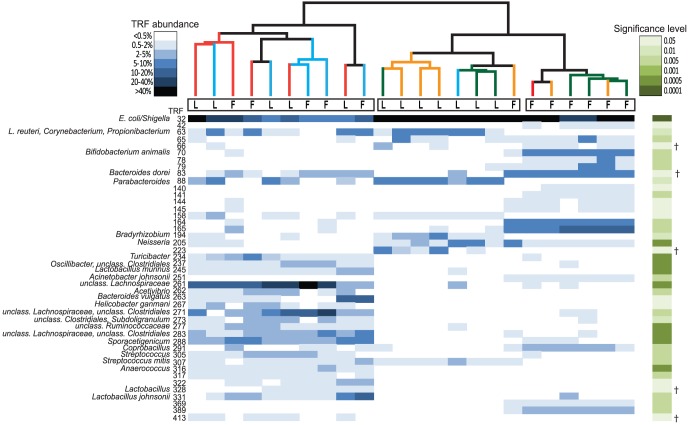
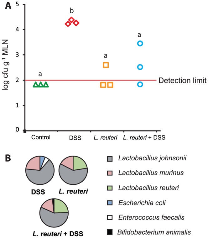
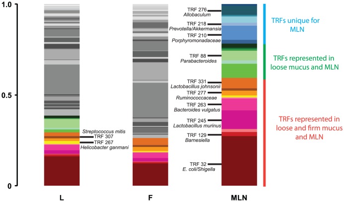
Treatment with the probiotic bacterium Lactobacillus reuteri has been shown to prevent dextran sodium sulfate (DSS)-induced colitis in rats. This is partly due to reduced P-selectin-dependent leukocyte- and platelet-endothelial cell interactions, however, the mechanism behind this protective effect is still unknown. In the present study a combination of culture dependent and molecular based T-RFLP profiling was used to investigate the influence of L. reuteri on the colonic mucosal barrier of DSS treated rats. It was first demonstrated that the two colonic mucus layers of control animals had different bacterial community composition and that fewer bacteria resided in the firmly adherent layer. During DSS induced colitis, the number of bacteria in the inner firmly adherent mucus layer increased and bacterial composition of the two layers no longer differed. In addition, induction of colitis dramatically altered the microbial composition in both firmly and loosely adherent mucus layers. Despite protecting against colitis, treatment with L. reuteri did not improve the integrity of the mucus layer or prevent distortion of the mucus microbiota caused by DSS. However, L. reuteri decreased the bacterial translocation from the intestine to mesenteric lymph nodes during DSS treatment, which might be an important part of the mechanisms by which L. reuteri ameliorates DSS induced colitis.


Conflict of interest statement

Competing Interests: The authors have declared that no competing interests exist.

Figures

Figure 1
Figure 1. Disease activity index in animals treated with DSS alone or Lactobacillus reuteri+DSS.
In the first group, DSS were given in the drinking water for 9 days. In the second, 109 L. reuteri/day were given by gavage for 16 days and DSS were given for the last 9 days of the treatment. *p<0.05 DSS vs. L. reuteri+DSS.
Figure 2
Figure 2. Numbers of total anaerobic bacteria and lactobacilli determined by cultivation.
For each treatment group (controls, DSS, L. reuteri, L. reuteri+DSS), data are shown for both loosely and firmly adherent colonic mucus. Different letters (a–c) above the bars indicate significant differences in bacterial counts between groups or mucus layers (p<0.05). # Lactobacilli were only detected in two of the three samples.
Figure 3
Figure 3. Profiling of the microbial structure in colonic mucus layers by T-RFLP.
The columns show representative profiles from the loose (L) and firm (F) colonic mucus layers and treatment groups (control, L. reuteri, L. reuteri+DSS and DSS).
Figure 4
Figure 4. Clustering of samples according to its microbial structure.
The heatmap shows the relative abundances of TRFs that differed significantly between clusters. The base pair (bp) sizes are indicated in the figure. Each column represents the abundance profile for a sample and each row the abundance profile for a TRF. The abundance interval is shown in a blue color scale and significance levels in a green color scale. †Indicates TRFs that were identified in false discovery rate analysis. Samples were sorted according to clustering using Bray Curtis metrics. Branch color represents: red, DSS; blue, L. reuteri+DSS; green, control; and yellow, L. reuteri. T-RFLP data of loosely adherent mucus (L) and firmly adherent mucus (F) showing one cluster of control and L. reuteri firm mucus samples (right cluster), one cluster of control and L. reuteri loose mucus samples (middle cluster) and one cluster of DSS-treated animals including a mix of both loosely and firmly mucus (left cluster).
Figure 5
Figure 5. L. reuteri reduces DSS associated translocation.
A) Culture data (log cfu g−1 mesenteric lymph node) from mesenteric lymph nodes from the different treatment groups. B) The identity of the dominant culturable bacteria that translocated. Different letters (a–b) above the bars indicate significant differences in bacterial counts between groups (p<0.05). ANOVA followed by Tukey's post hoc test was used for the statistical analysis.
Figure 6
Figure 6. Consensus T-RFLP profiles from different sampling sites in the DSS treated animals.
TRFs colored in a red color scale were found in loose (L), firm (F) mucus and the mesenteric lymph nodes (MLN), TRFs colored in a green color scale were found in loose mucus and MLN, TRFs colored in a blue scale were unique for the MLN and TRFs colored in grey were only found in mucus.

References

    1. Packey CD, Sartor RB (2009) Commensal bacteria, traditional and opportunistic pathogens, dysbiosis and bacterial killing in inflammatory bowel diseases. Curr Opin Infect Dis 22: 292–301. - PMC - PubMed
    1. Atuma C, Strugala V, Allen A, Holm L (2001) The adherent gastrointestinal mucus gel layer: thickness and physical state in vivo. Am J Physiol Gastrointest Liver Physiol 280: G922–929. - PubMed
    1. Petersson J, Schreiber O, Hansson GC, Gendler SJ, Velcich A, et al. (2011) Importance and regulation of the colonic mucus barrier in a mouse model of colitis. Am J Physiol Gastrointest Liver Physiol 300: G327–333. - PMC - PubMed
    1. Johansson ME, Phillipson M, Petersson J, Velcich A, Holm L, et al. (2008) The inner of the two Muc2 mucin-dependent mucus layers in colon is devoid of bacteria. Proc Natl Acad Sci U S A 105: 15064–15069. - PMC - PubMed
    1. Jager S, Stange EF, Wehkamp J (2010) Antimicrobial peptides in gastrointestinal inflammation. Int J Inflam 2010: 910283. - PMC - PubMed

Publication types

MeSH terms

LinkOut - more resources