. 2012;7(9):e46453.
doi: 10.1371/journal.pone.0046453. Epub 2012 Sep 28.

The directed differentiation of human iPS cells into kidney podocytes

Affiliations

The directed differentiation of human iPS cells into kidney podocytes

Bi Song et al. PLoS One. 2012.

Abstract

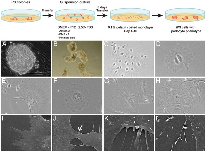
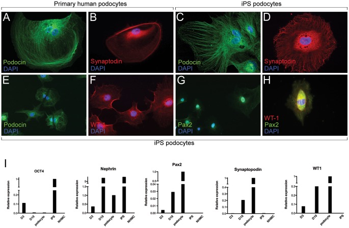
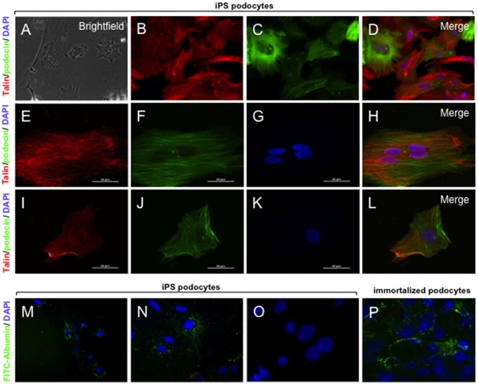
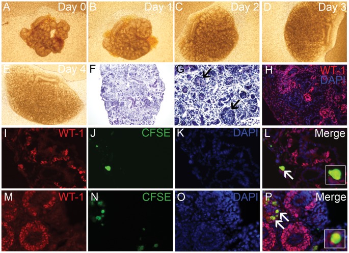
The loss of glomerular podocytes is a key event in the progression of chronic kidney disease resulting in proteinuria and declining function. Podocytes are slow cycling cells that are considered terminally differentiated. Here we provide the first report of the directed differentiation of induced pluripotent stem (iPS) cells to generate kidney cells with podocyte features. The iPS-derived podocytes share a morphological phenotype analogous with cultured human podocytes. Following 10 days of directed differentiation, iPS podocytes had an up-regulated expression of mRNA and protein localization for podocyte markers including synaptopodin, nephrin and Wilm's tumour protein (WT1), combined with a down-regulation of the stem cell marker OCT3/4. In contrast to human podocytes that become quiescent in culture, iPS-derived cells maintain a proliferative capacity suggestive of a more immature phenotype. The transduction of iPS podocytes with fluorescent labeled-talin that were immunostained with podocin showed a cytoplasmic contractile response to angiotensin II (AII). A permeability assay provided functional evidence of albumin uptake in the cytoplasm of iPS podocytes comparable to human podocytes. Moreover, labeled iPS-derived podocytes were found to integrate into reaggregated metanephric kidney explants where they incorporated into developing glomeruli and co-expressed WT1. This study establishes the differentiation of iPS cells to kidney podocytes that will be useful for screening new treatments, understanding podocyte pathogenesis, and offering possibilities for regenerative medicine.


Conflict of interest statement

Competing Interests: The authors have declared that no competing interests exist.

Figures

Figure 1
Figure 1. Podocyte Progenitors from iPS cells.
Diagrammatic depiction of the directed differentiation of iPS cells to podocyte progenitors (panel). A) iPS colonies derived from human kidney cells as assessed by phase contrast microscopy were B) mechanically dissociated and grown in suspension culture. C) By phase contrast microscopy the cell clusters attached and propagated in differentiation media devoid of a feeder layer. D) Following serial dilution into single cell cultures and 3 days of directed differentiation the cells appeared relatively unspecialised and immature with a rounded and often binucleated phenotype. E) At day 10 of differentiation the iPS-derived cells had cytoplasmic extensions with an arborized appearance resembling podocytes. F) The iPS podocytes were compared to cultured immortalised human podocytes that propagate at a permissive temperature of 33°C. G) At 37°C the immortalised podocytes enter growth arrest and display a typical arborized appearance. H) Primary cultures of human podocytes also contained large multinucleated cells displaying foot process extensions. I) and J) Scanning electron microscopy of iPS podocytes demonstrated a main cell body with cytoplasmic projections and tight junction-like structures connecting adjacent cells (arrow). K) Cytoplasmic projections could be observed extending from the cell body of the iPS podocyte. L) Tertiary projections were also common at the end processes of the cytoplasmic projections. Magnification A,B ×200; C,E,G,H ×100; D,F ×400; SEM I ×1.1K, J ×3.5K, K × 8K, L ×2.5K.
Figure 2
Figure 2. iPS Podocyte Characterisation and Gene Expression.
A) Immunofluorescence microscopy showed localisation of podocin (green) in human podocytes, counterstained with DAPI. B) Human podocytes localised with synaptopodin protein (red) throughout the cytoplasm surrounding the DAPI-stained nucleus (blue). C) At 10 days of directed differentiation iPS podocytes show localisation of podocin protein, counterstained with DAPI. D) Synaptopodin (red) was expressed in the cytoplasmic matrix of iPS podocytes. E) Unlike human podocytes, iPS podocytes proliferated in culture as observed by podocin expression and DAPI (blue) and are also WT1-positive (F). Differentiated iPS cells express Pax-2 protein (G) and show nuclear co-localisation of Pax-2 and WT1 proteins (H). Mag A–D, H ×400; E–G ×200. I) qPCR showed the upregulated mRNA expression of kidney metanephric mesenchymal and podocyte genes. From day 3 (D3) to day 10 (D10) of iPS podocyte directed differentiation there was an upregulated expression of Pax2 and WT1 and podocyte-specific markers synaptopodin and nephrin at comparable levels to primary human podocytes, but different from the starting normal human mesangial cells (NHMC).
Figure 3
Figure 3. Functional contractility and permeability.
Live cell imaging was used to record the response of iPS podocytes to the addition of AII (See Movie S1). A) Phase contrast imaging of iPS podocytes following the addition of AII at time 0. The iPS podocytes were transduced with RFP-actin (B) and immunostained with the contractile protein, podocin (C). D) Merge image of actin (red) and podocin (green) with DAPI-stained nuclei (blue). E) Confocal immunofluorescence shows that iPS-podocytes transduced with RFP-talin (E) co-expressed podocin (F) at time 0. G) DAPI-stained nuclei and merged image are shown (H). After 6 hours in culture, the iPS podocytes were viable and display a contracted morphology in response to AII (I–L) where RFP-talin (red), podocin (green) and DAPI (blue) are shown. (M-N) By fluorescence microscopy, the iPS podocytes were able to uptake FITC-albumin (green) into the cytoplasm when cultured at 37°C, compared to iPS podocytes cultured at 4°C (O) that served as a control. (P) Immortalized human podocytes also showed endocytosis of FITC-albumin in a similar morphological pattern. Mag A–D ×200; E–L, N-P ×1000; M ×100. Abbreviations: angiotensin II (AII); red fluorescent protein (RFP).
Figure 4
Figure 4. iPS podocyte integration into embryonic kidneys.
A–E) Bright field microscopy showing representative E13.5 mouse embryonic kidneys (n = 3) reaggregated with CFSE-labeled iPS podocytes and grown as an explant over 4 days in culture. Histology showed that the dissociated kidneys explants successfully reaggregated (F) that displayed definitive features including developing glomeruli (G; arrows) and tubular segments. H) Immunofluorescence microscopy showed WT1 (red) cells in glomerular aggregates with DAPI-stained nuclei (blue). I-L) Using immunofluorescence of WT1 protein (I; red), a co-localised expression was observed in CFSE-labeled iPS podocytes (J; green) counterstained with DAPI (K). The iPS cells were observed within glomerular aggregates of E13.5 kidney explants (Merge; L). M-P) E15.5 explants immunostained with WT1 (M; red) and CFSE-labeled iPS podocytes (N; green) counterstained with DAPI (O) showed further integration into WT1-positive glomerular structures (Merge; P). Counterstaining with DAPI (O). Mag A–E ×4; F ×100; G ×200; H ×200; I–P × 400.

References

    1. Wiggins RC (2007) The spectrum of podocytopathies: a unifying view of glomerular diseases. Kidney Int 71: 1205–1214. - PubMed
    1. Sato Y, Wharram BL, Lee SK, Wickman L, Goyal M, et al. (2009) Urine podocyte mRNAs mark progression of renal disease. Journal of the American Society of Nephrology : JASN 20: 1041–1052. - PMC - PubMed
    1. Abbate M, Zoja C, Remuzzi G (2006) How does proteinuria cause progressive renal damage? J Am Soc Nephrol 17: 2974–2984. - PubMed
    1. Mundel P, Shankland SJ (2002) Podocyte biology and response to injury. J Am Soc Nephrol 13: 3005–3015. - PubMed
    1. Humphreys BD, Bonventre JV (2007) The contribution of adult stem cells to renal repair. Nephrol Ther 3: 3–10. - PubMed

Publication types

MeSH terms