. 2012;7(9):e46479.
doi: 10.1371/journal.pone.0046479. Epub 2012 Sep 28.

Postconditioning with inhaled carbon monoxide counteracts apoptosis and neuroinflammation in the ischemic rat retina

Affiliations

Postconditioning with inhaled carbon monoxide counteracts apoptosis and neuroinflammation in the ischemic rat retina

Nils Schallner et al. PLoS One. 2012.

Abstract

Purpose: Ischemia and reperfusion injury (I/R) of neuronal structures and organs is associated with increased morbidity and mortality due to neuronal cell death. We hypothesized that inhalation of carbon monoxide (CO) after I/R injury ('postconditioning') would protect retinal ganglion cells (RGC).

Methods: Retinal I/R injury was performed in Sprague-Dawley rats (n = 8) by increasing ocular pressure (120 mmHg, 1 h). Rats inhaled room air or CO (250 ppm) for 1 h immediately following ischemia or with 1.5 and 3 h latency. Retinal tissue was harvested to analyze Bcl-2, Bax, Caspase-3, HO-1 expression and phosphorylation of the nuclear transcription factor (NF)-κB, p38 and ERK-1/2 MAPK. NF-κB activation was determined and inhibition of ERK-1/2 was performed using PD98059 (2 mg/kg). Densities of fluorogold prelabeled RGC were analyzed 7 days after injury. Microglia, macrophage and Müller cell activation and proliferation were evaluated by Iba-1, GFAP and Ki-67 staining.

Results: Inhalation of CO after I/R inhibited Bax and Caspase-3 expression (Bax: 1.9 ± 0.3 vs. 1.4 ± 0.2, p = 0.028; caspase-3: 2.0 ± 0.2 vs. 1.5 ± 0.1, p = 0.007; mean ± S.D., fold induction at 12 h), while expression of Bcl-2 was induced (1.2 ± 0.2 vs. 1.6 ± 0.2, p = 0.001; mean ± S.D., fold induction at 12 h). CO postconditioning suppressed retinal p38 phosphorylation (p = 0.023 at 24 h) and induced the phosphorylation of ERK-1/2 (p<0.001 at 24 h). CO postconditioning inhibited the expression of HO-1. The activation of NF-κB, microglia and Müller cells was potently inhibited by CO as well as immigration of proliferative microglia and macrophages into the retina. CO protected I/R-injured RGC with a therapeutic window at least up to 3 h (n = 8; RGC/mm(2); mean ± S.D.: 1255 ± 327 I/R only vs. 1956 ± 157 immediate CO treatment, vs. 1830 ± 109 1.5 h time lag and vs. 1626 ± 122 3 h time lag; p<0.001). Inhibition of ERK-1/2 did not counteract the CO effects (RGC/mm(2): 1956 ± 157 vs. 1931 ± 124, mean ± S.D., p = 0.799).

Conclusion: Inhaled CO, administered after retinal ischemic injury, protects RGC through its strong anti-apoptotic and anti-inflammatory effects.


Conflict of interest statement

Competing Interests: The authors have declared that no competing interests exist.

Figures

Figure 1
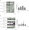
Figure 1. Effects of carbon monoxide postconditioning on retinal Bax and Bcl-2 mRNA and protein expression.
(A) Fold induction of Bax mRNA expression in ischemic retinal tissue compared to GAPDH in relation to the corresponding non-ischemic retinae analyzed by RT-PCR (n = 8 per group; mean±SD; * p = 0.028, 0.024 and 0.016 I/R vs. I/R+CO at 12, 48 and 72 h). (B) Representative Western blot images (of n = 4) analyzing the suppression of retinal Bax protein expression by carbon monoxide postconditioning. Densitometric analysis of n = 4 western blots (mean±SD; * p = 0.028, <0.001 and <0.001 I/R vs. I/R+CO at 12, 24 and 48 h). (C) Retinal expression of Bcl-2 mRNA (n = 8 per group; mean±SD; * p = 0.001, 0.011 and 0.038 I/R vs. I/R+CO at 12, 24 and 48 h). (D) Representative Western blot images (of n = 4) analyzing the induction of retinal Bcl-2 protein expression by carbon monoxide postconditioning. Densitometric analysis of n = 4 western blots (mean±SD; * p<0.001 I/R vs. I/R+CO at 48 h).
Figure 2
Figure 2. Effect of carbon monoxide postconditioning on retinal expression of caspase-3 mRNA and caspase-3 cleavage.
(A) Fold induction of caspase-3 mRNA expression in ischemic retinal tissue compared to GAPDH in relation to the corresponding non-ischemic retinae analyzed by RT-PCR (n = 8 per group; mean±SD; * p = 0.007, 0.002, <0.001 and 0.006 I/R vs. I/R+CO at 12, 24, 48 and 72 h). (B) Representative Western blot images (of n = 4) analyzing the suppression of retinal cleavage of caspase-3 protein by carbon monoxide postconditioning. Densitometric analysis of n = 4 western blots (mean±SD; p = 0.042 and 0.028 I/R vs. I/R+CO at 24 and 48 h).
Figure 3
Figure 3. Effects of carbon monoxide postconditioning, inhibiting p38 MAPK while activating ERK-1/2 MAPK phosphorylation.
(A) Representative Western blot images (of n = 4) analyzing the influence of carbon monoxide postconditioning on the phosphorylation of retinal p38 MAPK. Densitometric analysis of n = 4 western blots (mean±SD; * p = 0.023 and <0.001 I/R vs. I/R+CO at 24 and 48 h). (B) Representative Western blot images (of n = 4) analyzing the influence of carbon monoxide postconditioning on the phosphorylation of retinal ERK-1/2 MAPK. Densitometric analysis of n = 4 western blots (mean±SD; * p<0.001 I/R vs. I/R+CO at 24 and 48 h).
Figure 4
Figure 4. Effect of carbon monoxide postconditioning on phosphorylation of ERK-1/2 in Thy-1 positive RGC.
Representative images of immunhistochemical staining against p-ERK-1/2 and Thy-1 in the retina, showing increased phosphorylation of ERK-1/2 in the GCL after I/R and I/R+CO. Only after I/R+CO, ERK-1/2 is phosphorylated in the RGC (white arrows: Thy-1 positive RGC), whereas after I/R alone, p-ERK-1/2 is evident mainly in other cells of the GCL (*: p-ERK-1/2 positive cells). Scale bar 100 µm and 50 µm (in “Detail GCL” pictures). Abbreviations: GCL = ganglion cell layer, INL = inner nuclear layer, ONL = outer nuclear layer, DAPI = 4′,6-diamidino-2-phenylindole.
Figure 5
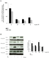
Figure 5. Effect of carbon monoxide postconditioning on retinal expression of HO-1 mRNA and HO-1 protein.
(A) Fold induction of HO-1 mRNA expression in ischemic retinal tissue compared to GAPDH in relation to the corresponding non-ischemic retinae analyzed by RT-PCR (n = 8 per group; mean±SD; * p<0.001 I/R vs. I/R+CO at 12, logarithmic scale). (B) Representative Western blot images (of n = 4) analyzing the suppression of retinal HO-1 protein expression by carbon monoxide postconditioning. Densitometric analysis of n = 4 western blots (mean±SD; * p<0.001 I/R vs. I/R+CO at 12 and 48 h).
Figure 6
Figure 6. Effects of carbon monoxide postconditioning on NF-κB protein expression, phosphorylation and NF-κB DNA-binding.
(A) Representative Western blot images (of n = 4) analyzing the influence of carbon monoxide postconditioning on expression and phosphorylation of retinal NF-κB p65. Densitometric analysis of n = 4 western blots (mean±SD; * p<0.001 I/R vs. I/R+CO at 24 h). (B) Representative EMSA (of n = 4) of NF-κB DNA binding. Lanes 1–16: individual experiments at 12, 24, 48 and 72 hours after carbon monoxide postconditioning, lanes 17 and 18: supershift analysis shows specificity of NF-κB, lane 19: self competition with unlabeled NF-κB, lane 20: non-self competition with unlabeled AP-1, lane 21: positive control, achieved by induction of SY5Y cell line exposed to PMA/Ionomycin. Densitometric analysis of n = 4 EMSA (mean±SD; * p = 0.016 I/R vs. I/R+CO at 48 h). (Abbreviations: NF-κB Ab = nuclear factor κB antibody, c-fos Ab = c-fos antibody, AP-1 = activator protein 1).
Figure 7
Figure 7. Effect of carbon monoxide postconditioning on glial cell activation in the retina.
Representative immunohistochemical GFAP (1st row; Müller cells, “macroglia”) and Iba-1 (2nd row; microglia, macrophages) staining in I/R injured eyes with (right column) or without (left column) CO postconditioning, indicating reduced glial cell activation in the CO-treated retina. Scale bar: 100 µm and 50 µm (in detail picture). Abbreviations: GFAP = Glial fibrillary acidic protein, DAPI = 4′,6-diamidino-2-phenylindole, Iba-1 = ionized calcium binding adaptor molecule 1.
Figure 8
Figure 8. Effect of carbon monoxide postconditioning on immigration of proliferating cells into the retina.
Representative immunohistochemical Ki-67 (1st row, dual staining 3rd to 5th row; proliferation marker), Iba-1 (2nd and 3rd row; microglia/macrophage marker), GFAP (4th row; glial cell marker) and p-ERK-1/2 (5th row) staining in I/R injured eyes with (right column) or without (left column) CO postconditioning. White box = Ki-67/Iba-1 positive microglia; White arrows = Ki67/Iba-1 positive macrophages; White * = absent colocalization of GFAP and Ki-67. Abbreviations: GFAP = Glial fibrillary acidic protein, Iba-1 = ionized calcium binding adaptor molecule 1.
Figure 9
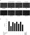
Figure 9. Effect of carbon monoxide postconditioning on ischemia reperfusion (I/R) injury in RGC.
(A) Representative images (of n = 8) from flat mounts with flourogold-labeled RGC 7 days after I/R injury and CO treatment immediately, 1.5 h and 3 h after initiation of reperfusion. (B) Quantification of retinal ganglion cell density [cells/mm2] 7 days after I/R injury (n = 8 per group; mean±S.D.; * p<0.001 I/R vs. I/R+CO immediate, vs. I/R+CO 1.5 h and vs. I/R+CO 3 h; IR+CO immediate vs. I/R+CO 3 h).
Figure 10
Figure 10. Diagram depicting the proposed mechanism of CO-mediated protective effects on RGC after I/R injury.
CO postconditioning after retinal I/R strongly inhibits the inflammatory response by suppressing NF-κB activation, abolishing the infiltration of proliferating microglia and macrophages and inhibiting the activation of Müller cells. Inflammatory oxidative cellular stress is reduced, indicated by inhibition of HO-1 expression. CO attenuates RGC apoptosis as indicated by reduced Caspase-3 activity and Bax expression and by increased Bcl-2 expression. Apoptosis- and inflammation-related MAPK pathways are regulated in favor of anti-apoptosis and anti-inflammation. Overall, these anti-inflammatory and anti-apoptotic effects lead to higher RGC survival and neuroprotection (→ = activation; ⊣ = inhibition).

References

    1. Wong GY, Warner DO, Schroeder DR, Offord KP, Warner MA, et al. (2000) Risk of surgery and anesthesia for ischemic stroke. Anesthesiology 92: 425–432. - PubMed
    1. Mashour GA, Shanks AM, Kheterpal S (2011) Perioperative stroke and associated mortality after noncardiac, nonneurologic surgery. Anesthesiology 114: 1289–1296. - PubMed
    1. O'Collins VE, Macleod MR, Donnan GA, Horky LL, van der Worp BH, et al. (2006) 1,026 experimental treatments in acute stroke. Ann Neurol 59: 467–477. - PubMed
    1. Otterbein LE, Bach FH, Alam J, Soares M, Tao Lu H, et al. (2000) Carbon monoxide has anti-inflammatory effects involving the mitogen-activated protein kinase pathway. Nat Med 6: 422–428. - PubMed
    1. Zhang X, Shan P, Otterbein LE, Alam J, Flavell RA, et al. (2003) Carbon monoxide inhibition of apoptosis during ischemia-reperfusion lung injury is dependent on the p38 mitogen-activated protein kinase pathway and involves caspase 3. J Biol Chem 278: 1248–1258. - PubMed

Publication types

MeSH terms