

Functional neural correlates of emotional expression processing deficits in behavioural variant frontotemporal dementia
- PMID: 23031250
- PMCID: PMC3633710
- DOI: 10.1503/jpn.120008
Functional neural correlates of emotional expression processing deficits in behavioural variant frontotemporal dementia
Abstract
Background: Frontotemporal dementia (FTD) is a neurodegenerative disorder resulting in social-cognitive deficits partially attributed to abnormalities processing social cues, such as facial expressions. However, to our knowledge, the functional neuroanatomy of deficient social cue processing in individuals with FTD has not been examined. The objective of this study was to delineate the functional abnormalities under lying altered facial expression processing in individuals with FTD using functional magnetic resonance imaging (fMRI).
Methods: Patients meeting Neary criteria for behavioural variant FTD (bvFTD) with supportive neuroimaging and 18 age-matched healthy controls completed an implicit facial expression task during fMRI. We conducted volumetric brain morphometry to correct functional imaging data for volume differences.
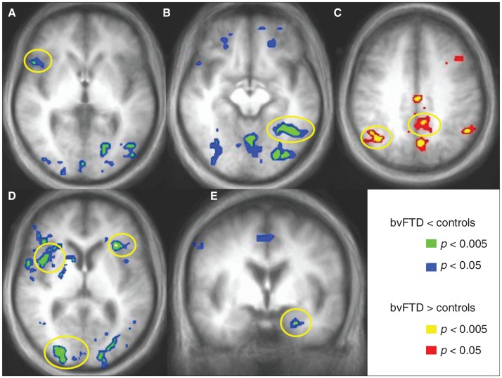
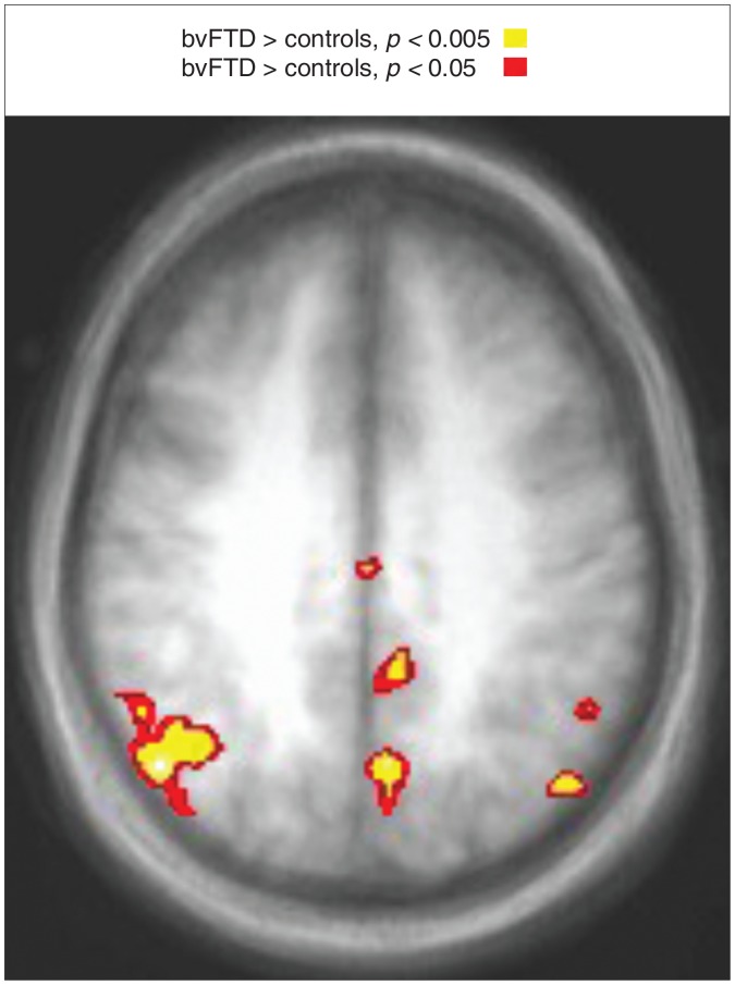
Results: We included 20 patients with bvFTD and 18 controls in our study. The results demonstrate emotion-specific functional abnormalities in frontal and limbic regions in patients with bvFTD. Patients also showed decreased activity in posterior ventral visual regions, specifically the fusiform cortex, possibly reflecting reduced afferent input from limbic regions. Finally, bvFTD was associated with increased activity in posterior regions, including the inferior parietal cortex.
Limitations: Autopsy validation of frontotemporal dementia is not yet available for this cohort.
Conclusion: Together, these findings suggest that fMRI combined with tasks targeting social-cognitive deficits is a powerful technique to objectively measure neural systems involved in emotion processing in individuals with bvFTD. As viewing emotional expressions is known to engage many of the same neural systems that are active when experiencing the emotion itself, fMRI during expression processing provides a novel window into the emotions of patients with FTD.
Figures

References
-
- Rankin KP, Kramer JH, Miller BL. Patterns of cognitive and emotional empathy in frontotemporal lobar degeneration. Cogn Behav Neurol. 2005;18:28–36. - PubMed
-
- Darwin C. The expression of the emotions in man and animals. London: Albemarle; 1872.
-
- Blair RJ, Cipolotti L. Impaired social response reversal. A case of ‘acquired sociopathy’. Brain. 2000;123:1122–41. - PubMed
MeSH terms
LinkOut - more resources
Full Text Sources
Other Literature Sources
Research Materials