. 2013 Apr;21(4):367-72.
doi: 10.1038/ejhg.2012.198. Epub 2012 Oct 3.

Genome-wide linkage analysis is a powerful prenatal diagnostic tool in families with unknown genetic defects

Affiliations

Genome-wide linkage analysis is a powerful prenatal diagnostic tool in families with unknown genetic defects

Maria Arélin et al. Eur J Hum Genet. 2013 Apr.

Abstract

Genome-wide linkage analysis is an established tool to map inherited diseases. To our knowledge it has not been used in prenatal diagnostics of any genetic disorder. We present a family with a severe recessive mental retardation syndrome, where the mother wished pregnancy termination to avoid delivering another affected child. By genome-wide scanning using the Affymetrix (Santa Clara, CA, USA) 10k chip we were able to establish the disease haplotype. Without knowing the exact genetic defect, we excluded the condition in the fetus. The woman finally gave birth to a healthy baby. We suggest that genome-wide linkage analysis--based on either SNP mapping or full-genome sequencing--is a very useful tool in prenatal diagnostics of diseases.


Figures
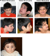
Figure 1
Figure 1
Facial appearance of the patients and their healthy brother who was born subsequent to prenatal gene mapping. (a) All the three affected siblings (784, 787 and 788) showed slightly downward-slanted palpebral fissures, heavy eyebrows, a prominent root of the nose, a short philtrum, an unusual preauricular hairline, posteriorly rotated ears and a prominent lower lip; in two of the patients (784 and 788) divergent strabismus was diagnosed. (b) After the final result of our indirect prenatal analysis, the parents decided to continue the pregnancy and the mother gave birth to a healthy baby. The boy started walking and talking at the age of 16 months, and until now his motor and mental development is normal.
Figure 2
Figure 2
Prenatal genome-wide linkage scan with subsequent finemapping and haplotype analyses excluded an affection of the fetus. (a) Initial LOD score after the genome-wide SNP mapping with linkage to chromosome 1p (LOD score of 2.56). The initial mapping included the parents (individuals 782 and 783), as well as their affected and unaffected children (784, 785, 786, 787 and 788). Subsequently, we analyzed the fetus (C052) at week 12 of gestation. (b) Final LOD score after additional finemapping of microsatellite markers, including the core family as well as one affected and one unaffected cousin from a second family branch (individuals 13389 and 9408), resulting in a significant LOD score of 3.98. (c) Haplotypes after final mapping results. The disease-linked region shown here is located on chromosome 1p36.12. The affected children (784, 787, 788 and 9408) inherited two disease-associated dark–green haplotypes from their parents. The risk region for the fetus (C052) is narrowed down to a 120 717-bp interval, which contains three exons (plus one alternative exon 1) of the EPHB2 gene. We sequenced all coding exons and did not find any mutations. Thus, an affection of the fetus was extremely unlikely. The remaining critical region of homozygosity between markers D1S2826 and rs1961413 in all the affected individuals (784, 787, 788 9408) contains 58 candidate genes (markers that were tested for this region are listed in order from the p-terminal end of the chromosome (NCBI build 36.1)). Haplotypes for all tested genetic markers and SNPs are shown in columns beneath family members who underwent linkage analysis. The disease-associated haplotype is marked in dark-green, haplotypes marked in blue or light orange are not disease-linked. Non-informative segments are marked in gray (non-informativity results from recombination events in regions where the parents are homozygous for the marker allele).
Figure 3
Figure 3
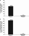
Development of the risk region in the fetus and the affected individuals throughout the different steps of the prenatal diagnosis. The comparison of the risk region for the fetus concerning cM at risk (a) and coding genes at risk (b) in phase 2 and 3 of the prenatal diagnosis compared with the affected region is shown here. Especially in the situation of a prenatal genetic counseling, this figure could help to demonstrate the power of mapping and finemapping results, and the possible reduction of the risk for the fetus from the initial 25% risk in phase 1 (autosomal recessive inheritance) to a minimized final risk of 4 coding exons (1 coding gene), which could also be excluded.

Similar articles

Cited by

References

    1. Alfonso-Sanchez MA, Pena JA. Effects of consanguinity on pre-reproductive mortality: does demographic transition matter. Am J Hum Biol. 2005;17:773–786. - PubMed
    1. Hussain R. The impact of consanguinity and inbreeding on perinatal mortality in Karachi, Pakistan. Paediatr Perinat Epidemiol. 1998;12:370–382. - PubMed
    1. Jorde LB. Consanguinity and prereproductive mortality in the Utah Mormon population. Hum Hered. 2001;52:61–65. - PubMed
    1. Modell B, Darr A. Science and society: genetic counselling and customary consanguineous marriage. Nat Rev Genet. 2002;3:225–229. - PubMed
    1. Bell CJ, Dinwiddie DL, Miller NA, et al. Carrier testing for severe childhood recessive diseases by next-generation sequencing. Sci Transl Med. 2011;3:65ra64. - PMC - PubMed

Publication types