

Dentine microhardness after different methods for detection and removal of carious dentine tissue
- PMID: 23032207
- PMCID: PMC3881824
- DOI: 10.1590/s1678-77572012000400010
Dentine microhardness after different methods for detection and removal of carious dentine tissue
Abstract
There are several methods for identifying carious dentinal tissue aiming to avoid removal of healthy dentinal tissue.
Objectives: The purpose of this study was to test different methods for the detection of carious dentinal tissue regarding the amount of carious tissue removed and the remaining dentin microhardness after caries removal.
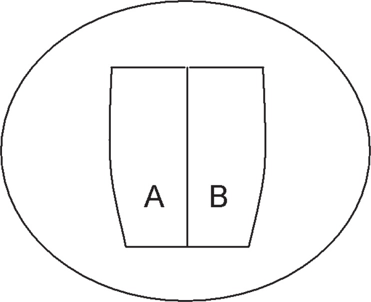
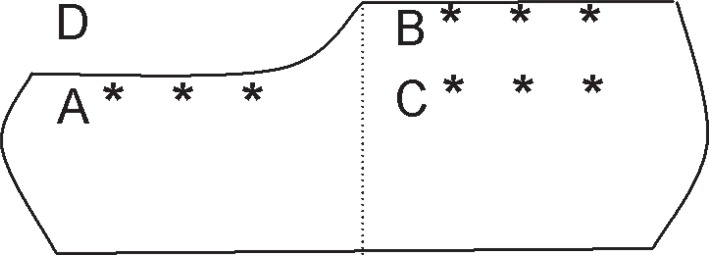
Material and methods: The dentin surfaces of 20 bovine teeth were exposed and half of the surface was protected with nail polish. Cariogenic challenge was performed by immersion in a demineralizing solution for 14 days. After transverse cross-section of the crown, the specimens were divided into four groups (n=10), according to the method used to identify and remove the carious tissue: "Papacárie", Caries-detector dye, DIAGNOdent and Tactile method. After caries removal, the cross-sectional surface was included in acrylic resin and polished. In a microhardness tester, the removed dentin thickness and the Vickers microhardness of the following regions were evaluated: remaining dentin after caries removal and superficial and deep healthy dentin.
Results: ANOVA and Tukey's test (α=0.05) were performed, except for DIAGNOdent, which did not detect the presence of caries. Results for removed dentin thickness were: "Papacárie" (424.7 ± 105.0; a), Caries-detector dye (370.5 ± 78.3; ab), Tactile method (322.8 ± 51.5; bc). Results for the remaining dentin microhardness were: "Papacárie" (42.2 ± 10.5; bc), Caries-detector dye (44.6 ± 11.8; abc), Tactile method (24.3 ± 9.0; d).
Conclusions: DIAGNOdent did not detect the presence of carious tissue; Tactile method and "Papacárie" resulted in the least and the most dentinal thickness removal, respectively; Tactile method differed significantly from "Papacárie" and Caries-detector dye in terms of the remaining dentin microhardness, and Tactile method was the one which presented the lowest microhardness values.
Figures
References
-
- Anderson MH, Loesche WJ, Charbeneau GT. Bacteriologic study of a basic fuchsin caries disclosing dye. J Prosthet Dent. 1985;54:51–55. - PubMed
-
- Angker L, Nockolds C, Swain MV, Kilpatrick N. Correlating the mechanical properties to the mineral content of carious dentine - a comparative study using an ultra-micro-indentation system (UMIS) and SEM-BSE signals. Arch Oral Bio. 2004;49:369–378. - PubMed
-
- Banerjee A, Sherriff M, Kidd EA, Watson TF. A confocal microscopic study relating the autofluorescence of carious dentine to its microhardness. Br Dent J. 1999;187:206–210. - PubMed
-
- Craig RG, Gehring PE, Peyton FA. Relation of structure to the microhardness of human dentin. J Dent Res. 1959;38:624–630. - PubMed
-
- Feagin F, Patel PR, Koulourides T, Pigman W. Study of the effect of calcium, phosphate, fluoride and hydrogen ion concentrations on the remineralization of partially demineralized human and bovine enamel surfaces. Arch Oral Bio. 1971;16:535–548. - PubMed
MeSH terms
Substances
LinkOut - more resources
Full Text Sources
Medical
