. 2013 Jan;27(1):151-62.
doi: 10.1096/fj.12-213017. Epub 2012 Oct 2.

αB-Crystallin regulates expansion of CD11b⁺Gr-1⁺ immature myeloid cells during tumor progression

Affiliations

αB-Crystallin regulates expansion of CD11b⁺Gr-1⁺ immature myeloid cells during tumor progression

Lothar C Dieterich et al. FASEB J. 2013 Jan.

Abstract

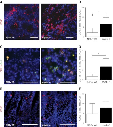
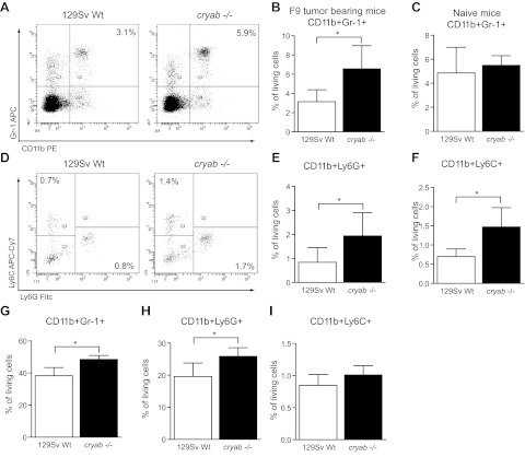
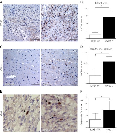
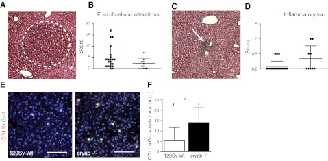
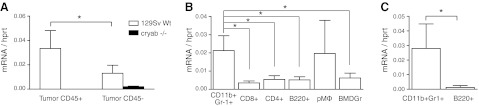
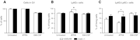
The molecular chaperone αB-crystallin has emerged as a target for cancer therapy due to its expression in human tumors and its role in regulating tumor angiogenesis. αB-crystallin also reduces neuroinflammation, but its role in other inflammatory conditions has not been investigated. Here, we examined whether αB-crystallin regulates inflammation associated with tumors and ischemia. We found that CD45(+) leukocyte infiltration is 3-fold increased in tumors and ischemic myocardium in αB-crystallin-deficient mice. Notably, αB-crystallin is prominently expressed in CD11b(+) Gr-1(+) immature myeloid cells (IMCs), known as regulators of angiogenesis and immune responses, while lymphocytes and mature granulocytes show low αB-crystallin expression. αB-Crystallin deficiency results in a 3-fold higher accumulation of CD11b(+) Gr-1(+) IMCs in tumors and a significant rise in CD11b(+) Gr-1(+) IMCs in spleen and bone marrow. Similarly, we noted a 2-fold increase in CD11b(+) Gr-1(+) IMCs in chronically inflamed livers in αB-crystallin-deficient mice. The effect of αB-crystallin on IMC accumulation is limited to pathological conditions, as CD11b(+) Gr-1(+) IMCs are not elevated in naive mice. Through ex vivo differentiation of CD11b(+) Gr-1(+) cells, we provide evidence that αB-crystallin regulates systemic expansion of IMCs through a cell-intrinsic mechanism. Our study suggests a key role of αB-crystallin in limiting expansion of CD11b(+) Gr-1(+) IMCs in diverse pathological conditions.


Figures

Figure 1.
Figure 1.
Infiltration of leukocytes and CD11b+ Gr-1+ IMCs is increased in F9 tumors in cryab−/− mice. A) Immunofluorescence images of CD45 staining (red) in F9 tumor sections of wild-type and cryab−/− mice. B) Quantification of CD45+ cells in F9 tumors (n=5). C) Immunofluorescence images of CD11b (red) and Gr-1 (green) staining in F9 tumor sections of wild-type and cryab−/− mice. D) Quantification of CD11b+ Gr-1+ cells relative to tumor area (n=6–7). E) Immunofluorescence images of CD68 staining (red) in F9 tumor sections of wild-type and cryab−/− mice. F) Quantification of CD68+ cells relative to tumor area (n=5–7). Scale bars = 100 μm (A); 50 μm (C); 200 μm (E). Bars represent means ± sd. *P < 0.05.
Figure 2.
Figure 2.
IMCs are highly increased in spleens and bone marrow of tumor-bearing cryab−/− mice. A) Representative FACS plots showing CD11b/Gr-1 staining in splenocytes of tumor-bearing wild-type and cryab−/− mice. B) Quantification of CD11b+ Gr-1+ IMCs in spleens of F9 tumor-bearing wild-type (open bars) and cryab−/− mice (solid bars) by FACS (n=9–10). C) Quantification of CD11b+ Gr-1+ IMCs in spleens of naive wild-type (open bars) and cryab−/− mice (solid bars) by FACS (n=4–5). D) Representative FACS plots of Ly6G and Ly6C expression on splenocytes (pregated for CD11b) of tumor-bearing wild-type and cryab−/− mice. E, F) Quantification of CD11b+ Ly6G+ (E) and CD11b+ Ly6C+ cells (F) in spleens of F9 tumor-bearing mice (n=9–10). G–I) FACS-based quantification of CD11b+ Gr-1+ cells (G), CD11b+ Ly6G+ cells (H) and CD11b+ Ly6C+ cells (I) in the bone marrow of tumor-bearing wild-type (open bars) and cryab−/− (solid bars) mice (n=8). Bars represent means ± sd. *P < 0.05.
Figure 3.
Figure 3.
Recruitment of CD45+ leukocytes and Gr-1+ myeloid cells is increased in ischemic myocardium in cryab−/− mice. A) Immunohistochemistry images of CD45 staining in infarct areas 7 d after ligation of the LAD coronary artery in wild-type and cryab−/− mice. B) Quantification of CD45+ area in the entire infarct area (n=6–7). C) Immunohistochemistry images of CD45 staining in healthy myocardium of ischemic hearts. D) CD45+ area was quantified in 5 randomly chosen ×20 fields of healthy myocardium (n=7–8). E) Immunohistochemistry images of infarct areas stained for Gr-1. F) Quantification of Gr-1+ cells residing within the infarct areas (n=5 cryab−/−; n=9 wild type). Scale bars = 50 μm. Bars represent means ± sd. *P < 0.05.
Figure 4.
Figure 4.
Intrahepatic CD11b+ Gr-1+ IMCs are increased in cryab−/− mice during chronic liver inflammation. A) DEN-induced liver damage develops over a period of 30 wk after injection and results in foci of benign cellular alterations (dotted line). B) Semiquantitative scoring of foci of cellular alteration in the liver, as outlined in Materials and Methods (n=9 cryab−/−; n=21 wild type). C) Microscopic image of an inflammatory focus in the liver 30 wk after DEN injection (arrow). D) Scoring of inflammatory foci in the liver as outlined in Materials and Methods (n=9 cryab−/−; n=21 wild type). E) Immunofluorescence images of CD11b (red) and Gr-1 (green) staining in liver sections of wild-type and cryab−/− mice. Scale bars = 100 μm. F) Quantification of CD11b+ Gr-1+ cells relative to liver area (n=9 cryab−/−; n=21 wild type). Bars represent means ± sd. *P < 0.05.
Figure 5.
Figure 5.
Cryab is expressed in CD45+ tumor-infiltrating cells and CD11b+ Gr-1+ splenocytes in tumor-bearing mice. A) Expression of cryab in CD45+ and CD45 cells isolated from F9 tumors grown in wild-type (open bars) or cryab−/− mice (solid bars) was determined by qPCR (n=4-5 wild type; n=2-3 cryab−/−). B) qPCR results showing expression of cryab in FACS-sorted CD11b+ Gr-1+, CD8+, CD4+, and B220+ splenocytes, peritoneal macrophages (pMΦ) and bone marrow-derived granulocytes (BMDGr) from F9 tumor-bearing 129Sv wild-type mice (n=6). C) Expression of cryab in FACS-sorted CD11b+Gr-1+ and B220+ splenocytes from T241 tumor-bearing C57BL/6 wild-type mice (n=4). Bars represent means ± sd. *P < 0.05.
Figure 6.
Figure 6.
Lack of αB-crystallin alters ATRA-induced differentiation of bone marrow-derived CD11b+ Gr-1+ cells. CD11b+ Gr-1+ cells were isolated from the bone marrow of wild-type and cryab−/− mice and cultured for 72 h in the presence of ATRA or GM-CSF. A) Cell cycle analysis shows complete G1 arrest of bone marrow-derived CD11b+ Gr-1+ cells after 72-h culture in presence of ATRA or control medium, while ∼20% of cells are proliferating in presence of GM-CSF (n=3). B, C) Percentage of Ly6G+ (B) and Ly6GLy6C+ cells (C) determined by FACS (n=3). Bars represent means ± sd. *P < 0.05.

References

    1. Mantovani A., Allavena P., Sica A., Balkwill F. (2008) Cancer-related inflammation. Nature 454, 436–444 - PubMed
    1. Schafer M., Werner S. (2008) Cancer as an overhealing wound: an old hypothesis revisited. Nat. Rev. Mol. Cell. Biol. 9, 628–638 - PubMed
    1. Murdoch C., Muthana M., Coffelt S. B., Lewis C. E. (2008) The role of myeloid cells in the promotion of tumour angiogenesis. Nat. Rev. Cancer 8, 618–631 - PubMed
    1. Shojaei F., Wu X., Malik A. K., Zhong C., Baldwin M. E., Schanz S., Fuh G., Gerber H. P., Ferrara N. (2007) Tumor refractoriness to anti-VEGF treatment is mediated by CD11b+Gr1+ myeloid cells. Nat. Biotechnol. 25, 911–920 - PubMed
    1. Condamine T., Gabrilovich D. I. (2011) Molecular mechanisms regulating myeloid-derived suppressor cell differentiation and function. Trends Immunol. 32, 19–25 - PMC - PubMed