. 2012 Dec;67(12):1032-9.
doi: 10.1136/thoraxjnl-2012-201781. Epub 2012 Oct 2.

Low levels of tissue factor lead to alveolar haemorrhage, potentiating murine acute lung injury and oxidative stress

Affiliations

Low levels of tissue factor lead to alveolar haemorrhage, potentiating murine acute lung injury and oxidative stress

Julie A Bastarache et al. Thorax. 2012 Dec.

Abstract

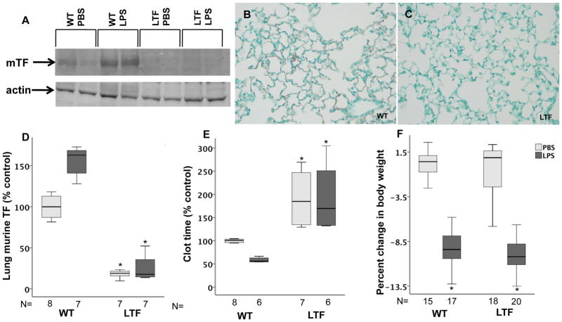
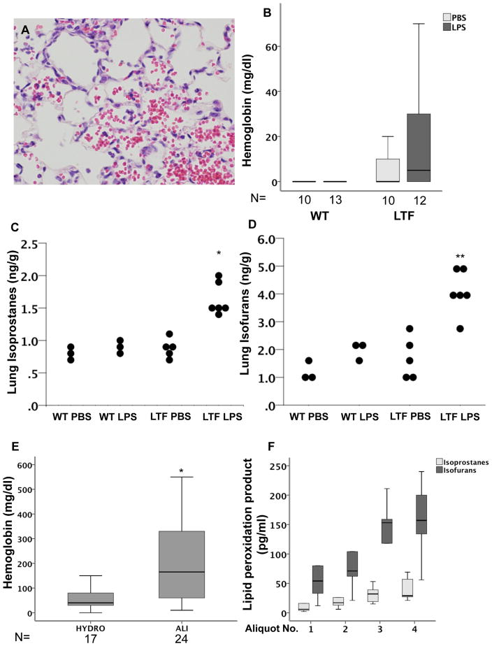
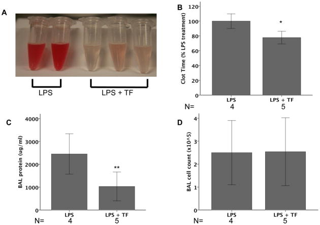
Background: Systemic blockade of tissue factor (TF) attenuates acute lung injury (ALI) in animal models of sepsis but the effects of global TF deficiency are unknown. We used mice with complete knockout of mouse TF and low levels (∼1%) of human TF (LTF mice) to test the hypothesis that global TF deficiency attenuates lung inflammation in direct lung injury.

Methods: LTF mice were treated with 10 μg of lipopolysaccharide (LPS) or vehicle administered by direct intratracheal injection and studied at 24 h.

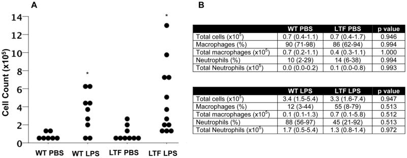
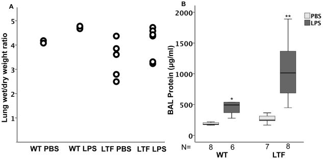
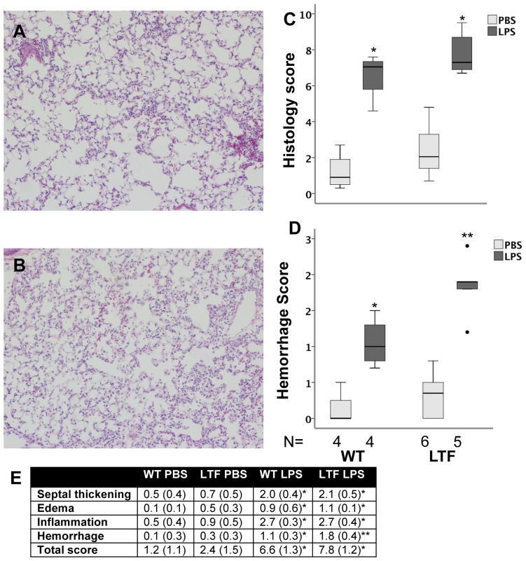
Results: Contrary to our hypothesis, LTF mice had increased lung inflammation and injury as measured by bronchoalveolar lavage cell count (3.4×10(5) wild-type (WT) LPS vs 3.3×10(5) LTF LPS, p=0.947) and protein (493 μg/ml WT LPS vs 1014 μg/ml LTF LPS, p=0.006), proinflammatory cytokines (TNF-α, IL-10, IL-12, p<0.035 WT LPS vs LTF LPS) and histology compared with WT mice. LTF mice also had increased haemorrhage and free haemoglobin in the airspace accompanied by increased oxidant stress as measured by lipid peroxidation products (F(2) isoprostanes and isofurans).

Conclusions: These findings indicate that global TF deficiency does not confer protection in a direct lung injury model. Rather, TF deficiency causes increased intra-alveolar haemorrhage following LPS leading to increased lipid peroxidation. Strategies to globally inhibit TF may be deleterious in patients with ALI.


Conflict of interest statement

CONFLICT OF INTEREST DISCLOSURES

The authors have no conflicts of interest to disclosures

Figures

Figure 1
Figure 1
TF expression. Wild type mice have low but detectable levels of murine TF expression at base line that increases following IT LPS treatment while murine TF levels are undetectable in LTF mice. Panel A shows a representative western blot for murine TF and Panel D shows densitometric quantification from 4 independent experiments. *p=<0.001 versus both wild type groups. Panel B and C show immunostaining for murine TF (red/brown) in WT (B) and LTF (C) mice 24 hours after IT LPS. While wild type mice have a shortened BAL clot time in response to IT LPS (Panel E), LTF mice have a prolonged clot time that is not shortened by IT LPS. *p<0.001 versus WT. Panel F shows weight change 24 hours after treatment with LPS. *p<0.001 versus PBS treatment.
Figure 2
Figure 2
Intratracheal LPS induces lung inflammation. Dot plot of BAL cell counts 24 hours after IT LPS administration. BAL total cell count was increased in both wild type and low TF mice compared to saline treated animals (Panel A). *p<0.005 versus PBS treatment by ANOVA of log transformed data with post hoc Tukey test. Panel B shows median (IQR) total BAL macrophage and neutrophil counts and percent counts.
Figure 3
Figure 3
IT LPS increases lung edema. IT LPS causes a modest but non-significant increase in lung wet to dry weight ratio (Panel A) but there are no differences between WT and LTF. In contrast, BAL total protein is significantly increased by IT LPS (Panel B). *p<0.007 versus WT PBS and LTF LPS groups, **p<0.007 versus all other groups.
Figure 4
Figure 4
Lung histology 24 hours after IT LPS. IT LPS induced lung inflammation and injury in both WT (Panel A) and LTF (Panel B) mice. LPS treatment increased lung injury in both genotypes (Panel C) [*p<0.002 versus PBS treatment]. Lung hemorrhage was more severe in LPS treated LTF mice compared to WT (Panel D) [*p<0.027 versus all other groups,** p<0.019 versus all other groups]. Panel E shows the individual components of the score [* p<0.05 vs PBS treatment in both genotypes, ** p<0.05 vs PBS treatment in both genotypes and WT LPS treatment].
Figure 5
Figure 5
Free hemoglobin and lipid peroxidation. LTF mice have increased alveolar hemorrhage (Panel A) and increased free hemoglobin in BAL fluid (Panel B) both basally and in response to IT LPS. LTF mice also have an increase in total lung isporostanes (Panel C) and isofurans (Panel D) in response to IT LPS (*p<0.001 versus all other groups and **p<0.021 versus all other groups). Patients with ALI also have increased levels of free hemoglobin in the lung compared to patients with hydrostatic edema (Panel E) (*p=0.007). In patients with a diagnosis of alveolar hemorrhage, BAL isoprostanes and isofurans increase with increasing aliqout number (Panel F).
Figure 6
Figure 6
Effects of TF replacement in LTF mice. LTF mice were treated with 10μg LPS IT (N=4) or 10μg LPS + 100 nM recombinant mTF IT (N=5) and harvested at 24 hours. With the administration of mTF, LTF mice had visibly reduced BAL hemorrhage (Panel A) and significantly shortened BAL clot time (Panel B) compared to LPS treated mice that did not receive recombinant TF. BAL total protein was higher in mice given mTF (Panel C) but BAL cell counts did not differ (Panel D). (*p=0.011, **p=0.031).

References

    1. Gando S, Kameue T, Matsuda N, Hayakawa M, Morimoto Y, Ishitani T, et al. Imbalances between the levels of tissue factor and tissue factor pathway inhibitor in ARDS patients. Thromb Res. 2003;109(2–3):119–24. - PubMed
    1. Bastarache JA, Wang L, Geiser T, Wang Z, Albertine KH, Matthay MA, et al. The alveolar epithelium can initiate the extrinsic coagulation cascade through expression of tissue factor. Thorax. 2007;62(7):608–16. - PMC - PubMed
    1. He X, Han B, Mura M, Li L, Cypel M, Soderman A, et al. Anti-human tissue factor antibody ameliorated intestinal ischemia reperfusion-induced acute lung injury in human tissue factor knock-in mice. PLoS One. 2008;3(1):e1527. - PMC - PubMed
    1. Welty-Wolf KE, Carraway MS, Ortel TL, Ghio AJ, Idell S, Egan J, et al. Blockade of Tissue Factor-Factor X binding attenuates sepsis-induced respiratory and renal failure. Am J Physiol Lung Cell Mol Physiol. 2005 - PubMed
    1. Welty-Wolf KE, Carraway MS, Miller DL, Ortel TL, Ezban M, Ghio AJ, et al. Coagulation blockade prevents sepsis-induced respiratory and renal failure in baboons. Am J Respir Crit Care Med. 2001;164(10 Pt 1):1988–96. - PubMed

Publication types

MeSH terms