. 2012 Oct 2;3(5):e00312-12.
doi: 10.1128/mBio.00312-12. Print 2012.

Inhibition of LpxC protects mice from resistant Acinetobacter baumannii by modulating inflammation and enhancing phagocytosis

Affiliations

Inhibition of LpxC protects mice from resistant Acinetobacter baumannii by modulating inflammation and enhancing phagocytosis

Lin Lin et al. mBio. .

Abstract

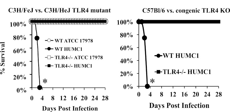
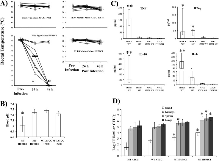
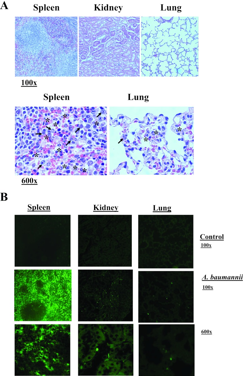
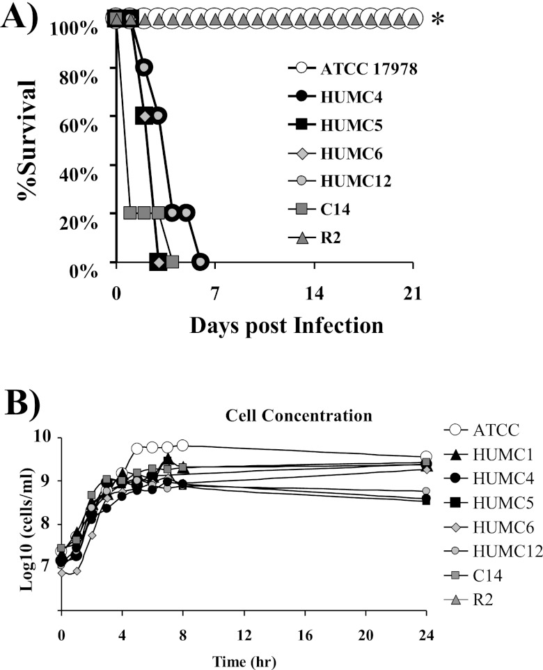
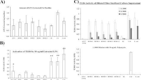
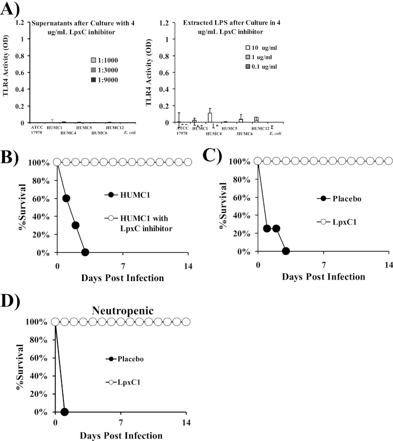
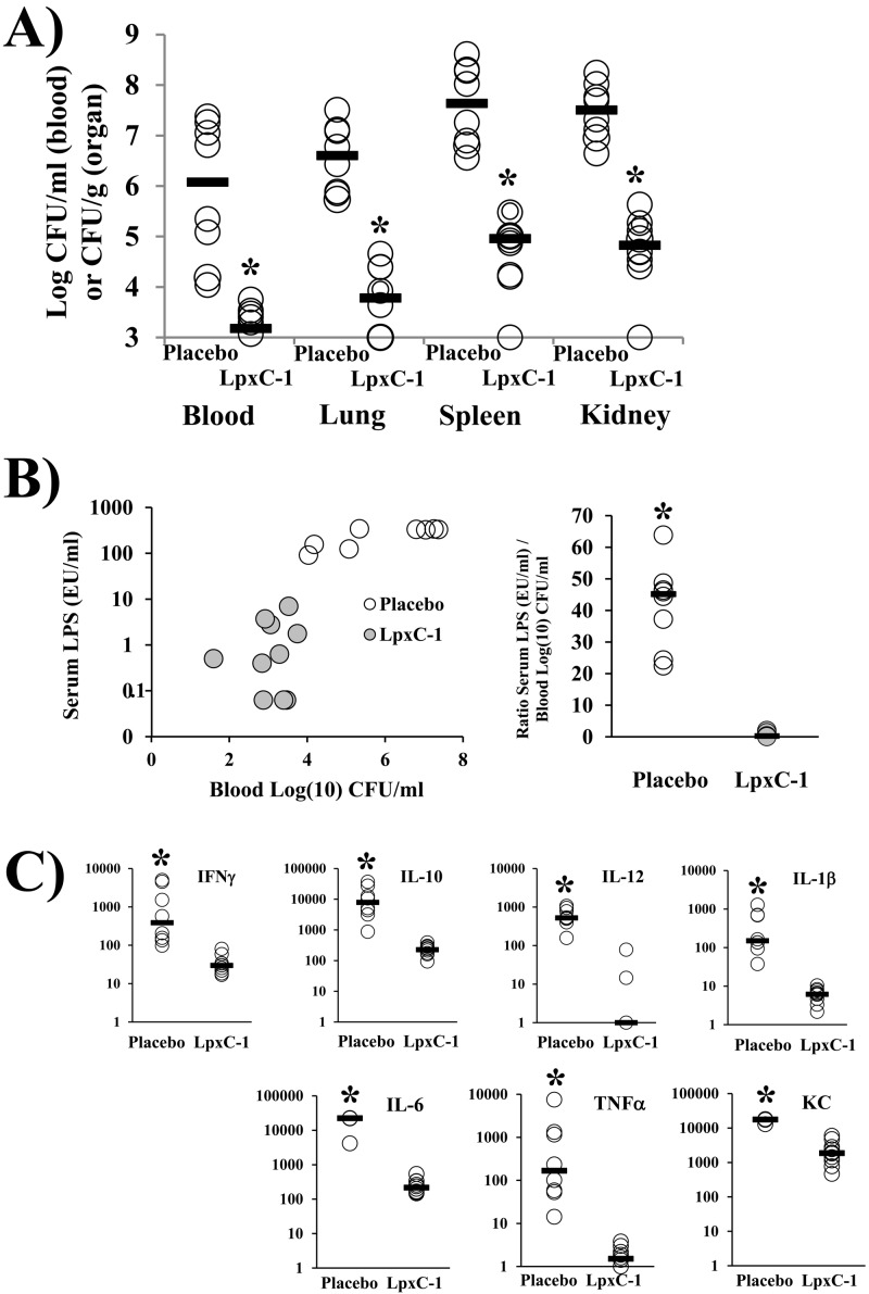
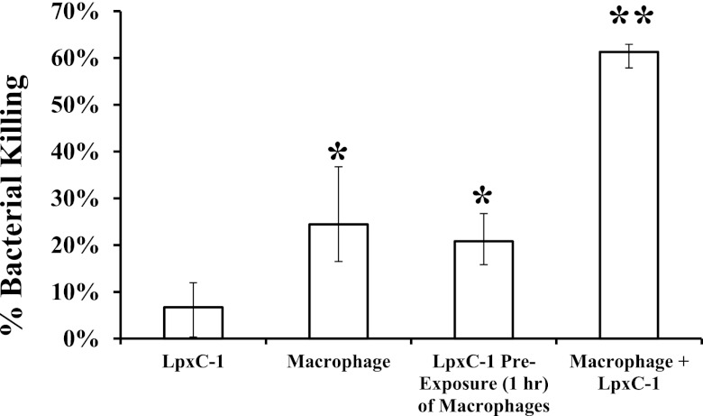
New treatments are needed for extensively drug-resistant (XDR) Gram-negative bacilli (GNB), such as Acinetobacter baumannii. Toll-like receptor 4 (TLR4) was previously reported to enhance bacterial clearance of GNB, including A. baumannii. However, here we have shown that 100% of wild-type mice versus 0% of TLR4-deficient mice died of septic shock due to A. baumannii infection, despite having similar tissue bacterial burdens. The strain lipopolysaccharide (LPS) content and TLR4 activation by extracted LPS did not correlate with in vivo virulence, nor did colistin resistance due to LPS phosphoethanolamine modification. However, more-virulent strains shed more LPS during growth than less-virulent strains, resulting in enhanced TLR4 activation. Due to the role of LPS in A. baumannii virulence, an LpxC inhibitor (which affects lipid A biosynthesis) antibiotic was tested. The LpxC inhibitor did not inhibit growth of the bacterium (MIC>512 µg/ml) but suppressed A. baumannii LPS-mediated activation of TLR4. Treatment of infected mice with the LpxC inhibitor enhanced clearance of the bacteria by enhancing opsonophagocytic killing, reduced serum LPS concentrations and inflammation, and completely protected the mice from lethal infection. These results identify a previously unappreciated potential for the new class of LpxC inhibitor antibiotics to treat XDR A. baumannii infections. Furthermore, they have far-reaching implications for pathogenesis and treatment of infections caused by GNB and for the discovery of novel antibiotics not detected by standard in vitro screens.

Importance: Novel treatments are needed for infections caused by Acinetobacter baumannii, a Gram-negative bacterium that is extremely antibiotic resistant. The current study was undertaken to understand the immunopathogenesis of these infections, as a basis for defining novel treatments. The primary strain characteristic that differentiated virulent from less-virulent strains was shedding of Gram-negative lipopolysaccharide (LPS) during growth. A novel class of antibiotics, called LpxC inhibitors, block LPS synthesis, but these drugs do not demonstrate the ability to kill A. baumannii in vitro. We found that an LpxC inhibitor blocked the ability of bacteria to activate the sepsis cascade, enhanced opsonophagocytic killing of the bacteria, and protected mice from lethal infection. Thus, an entire new class of antibiotics which is already in development has heretofore-unrecognized potential to treat A. baumannii infections. Furthermore, standard antibiotic screens based on in vitro killing failed to detect this treatment potential of LpxC inhibitors for A. baumannii infections.


Figures

FIG 1
FIG 1
TLR4 is antiprotective against A. baumannii bloodstream infection. C3H/FeJ wild-type or C3H/HeJ TLR4-mutant mice (n = 10 mice per group, except for 9 mice in the wild-type HUMC1-infected group) or C57BL/6 or congenic TLR4-knockout (KO) mice were infected via the tail vein with 2 × 10⁷ bacteria and followed for 28 days. All remaining mice at day 28 appeared clinically well. *, P < 0.05 versus results for all other groups.
FIG 2
FIG 2
Lethally infected wild-type mice had septic shock, whereas TLR4-mutant mice did not. (A) Rectal temperatures taken from mice infected with A. baumannii HUMC1 or ATCC 17978 (the same mice in Fig. 1; n = 10 mice per group, except for 9 in the wild-type HUMC1-infected group). Temperatures were taken at the same time (8 to 9 AM) daily before and for 2 days after infection (wild-type mice began dying on day 2). *, P < 0.05 versus preinfection findings. (B) Blood pH at 48 h of infection was measured by using i-STAT cartridges from separate C3H/FeJ or C3H/HeJ (TLR4-mutant) mice infected with 2 × 10⁷ A. baumannii HUMC1 or ATCC 17978 bacteria (n = 16 mice per group from 2 experiments, except for 8 mice for HUMC1-infected wild-type mice). *, P < 0.05 versus results for other groups. (C) Serum inflammatory and anti-inflammatory cytokine levels were measured at 48 h of infection in C3H/FeJ or C3H/HeJ (TLR4-mutant) mice. n = 8 mice per group. **, P < 0.05 versus results for all other groups; *, P < 0.05 versus results for ATCC 17978 groups. (D) Blood and tissue bacterial burden at 48 h of infection differed between mice infected with 2 × 10⁷ HUMC1 and ATCC 17978 bacteria (n = 12 mice per group from 2 experiments) but not between mutant and wild-type mice. *, P < 0.05 versus results for mice infected with ATCC 17978. Median and interquartile ranges are graphed.
FIG 3
FIG 3
Histopathology and immunohistochemistry of A. baumannii during infection in mice. (A) Histopathology of spleens, lungs, and kidneys from C3H/FeJ mice given lethal infection with A. baumannii HUMC1 (2 × 10⁷ inoculum) demonstrated normal parenchymal anatomy, with no evidence of bacterial invasion (100× power shown). At higher power (600×), the only abnormalities found were accumulation of neutrophils (asterisks), including pyknotic neutrophils (arrows) undergoing nucleolysis consistent with apoptosis, in the splenic perifollicular red pulp area and in the pulmonary capillaries, consistent with Gram-negative LPS-induced sepsis. The kidney appeared histopathologically normal at higher power (not shown). (B) Immunohistochemistry was used to localize A. baumannii in parenchymal organs. In the spleen, the bacteria accumulated in the perifollicular red pulp areas and spared lymph node follicles (note green spots surrounding dark follicles). In the kidneys, the organisms were found scatted in capillaries surrounding renal tubules, and there was no evidence of parenchymal organ invasion. In the lung, the organisms were found in capillaries in the interstitium, and again there was no evidence of alveolar or parenchymal invasion from the capillaries. The control was stained with normal mouse serum as the primary antibody.
FIG 4
FIG 4
Various virulences of clinical isolates, including colistin-resistant strains. (A) C3H/FeJ mice were infected iv with 5 × 10⁷ HUMC4, -5, -6, and -12 and R2 and C14 bacteria, all of which caused 100% mortality (n = 5 to 8 mice per group). ATCC 17978 and R2 were avirulent. (B) In vitro growth rates did not differ substantially among strains irrespective of virulence in vivo.
FIG 5
FIG 5
In vitro correlate of in vivo virulence. (A) LPS content (ng/bacillus) was similar across strains of highly varying virulences (e.g., HUMC1 versus ATCC 17978 versus R2). Results are from at least two separate extractions, each done in duplicate. (B) TLR4-activating potency of extracted LPS was higher for R2 and C14 than all other A. baumannii strains (**, P < 0.05 versus results for all other strains except E. coli). Of the colistin-susceptible A. baumannii strains, TLR4-activating potency was highest among A. baumannii ATCC 17978, which was avirulent (*, P < 0.05 versus HUMC strains). Results are from a minimum of two assays per strain, each done in duplicate. (C) Filter-sterilized culture supernatant induced a much stronger TLR4 signal from strains that caused lethal infections in vivo than from avirulent strains. Also, addition of polymyxin blocked the TLR4 activation from all strains except those highly resistant to colistin (C8 and C14), which were not affected by polymyxin. Results are from a minimum of three assays per strain, each done in duplicate. For all panels, median and interquartile ranges are graphed.
FIG 6
FIG 6
Inhibition of LPS biosynthesis with an inhibitor of LpxC blocked TLR4 activation in vitro and abrogated virulence in vivo. (A) TLR4-activating potency of filtered culture supernatant and extracted LPS from A. baumannii strains passaged to log phase in the presence of 4 µg/ml of LpxC inhibitor (LpxC-1). The results with LpxC-1 were run concurrently with those without LpxC-1 (compare signal with and without LpxC-1 inhibitor in Fig. 5C versus Fig. 6A). (B) Survival of wild-type C3H/FeJ mice (n = 10 per group) which were either infected with normal A. baumannii HUMC1 and treated with a placebo (40% cyclodextrin in water i.v. once daily) for 3 days starting on the day of infection or infected with A. baumannii HUMC1 that was cultured overnight and during log passage in the presence of 4 µg/ml of LpxC inhibitor and treated with LpxC-1 (100 mg/kg in 40% cyclodextrin i.v.) for 3 days postinfection. (C) Survival of wild-type C3H/FeJ mice (n = 10 per group) which were infected with A. baumannii HUMC1 and treated with a placebo (40% cyclodextrin in water) or LpxC-1 (100 mg/kg in 40% cyclodextrin i.v.) starting 1 h after infection and for 3 days postinfection. (D) Survival of BALB/c mice (n = 11 in the placebo group; n = 10 in the LpxC1-treated group) made neutropenic with cyclophosphamide, infected with A. baumannii HUMC1, and treated with placebo or LpxC-1 starting after infection and for 3 days postinfection.
FIG 7
FIG 7
Bacterial densities in blood and tissue and serum LPS and cytokine concentrations for mice treated with LpxC-1 or a placebo. C3H/FeJ mice (n = 15 per group) were infected with A. baumannii HUMC1. At 1 h and 24 h, infected mice were treated i.v. with LpxC-1 (100 mg/kg). Five control mice died before the 24-h time point; no treated mice died. (A) Bacterial densities in blood and tissue for treated versus control mice. (B) Serum LPS levels for treated versus control mice. (C) Serum cytokine levels for treated versus control mice. *, P < 0.01 versus results for the control.
FIG 8
FIG 8
Macrophage killing of A. baumannii is enhanced by exposure to LpxC-1. Reduction of CFU at 1 h of A. baumannii incubation with LpxC-1 alone, macrophages alone, macrophages that had been preexposed to LpxC-1 for 1 h followed by rinsing away the LpxC-1, and macrophages plus LpxC-1. Median and interquartile ranges of killing are shown. Results from are from eight samples per group. *, P < 0.05 versus results for LpxC-1 alone; **, P < 0.05 versus results for all other groups.

References

    1. Chow JC, Young DW, Golenbock DT, Christ WJ, Gusovsky F. 1999. Toll-like receptor-4 mediates lipopolysaccharide-induced signal transduction. J. Biol. Chem. 274:10689–10692 - PubMed
    1. Hoshino K, et al. 1999. Cutting edge: toll-like receptor 4 (TLR4)-deficient mice are hyporesponsive to lipopolysaccharide: evidence for TLR4 as the Lps gene product. J. Immunol. 162:3749–3752 - PubMed
    1. Poltorak A, et al. 1998. Defective LPS signaling in C3H/HeJ and C57BL/10ScCr mice: mutations in Tlr4 gene. Science 282:2085–2088 - PubMed
    1. Bihl F, et al. 2003. Overexpression of Toll-like receptor 4 amplifies the host response to lipopolysaccharide and provides a survival advantage in transgenic mice. J. Immunol. 170:6141–6150 - PubMed
    1. Branger J, et al. 2004. Role of Toll-like receptor 4 in gram-positive and gram-negative pneumonia in mice. Infect. Immun. 72:788–794 - PMC - PubMed

Publication types

MeSH terms