. 2012 Nov 16;287(47):39613-25.
doi: 10.1074/jbc.M112.380162. Epub 2012 Oct 2.

Identification and characterization of a compound that protects cardiac tissue from human Ether-à-go-go-related gene (hERG)-related drug-induced arrhythmias

Affiliations

Identification and characterization of a compound that protects cardiac tissue from human Ether-à-go-go-related gene (hERG)-related drug-induced arrhythmias

Franck Potet et al. J Biol Chem. .

Abstract

The human Ether-à-go-go-related gene (hERG)-encoded K(+) current, I(Kr) is essential for cardiac repolarization but is also a source of cardiotoxicity because unintended hERG inhibition by diverse pharmaceuticals can cause arrhythmias and sudden cardiac death. We hypothesized that a small molecule that diminishes I(Kr) block by a known hERG antagonist would constitute a first step toward preventing hERG-related arrhythmias and facilitating drug discovery. Using a high-throughput assay, we screened a library of compounds for agents that increase the IC(70) of dofetilide, a well characterized hERG blocker. One compound, VU0405601, with the desired activity was further characterized. In isolated, Langendorff-perfused rabbit hearts, optical mapping revealed that dofetilide-induced arrhythmias were reduced after pretreatment with VU0405601. Patch clamp analysis in stable hERG-HEK cells showed effects on current amplitude, inactivation, and deactivation. VU0405601 increased the IC(50) of dofetilide from 38.7 to 76.3 nM. VU0405601 mitigates the effects of hERG blockers from the extracellular aspect primarily by reducing inactivation, whereas most clinically relevant hERG inhibitors act at an inner pore site. Structure-activity relationships surrounding VU0405601 identified a 3-pyridiyl and a naphthyridine ring system as key structural components important for preventing hERG inhibition by multiple inhibitors. These findings indicate that small molecules can be designed to reduce the sensitivity of hERG to inhibitors.


Figures

REACTION 1
REACTION 1
REACTION 2
REACTION 2
FIGURE 1.
FIGURE 1.
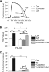
Optical measurements of action potentials in ex vivo rabbit hearts using voltage-sensitive dyes. A, perfusion with 100 nm dofetilide (Dof) prolonged AP compared with untreated controls (Con). 5 μm VU0405601 did not change APD compared with controls. B, shown is a statistical comparison of APD measured at 70% repolarization in controls, 100 nm dofetilide, and VU0405601-perfused hearts. VU0405601>Dof, pretreated for 30 min with 5 μm VU0405601 before 100 nm dofetilide was perfused. C, the hERG-specific inhibitor dofetilide (100 nm) caused increased arrhythmia incidence. Compare black versus clear bars. VU0405601 (gray bars) caused no arrhythmias above control levels. Pretreatment for 30 min with 5 μm VU0405601 (striped bars) protected hearts from dofetilide-induced, paced arrhythmias. **, p < 0.005; *, p < 0.01, Fisher's exact test. a.u., absorbance units.
FIGURE 2.
FIGURE 2.
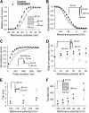
hERG currents recorded in hERG-HEK cells. A, shown is the time course of hERG current before (control) and after 50 μm VU0405601. Whole-cell currents were generated from a holding voltage of −80 mV and stepped to 10 mV for 2 s (▴) followed by a tail pulse at −50 mV for 2 s (○). Dofetilide was added as indicated. Representative current traces are shown in the inset. B, shown is dose response of VU0405601 measured at 10 mV (test pulse) and the ensuing peak tail current currents (−50 mV). Current amplitudes were normalized to untreated control conditions and plotted against VU0405601 concentrations. A vertical dashed line is drawn at 5 μm. C, pulse protocol was used to assess time course of hERG inhibition. Whole-cell currents were generated from a holding voltage of −80 mV and stepped to +10 mV for 2 s followed by a tail pulse at −50 mV for 2 s. Continued pulsing with dofetilide (Dof) results in a gradual onset of hERG inhibition due to open-state block. D, hERG-HEK cells were stimulated until steady-state effects of VU0405601 or PD-119057 was achieved and inhibitor was applied subsequently. Tail currents were normalized for maximally stimulated current (pre-inhibitor) and measured at multiple time points with 100 nm dofetilide alone (open circles, n = 23) with 10 μm PD-118057 followed by 100 nm dofetilide (green triangles, n = 6), with 5 and 50 μm VU0405601 (red circles and triangles, n = 6 and 12, respectively) followed by 100 nm dofetilide, and with intracellular application of 50 μm VU0405601 followed by 100 nm dofetilide (black triangles, n = 5).
FIGURE 3.
FIGURE 3.
Dose-response curves of VU0405601 with hERG inhibitors. VU0405601 shifts the IC50 of dofetilide from 38.7 to 76.3 nm (A) and the IC50 of droperidol from 0.35 to 48 μm (B). The number or cells per concentration is indicated in parentheses. The pulse protocol was as in Fig. 2C. C, comparison of PD-118057 and VU0405601 effects on current amplitude are shown. The y axis plots % current remaining after 15 min of pulsing in the presence of 100 nm dofetilide utilizing the pulse protocol as described in Fig. 2C. The x axis plots increases in tail currents measured in response to individual agonist. D, VU0405601 selectively increases outward currents. Currents were measured in hERG-HEK cells using the voltage protocol shown in the inset. 50 μm VU0405601 (solid symbols) increased the outward but not the inward currents through the hERG channel. For each cell current amplitudes were measured at peak (defined as fully activated current). The fully activated I-V relationship was normalized relative to peak outward in control. Error bars denote S.E. (n = 7).
FIGURE 4.
FIGURE 4.
Response to VU0405601 of hERG currents recorded in hERG-HEK cells. A, voltage dependence of activation is shown. Tail currents were measured at −50 mV after pulses from −80 to 60 mV. Values were normalized to the peak value of each condition. For control, V½ of activation was −9.8 ± 3.4 mV, and k was 10.9 ± 0.6 mV (○). With VU0405601 the V½ was −23.6 ± 2.5 mV (▴; p < 0.001; n = 8). B, voltage dependence of inactivation is shown. Peak tail currents were normalized to the maximal peak tail current and plotted against voltages. The V½ in controls was −64.6 ± 3.7 mV, and the slope factor was −19.9 ± 1.2 mV (○). With VU0405601, the V½ and the slope factor were −48.8 ± 1.7 mV (▴, p < 0.005, n = 8) and −18.9 ± 1.1 mV (p = not significant), respectively. The curves were fitted with the Boltzmann function. C, VU0405601 accelerated the activation kinetics by decreasing the time constant from 899.7 ± 294.2 (○) to 258.2 ± 47.1 ms (▴, p < 0.005, n = 7). D, VU0405601 slowed the rate of hERG inactivation. The decay of current traces during the third pulse was fit with a single exponential function to generate the time constants (n = 6); *, p < 0.05. E, VU0405601 accelerated recovery from inactivation. Cells were depolarized to 60 mV for 2 s and then returned to potentials from −120 to −100 mV for 2 s to generate inward tail currents. Recovery from inactivation was determined by fitting the initial rising phase of these tail currents to a single exponential function and are plotted as function of voltage (p < 0.05, n = 6). F, tail currents were fitted with a double-exponential function to obtain the fast time constants. (n = 7); *, p < 0.05. Error bars denote S.E.
FIGURE 5.
FIGURE 5.
Response of inactivation-impaired hERG variants, inhibitor binding site variants, and intracellular VU0405601 application. A, inactivation-impaired hERG S631A shows reduced sensitivity to 50 μm 601. n = 23 for WT and n = 13 for S631A. F656T, a drug binding site mutant, maintains sensitivity to VU0405601; n = 6. Intracellular application of 50 μm VU0405601 (In) through the patch pipette had no effect on hERG current amplitude. Error bars denote S.E. ***, p < 0.001. B, an outer pore blocker, BeKm-1, but not an inner cavity blocker, dofetilide (Dof), suppresses VU0405601 effects. % increase in current magnitude induced by 50 μm VU0405601 after partial inhibition by 50 nm dofetilide or BeKm-1 is shown. Error bars denote S.E. *, p < 0.05. Voltage protocol was as in Fig. 2C.
FIGURE 6.
FIGURE 6.
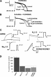
Effects of VU0405601 on AP measured in isolated rabbit myocytes are small when IKr is blocked. Evoked APs were measured from freshly isolated rabbit ventricular cardio-myocytes (A) in the absence or presence of VU0405601 (5 and 50 μm). The percentage reductions in APD50 and APD90 with 5 μm VU0405601were 4 ± 3 and 5 ± 2%, respectively (p = not significant, paired t test; n = 7). With 50 μm VU0405601 APD50 and APD90 were reduced by 35 ± 6 and 32 ± 4%, respectively (p < 0.05, paired t test; n = 5). B, dofetilide (Dof; 1 μm) attenuates the AP-shortening effects of 50 μm VU0405601. In the presence of dofetilide alone, APD50 and APD90 were prolonged by 22 ± 6 and 18 ± 7%, respectively (p < 0.05, paired t test; n = 3). Co-application of 50 μm VU0405601 with dofetilide resulted in a modest effect on APD compared with VU0405601 alone: 12 ± 4 and 10 ± 2% for APD 50 and APD90, respectively (p < 0.05, t test, n = 3. C and D, VU0405601 effects on individual cardiac ion channels were recorded in transfected CHO cells. C, raw current traces are shown. Voltage protocols as indicated. Current was measured at the arrow. Gray lines are with VU0405601. D, effects of 50 μm VU0405601 on current amplitudes are shown.
FIGURE 7.
FIGURE 7.
Structure-activity relationships. A, chemical structures of VU0405601 and its derivatives are shown. The 3-pyridyl group (red) and the halogenated naphthyridine ring (blue) are essential for activity. Effects of VU0405601 and its derivatives on hERG current amplitudes (B) and dofetilide sensitivity (C) are shown. con, control. Measurements in panel B were performed as in Fig. 2B. Measurements in panel C were performed as described in Fig. 2D.

References

    1. Sanguinetti M. C., Jiang C., Curran M. E., Keating M. T. (1995) A mechanistic link between an inherited and an acquired cardiac arrhythmia. HERG encodes the IKr potassium channel. Cell 81, 299–307 - PubMed
    1. Trudeau M. C., Warmke J. W., Ganetzky B., Robertson G. A. (1995) HERG, a human inward rectifier in the voltage-gated potassium channel family. Science 269, 92–95 - PubMed
    1. Curran M. E., Splawski I., Timothy K. W., Vincent G. M., Green E. D., Keating M. T. (1995) A molecular basis for cardiac arrhythmia. HERG mutations cause long QT syndrome. Cell 80, 795–803 - PubMed
    1. Hedley P. L., Jørgensen P., Schlamowitz S., Wangari R., Moolman-Smook J., Brink P. A., Kanters J. K., Corfield V. A., Christiansen M. (2009) The genetic basis of long QT and short QT syndromes. A mutation update. Hum. Mutat. 30, 1486–1511 - PubMed
    1. Spector P. S., Curran M. E., Keating M. T., Sanguinetti M. C. (1996) Class III antiarrhythmic drugs block HERG, a human cardiac delayed rectifier K+ channel. Open-channel block by methanesulfonanilides. Circ. Res. 78, 499–503 - PubMed

Publication types

MeSH terms