. 2012 Oct 3:9:230.
doi: 10.1186/1742-2094-9-230.

Neuronal Toll-like receptor 4 signaling induces brain endothelial activation and neutrophil transmigration in vitro

Affiliations

Neuronal Toll-like receptor 4 signaling induces brain endothelial activation and neutrophil transmigration in vitro

Sophie Leow-Dyke et al. J Neuroinflammation. .

Abstract

Background: The innate immune response in the brain is initiated by pathogen-associated molecular patterns (PAMPS) or danger-associated molecular patterns (DAMPS) produced in response to central nervous system (CNS) infection or injury. These molecules activate members of the Toll-like receptor (TLR) family, of which TLR4 is the receptor for bacterial lipopolysaccharide (LPS). Although neurons have been reported to express TLR4, the function of TLR4 activation in neurons remains unknown.

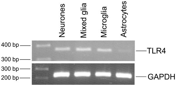
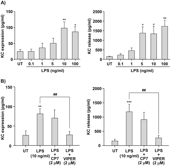
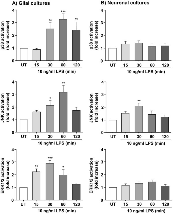
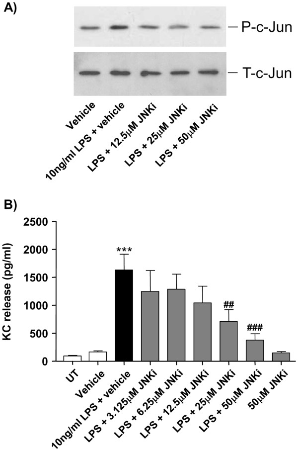
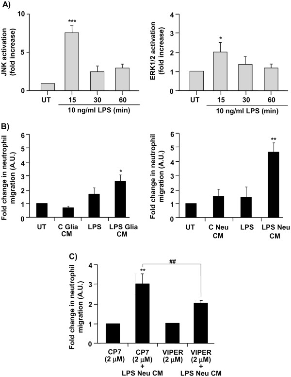
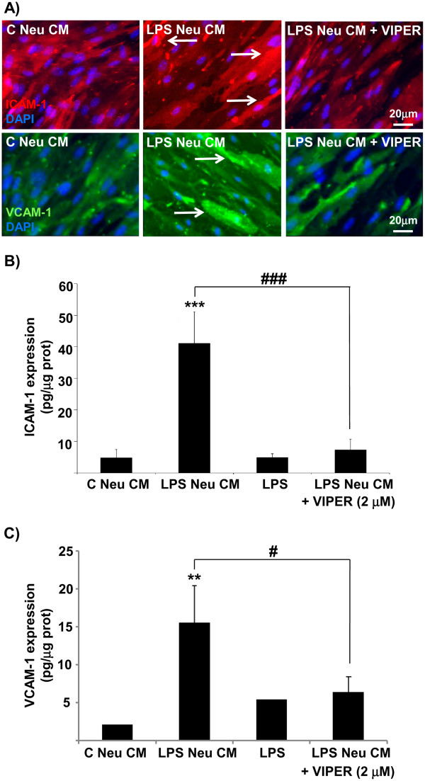
Methods: TLR4 mRNA expression in primary mouse glial and neuronal cultures was assessed by RT-PCR. Mouse mixed glial, neuronal or endothelial cell cultures were treated with LPS in the absence or the presence of a TLR4 specific antagonist (VIPER) or a specific JNK inhibitor (SP600125). Expression of inflammatory mediators was assayed by cytometric bead array (CBA) and ELISA. Activation of extracellular-signal regulated kinase 1/2 (ERK1/2), p38, c-Jun-N-terminal kinase (JNK) and c-Jun was assessed by Western blot. The effect of conditioned media of untreated- versus LPS-treated glial or neuronal cultures on endothelial activation was assessed by neutrophil transmigration assay, and immunocytochemistry and ELISA were used to measure expression of intercellular cell adhesion molecule (ICAM-1) and vascular cell adhesion molecule (VCAM-1).

Results: LPS induces strong release of the chemokines RANTES and CXCL1 (KC), tumor necrosis factor-α (TNFα) and IL-6 in primary mouse neuronal cultures. In contrast, LPS induced release of IL-1α, IL-1β and granulocyte-colony stimulating factor (G-CSF) in mixed glial, but not in neuronal cultures. LPS-induced neuronal KC expression and release were completely blocked by VIPER. In glial cultures, LPS induced activation of ERK1/2, p38 and JNK. In contrast, in neuronal cultures, LPS activated JNK but not ERK1/2 or p38, and the specific JNK inhibitor SP600125 significantly blocked LPS-induced KC expression and release. Finally, conditioned medium of LPS-treated neuronal cultures induced strong expression of ICAM-1 and VCAM-1 on endothelial cells, and induced infiltration of neutrophils across the endothelial monolayer, which was inhibited by VIPER.

Conclusion: These data demonstrate for the first time that neurons can play a role as key sensors of infection to initiate CNS inflammation.


Figures

Figure 1
Figure 1
Expression of TLR4 mRNA in neuronal and glial cell cultures. Neuronal, mixed glial, microglial and astrocytic primary cultures were analyzed by RT-PCR for expression of TLR4 and GAPDH mRNAs. cDNA amplification was visualized by agarose gel electrophoresis. Images shown are representative of three experiments carried out on separate cultures. GAPDH, glyceraldehyde 3-phosphate dehydrogenase; TLR4, Toll-like receptor 4.
Figure 2
Figure 2
Effect of LPS on KC expression and release in neuronal cultures. Neuronal primary cultures were left untreated (UT) or were treated with LPS (0.1 to 100 ng/ml) for 24 hours (a). Neuronal cultures were left untreated (UT) or were treated with LPS (10 ng/ml) for 24 hours, in the absence or the presence of the TLR4 antagonist VIPER or control peptide CP7 (2 μM) (b). Cell lysates and culture supernatants were assayed for KC levels by ELISA. Data are presented as mean ± SD of at least three independent experiments carried out on separate cultures. *P <0.05, **P <0.01, ***P <0.001, LPS-treated versus untreated cultures, ##P <0.01 LPS + VIPER versus. LPS alone, using one-way ANOVA and Tukey’s multiple comparison post-hoc test. ANOVA, analysis of variance; KC, CXCL1; LPS, lipopolysaccharide; TLR4, Toll-like receptor 4.
Figure 3
Figure 3
Effect of LPS on activation of p38, ERK1/2 and JNK MAPKs in neuronal and mixed glial cultures. Glial (a) or neuronal (b) primary cultures were left untreated (UT) or were treated with LPS (10 ng/ml) for 15, 30, 60 or 120 min. Cell lysates were assayed for p38, ERK1/2 and JNK activation by Western blot analysis. Levels of phosphorylated MAPKs were analyzed semi-quantitatively, normalized relative to total MAPKs, and presented as fold increase compared to untreated cultures. Data are presented as mean ± SD of at least three independent experiments carried out on separate cultures. *P <0.05, **P <0.01, ***P <0.001, LPS-treated versus untreated cultures, using one-way ANOVA and Tukey’s multiple comparison post-hoc test. ANOVA, analysis of variance; ERK1/2, extracellular-signal regulated kinase 1/2; JNK, c-Jun-N-terminal kinase; LPS, lipopolysaccharide; MAPK, mitogen-activated protein kinase.
Figure 4
Figure 4
Effect of JNK inhibition on c-Jun activity and LPS-induced KC release in neuronal cultures. Neuronal primary cultures were left untreated (UT), were treated with vehicle (DMSO), or were treated with LPS (10 ng/ml prepared in DMSO) for 30 min (for c-Jun activity, levels of phosphorylated (P-c-Jun) compared to total (T-c-Jun) c-Jun, a) or for 24 hours (for KC release, b) in the absence or the presence of increasing concentrations of a specific JNK inhibitor (SP600125, JNKi) added 30 min prior to treatment with LPS. Cell lysates were assayed for c-Jun activity by Western blot analysis. Image shown is representative of three experiments carried out on separate cultures (a). Culture supernatants were assayed for KC levels by ELISA (b). Data are presented as mean ± SD of at least three independent experiments carried out on separate cultures. **P <0.01, ***P <0.001, LPS + JNKi-treated versus LPS-treated cultures, using one-way ANOVA and Tukey’s multiple comparison post-hoc test. ANOVA, analysis of variance; DMSO, dimethyl sulfoxide; JNK, c-Jun-N-terminal kinase; KC, CXCL1; LPS, lipopolysaccharide.
Figure 5
Figure 5
Effect of LPS and conditioned medium of LPS-treated glial or neuronal cultures on MAPKs activation in endothelial cells and neutrophil transmigration. Endothelial primary cultures were left untreated (UT) or were treated with LPS (10 ng/ml) for 15, 30 or 60 min. Cell lysates were assayed for JNK and ERK1/2 activation by Western blot analysis. Levels of phosphorylated MAPKs were analyzed semi-quantitatively, normalized relative to total MAPKs, and represented as fold increase compared to untreated cultures (a). Endothelial primary cultures were left untreated (UT), were treated with LPS, or were treated with conditioned medium of untreated glial (C Glia CM) or neuronal cultures (C Neu CM), or conditioned medium of LPS (10 ng/ml for 24 hours)-treated glial (LPS Glia CM) or LPS (10 ng/ml for 24 hours)-treated neurons (LPS Neu CM), and neutrophil trans-endothelial migration was assayed (b). Endothelial primary cultures were treated with conditioned medium of neuronal cultures treated with VIPER or CP7 (2 μM) in the absence or the presence of LPS (10 ng/ml) (LPS Neu CM), and neutrophil trans-endothelial migration was assayed (c). Data are presented as mean ± SD of three independent experiments carried out on separate cultures. *P <0.05, **P <0.01, ***P <0.001 LPS-treated versus untreated cultures, ##P <0.01 LPS + VIPER versus LPS + CP7, using one-way ANOVA and Tukey’s multiple comparison post-hoc test. ANOVA, analysis of variance; ERK1/2, extracellular-signal regulated kinase1/2; JNK, c-Jun-N-terminal kinase; LPS, lipopolysaccharide; MAPK, mitogen-activated protein kinase.
Figure 6
Figure 6
Effect of neuronal LPS signaling on endothelial adhesion molecules expression. Endothelial primary cultures were treated with LPS (10 ng/ml), conditioned medium of untreated (C Neu CM) or LPS-treated (LPS Neu CM) neuronal cultures, in the absence or the presence of VIPER (2 μM). Cells were imaged for ICAM-1 and VCAM-1 expression by immunocytochemistry (a). Arrows show increased cellular expression of ICAM-1 and VCAM-1. Cell lysates were assayed for ICAM-1 expression and VCAM-1 expression (b) by ELISA. Data are presented as mean ± SD of three independent experiments carried out on separate cultures. **P <0.01, ***P <0.001, LPS-treated versus untreated cultures, #P <0.05, ###P <0.001, LPS + VIPER versus LPS alone, using one-way ANOVA and Tukey’s multiple comparison post-hoc test. ANOVA, analysis of variance; ICAM-1, intercellular cell adhesion molecule; LPS, lipopolysaccharide; VCAM-1, vascular cell adhesion molecule.

References

    1. Creagh EM, O'Neill LA. TLRs, NLRs and RLRs: a trinity of pathogen sensors that co-operate in innate immunity. Trends Immunol. 2006;27:352–357. doi: 10.1016/j.it.2006.06.003. - DOI - PubMed
    1. Bianchi ME. DAMPs, PAMPs and alarmins: all we need to know about danger. J Leukoc Biol. 2007;81:1–5. - PubMed
    1. Piccinini AM, Midwood KS. DAMPening inflammation by modulating TLR signalling. Mediators Inflamm. 2010. - DOI - PMC - PubMed
    1. Roach JC, Glusman G, Rowen L, Kaur A, Purcell MK, Smith KD, Hood LE, Aderem A. The evolution of vertebrate Toll-like receptors. Proc Natl Acad Sci U S A. 2005;102:9577–9582. doi: 10.1073/pnas.0502272102. - DOI - PMC - PubMed
    1. Bowie A, O'Neill LA. The interleukin-1 receptor/Toll-like receptor superfamily: signal generators for pro-inflammatory interleukins and microbial products. J Leukoc Biol. 2000;67:508–514. - PubMed

Publication types

Supplementary concepts