. 2012 Oct 11;490(7419):262-6.
doi: 10.1038/nature11466. Epub 2012 Oct 3.

Dopaminergic neurons inhibit striatal output through non-canonical release of GABA

Affiliations

Dopaminergic neurons inhibit striatal output through non-canonical release of GABA

Nicolas X Tritsch et al. Nature. .

Abstract

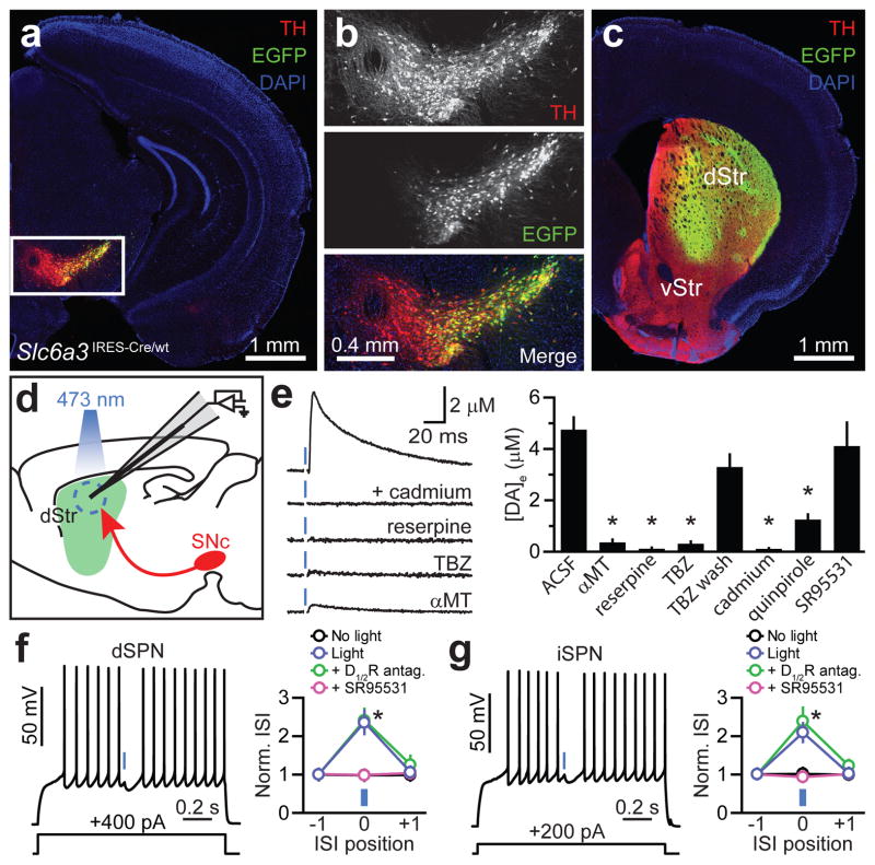
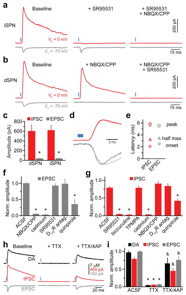
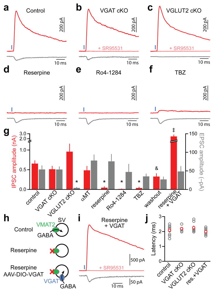
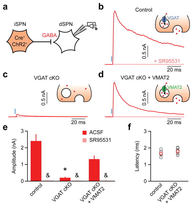
The substantia nigra pars compacta and ventral tegmental area contain the two largest populations of dopamine-releasing neurons in the mammalian brain. These neurons extend elaborate projections in the striatum, a large subcortical structure implicated in motor planning and reward-based learning. Phasic activation of dopaminergic neurons in response to salient or reward-predicting stimuli is thought to modulate striatal output through the release of dopamine to promote and reinforce motor action. Here we show that activation of dopamine neurons in striatal slices rapidly inhibits action potential firing in both direct- and indirect-pathway striatal projection neurons through vesicular release of the inhibitory transmitter GABA (γ-aminobutyric acid). GABA is released directly from dopaminergic axons but in a manner that is independent of the vesicular GABA transporter VGAT. Instead, GABA release requires activity of the vesicular monoamine transporter VMAT2, which is the vesicular transporter for dopamine. Furthermore, VMAT2 expression in GABAergic neurons lacking VGAT is sufficient to sustain GABA release. Thus, these findings expand the repertoire of synaptic mechanisms used by dopamine neurons to influence basal ganglia circuits, show a new substrate whose transport is dependent on VMAT2 and demonstrate that GABA can function as a bona fide co-transmitter in monoaminergic neurons.


Figures

Figure 1
Figure 1. DA neuron stimulation inhibits SPNs
a. Coronal midbrain section from a Slc6a3IRES-Cre/wt mouse transduced with a Cre-dependent EGFPAAV (green). DA neurons immunolabeled for TH (red). DAPI(blue), nuclear stain. b. Higher magnification of boxed area in a. Although expression levels vary greatly, the overwhelming majority of EGFP+ cells are TH+. c. EGFP+ SNc neurons densely innervate dorsal striatum (dStr). vStr; ventral striatum. d. Schematic of carbon-fiber recording configuration in a sagittal brain slice. ChR2+ DA neurons depicted red, laser illumination area blue. e. Left, light stimulation (blue) of DA terminals in dorsal striatum evoked cadmium-, reserpine-, TBZ-and αMT-sensitive DA release measured by amperometry. Stimulation artifacts blanked for clarity. Right, mean (n=4–19) peak extracellular DA concentration following optogenetic stimulation of nigrostriatal axons. DA transients recovered upon TBZ washout (wash). Asterisk, P<0.05vs. ACSF(Mann-Whitney). f, g. Left, membrane potential responses of a dorsal striatum dSPN(f) or iSPN(g) to current injection (1.3 s, bottom) and ChR2-mediated stimulation of DA axons(1 ms, blue). Right, average normalized duration of 3 consecutive interspike intervals (norm. ISI) that precede, straddle or follow the light flash(positions ‘−1’, ‘0’ and ‘+1’, respectively) upon ChR2 stimulation in ACSF (blue), DA receptor blockers (D1/2R antagonists = SCH23390 +SKF83566 + sulpiride +L-741,626; green) or SR95531(GABAA receptor blocker; purple). Black, light prevented from entering the sample. n=14dSPNs, 11iSPNs. Asterisk, P<0.05(two-way ANOVA). Data in e, f and g represent mean±s.e.m.
Figure 2
Figure 2. DA neurons directly release GABA onto SPNs
a. Evoked current responses from an iSPN held at indicated potentials (Vh) to optogenetic activation of nigrostriatal axons(1 ms, blue)upon sequential bath application ofSR95531 and NBQX/CPP. b. As in a for a dSPN with antagonists applied in reverse order. c. Mean IPSC (red) and EPSC (gray) absolute amplitudes in dSPNs (n=8) and iSPNs (n=21). Asterisk, P<0.05 vs. IPSC (Mann-Whitney). d. Normalized IPSCs and EPSCs from a (dark) and b (light) shown on an expanded time scale. Blue, light presentation. e. Average (n=29 SPNs) IPSC(red) and EPSC (gray) latencies from light onset to current onset (circle), half maximal amplitude (triangle) and peak amplitude (square). IPSCs are not delayed compared to EPSCs. f, g. Mean(n=3–18) EPSC(f) and IPSC(g) amplitudes under control conditions (ACSF) or in indicated antagonists normalized to baseline. Asterisk, P<0.05vs. ACSF (Mann-Whitney). h. Amperometric DA transients (black) and current responses of a voltage-clamped iSPN (Vh=0 mV, red; Vh=−70 mV, gray) to DA neuron stimulation under baseline conditions (left), in TTX (middle), and following co-application of TTX and 4AP (right). i. Extracellular DA concentration (black), IPSC amplitude (red) and EPSC amplitude (gray) evoked by ChR2 stimulation across conditions normalized to baseline. Data from ACSF condition same as in Figs. 1e and 2f, g. Asterisk, P<0.05vs. ACSF; ampersand, P<0.05 vs. TTX (Mann-Whitney). Error bars denote s.e.m.
Figure 3
Figure 3. GABA release from DA neurons requires VMAT2butnot VGAT
ac. Representative ChR2-evoked (1 ms, blue) IPSCs (red, Vh=0 mV) and EPSCs (gray, Vh=−70 mV) from SPNs in Slc6a3IRES-Cre/wt (control, a), Slc6a3IRES-Cre/wt;Slc32a1lox/lox (VGAT cKO, b), and Slc6a3IRES-Cre/wt;Slc17a6lox/lox (VGLUT2 cKO, c) mice. de. As in a for control Slc6a3IRES-Cre/wt mice treated in vivo and in vitro with the VMAT2 antagonists reserpine (d), Ro4-1284 (e) or TBZ (f). g. Mean IPSC (red) and EPSC (gray) amplitudes across conditions (n=4–33). Washout, slices obtained from mice treated in vivo with TBZ or Ro4-1284 and subsequently allowed to recover in vitro for >1 h in ACSF. Asterisk, P<0.05vs. ACSF; ampersand, P<0.05vs. TBZ/Ro4-1284; double dagger, P<0.05 vs. reserpine (Kruskal-Wallis). h. Schematic of working hypothesis: Provided DA neurons contain cytosolic GABA, viral expression of reserpine-resistant VGAT in DA neurons should rescue GABA transport into synaptic vesicles (SVs) and IPSCs in reserpine-treated mice. Note that VGAT might also incorporate into VMAT2 vesicles. i. Voltage-clamp recording (Vh=0 mV, red; Vh=−70 mV, gray) from a reserpine-treated iSPN upon optogenetic stimulation (1 ms, blue) of VGAT-expressing DA axons. j. IPSC onset latencies (gray, individual cells; red, average)did not differ across conditions. res, reserpine. GABAergic nature of outward currents (Vh=0 mV) in b, c and i was confirmed with SR95531 (pink). Error bars denote s.e.m.
Figure 4
Figure 4. VMAT2 functions as a vesicular GABA transporter
a. Experimental setup: ChR2 was selectively expressed in Cre-containing iSPNs of mice with one (control; Adora2A-Cre;Slc32a1lox/wt;Drd2-EGFP mice) or both alleles of the gene encoding VGAT flanked by lox sites (VGAT cKO; Adora2A-Cre;Slc32a1lox/lox;Drd2-EGFP mice). VGAT cKO + VMAT2, an AAV encoding Cre-dependent VMAT2 (AAV-DIO-VMAT2) was co-injected with AAV-DIO-ChR2 in the striatum of VGAT cKO mice to rescue GABA release from iSPNs. bd. Voltage-clamp recordings (Vh=0 mV) of axon-collateral IPSCs in dSPNs evoked by optogenetic stimulation (1 ms, blue) of iSPNs in the absence (red) or presence (pink) ofSR95531 in control (b), VGAT cKO (c) and VGAT cKO + VMAT2 (d) mice. Insets: iSPN presynaptic terminal schematic illustrating experimental conditions. Red triangles, GABA. e. Summary histogram (mean±s.e.m.) of experiments in bd (n=10–15 dSPNs). Asterisk, P<0.05vs. control and VGAT cKO+VMAT2 (Kruskal-Wallis); ampersand, P<0.05 vs. IPSC without SR95531 (Mann-Whitney). f. IPSC onset latencies from light presentation onset (gray, individual cells; red, mean±s.e.m.) in control (Adora2A-Cre;Slc32a1lox/wt;Drd2-EGFP mice) and VGAT cKO+VMAT2 mice (Adora2A-Cre;Slc32a1lox/lox;Drd2-EGFP mice transduced with AAV-DIO-VMAT2 in striatum).

Comment in

References

    1. Schultz W. Predictive reward signal of dopamine neurons. J Neurophysiol. 1998;80 (1):1–27. - PubMed
    1. Wickens JR, Reynolds JN, Hyland BI. Neural mechanisms of reward-related motor learning. Curr Opin Neurobiol. 2003;13 (6):685–690. - PubMed
    1. Gerfen CR, Surmeier DJ. Modulation of striatal projection systems by dopamine. Annu Rev Neurosci. 2011;34:441–466. - PMC - PubMed
    1. Palmiter RD. Dopamine signaling in the dorsal striatum is essential for motivated behaviors: lessons from dopamine-deficient mice. Ann N Y Acad Sci. 2008;1129:35–46. - PMC - PubMed
    1. Albin RL, Young AB, Penney JB. The functional anatomy of basal ganglia disorders. Trends Neurosci. 1989;12 (10):366–375. - PubMed

Publication types

MeSH terms

Substances